Podcast
Questions and Answers
What is the primary function of the myocardium in the heart?
What is the primary function of the myocardium in the heart?
- To provide the pumping power of the heart (correct)
- To house the coronary arteries and nerves
- To protect the heart from external impacts
- To facilitate the smooth flow of blood
Which layer of the heart is responsible for providing a smooth surface that directs blood flow?
Which layer of the heart is responsible for providing a smooth surface that directs blood flow?
- Epicardium
- Myocardium
- Pericardium
- Endocardium (correct)
What structure is unique to cardiac muscle cells that aids in electrical communication?
What structure is unique to cardiac muscle cells that aids in electrical communication?
- Intercalated discs (correct)
- Cisternae
- T tubules
- Sarcoplasmic reticulum
Which of the following correctly describes cardiomyocytes?
Which of the following correctly describes cardiomyocytes?
What advantage does the DIAD structure provide to cardiac muscle cells?
What advantage does the DIAD structure provide to cardiac muscle cells?
Which component of the intercalated discs provides mechanical strength to cardiac muscle cells?
Which component of the intercalated discs provides mechanical strength to cardiac muscle cells?
What type of muscle cell structure does cardiac muscle share with skeletal muscle?
What type of muscle cell structure does cardiac muscle share with skeletal muscle?
Which component serves as the calcium-storing part in the DIAD structure of cardiac muscle cells?
Which component serves as the calcium-storing part in the DIAD structure of cardiac muscle cells?
What is the primary function of the myocardium?
What is the primary function of the myocardium?
What role do intercalated discs play among cardiomyocytes?
What role do intercalated discs play among cardiomyocytes?
Which structure separates the atrial syncytium from the ventricular syncytium?
Which structure separates the atrial syncytium from the ventricular syncytium?
What is the firing frequency of the AV node?
What is the firing frequency of the AV node?
How does the heart's electrical conduction system initiate contraction?
How does the heart's electrical conduction system initiate contraction?
Which component of the heart conduction system has the slowest conduction speed?
Which component of the heart conduction system has the slowest conduction speed?
What type of muscle tissue does the myocardium resemble, and how does it function?
What type of muscle tissue does the myocardium resemble, and how does it function?
What is the primary rhythm generated by the SA node called?
What is the primary rhythm generated by the SA node called?
What occurs during the isovolumic relaxation stage?
What occurs during the isovolumic relaxation stage?
Which stage includes atrial contraction forcing blood into the ventricles?
Which stage includes atrial contraction forcing blood into the ventricles?
What defines tachycardia?
What defines tachycardia?
During which phase of the cardiac cycle do the semilunar valves open?
During which phase of the cardiac cycle do the semilunar valves open?
What typically happens to heart rate during sleep?
What typically happens to heart rate during sleep?
What is true about bradycardia?
What is true about bradycardia?
What happens to both systolic and diastolic durations when the heart rate increases?
What happens to both systolic and diastolic durations when the heart rate increases?
Which valves are closed during isovolumic contraction?
Which valves are closed during isovolumic contraction?
What effect does an increase in heart rate have on ventricular filling?
What effect does an increase in heart rate have on ventricular filling?
What is the formula for calculating cardiac output (CO)?
What is the formula for calculating cardiac output (CO)?
What sound is associated with the closure of the atrioventricular valves?
What sound is associated with the closure of the atrioventricular valves?
Which artery provides blood supply to the front and a significant part of the septum of the left ventricle?
Which artery provides blood supply to the front and a significant part of the septum of the left ventricle?
What change occurs to stroke volume when heart rate increases?
What change occurs to stroke volume when heart rate increases?
What is the resting membrane potential of cardiomyocytes during phase 4 of the cardiac action potential?
What is the resting membrane potential of cardiomyocytes during phase 4 of the cardiac action potential?
Which phase of the cardiac cycle is associated with the sound commonly referred to as 'lub'?
Which phase of the cardiac cycle is associated with the sound commonly referred to as 'lub'?
What is the primary function of the coronary arteries?
What is the primary function of the coronary arteries?
What happens to the pressure in the foot veins of a person who stands motionless for 30 seconds?
What happens to the pressure in the foot veins of a person who stands motionless for 30 seconds?
What is the primary function of the valves in the veins?
What is the primary function of the valves in the veins?
Which factor contributes to the development of varicose veins?
Which factor contributes to the development of varicose veins?
What is the venous pump, and how does it function?
What is the venous pump, and how does it function?
Which of the following systems is involved in long-term blood pressure regulation?
Which of the following systems is involved in long-term blood pressure regulation?
What occurs during Phase 0 of the action potential in cardiomyocytes?
What occurs during Phase 0 of the action potential in cardiomyocytes?
During which phase do calcium channels open in cardiomyocytes?
During which phase do calcium channels open in cardiomyocytes?
What is the primary role of the plateau phase in the cardiac action potential?
What is the primary role of the plateau phase in the cardiac action potential?
What characterizes the resting membrane potential of SA node cells compared to normal cardiomyocyte cells?
What characterizes the resting membrane potential of SA node cells compared to normal cardiomyocyte cells?
What is the main function of the funny Na+ leak channels in SA node cells?
What is the main function of the funny Na+ leak channels in SA node cells?
Which statement about veins is correct?
Which statement about veins is correct?
What is the threshold potential value's importance regarding cells?
What is the threshold potential value's importance regarding cells?
Which phase immediately follows the plateau phase in cardiomyocytes?
Which phase immediately follows the plateau phase in cardiomyocytes?
Flashcards
Myocardium
Myocardium
The middle layer of the heart responsible for pumping blood.
Cardiomyocytes
Cardiomyocytes
Specialized cells that make up the heart muscle.
Intercalated Discs
Intercalated Discs
Structures that connect cardiomyocytes, providing both electrical communication and mechanical strength.
Epicardium
Epicardium
Signup and view all the flashcards
Endocardium
Endocardium
Signup and view all the flashcards
DIAD (Dyadic Structure)
DIAD (Dyadic Structure)
Signup and view all the flashcards
T Tubule (Transverse Tubule)
T Tubule (Transverse Tubule)
Signup and view all the flashcards
Terminal Cisternae
Terminal Cisternae
Signup and view all the flashcards
Syncytium
Syncytium
Signup and view all the flashcards
Atria
Atria
Signup and view all the flashcards
Ventricles
Ventricles
Signup and view all the flashcards
SA Node (Sinoatrial Node)
SA Node (Sinoatrial Node)
Signup and view all the flashcards
AV Node (Atrioventricular Node)
AV Node (Atrioventricular Node)
Signup and view all the flashcards
Diastole
Diastole
Signup and view all the flashcards
Systole
Systole
Signup and view all the flashcards
Atrioventricular (AV) Valves
Atrioventricular (AV) Valves
Signup and view all the flashcards
Semilunar Valves
Semilunar Valves
Signup and view all the flashcards
Isovolumic Relaxation
Isovolumic Relaxation
Signup and view all the flashcards
Isovolumic Contraction
Isovolumic Contraction
Signup and view all the flashcards
Ejection Phase
Ejection Phase
Signup and view all the flashcards
Ventricular Filling
Ventricular Filling
Signup and view all the flashcards
Cardiac Output (CO)
Cardiac Output (CO)
Signup and view all the flashcards
Stroke Volume (SV)
Stroke Volume (SV)
Signup and view all the flashcards
Coronary Circulation
Coronary Circulation
Signup and view all the flashcards
First Heart Sound (S1)
First Heart Sound (S1)
Signup and view all the flashcards
Second Heart Sound (S2)
Second Heart Sound (S2)
Signup and view all the flashcards
Cardiac Action Potential
Cardiac Action Potential
Signup and view all the flashcards
Vein Pressure Increase While Standing
Vein Pressure Increase While Standing
Signup and view all the flashcards
Vein Valve Function
Vein Valve Function
Signup and view all the flashcards
Venous Pump
Venous Pump
Signup and view all the flashcards
Varicose Veins
Varicose Veins
Signup and view all the flashcards
Autonomic Nervous System
Autonomic Nervous System
Signup and view all the flashcards
Phase 0 (Depolarization)
Phase 0 (Depolarization)
Signup and view all the flashcards
Phase 1 (Early Repolarization)
Phase 1 (Early Repolarization)
Signup and view all the flashcards
Phase 2 (Plateau Phase)
Phase 2 (Plateau Phase)
Signup and view all the flashcards
Phase 3 (Repolarization)
Phase 3 (Repolarization)
Signup and view all the flashcards
SA Node Cells
SA Node Cells
Signup and view all the flashcards
Funny Channels (Na+ Leak Channels)
Funny Channels (Na+ Leak Channels)
Signup and view all the flashcards
Capacitance Vessels
Capacitance Vessels
Signup and view all the flashcards
Veins' Contractility & Relaxation
Veins' Contractility & Relaxation
Signup and view all the flashcards
Study Notes
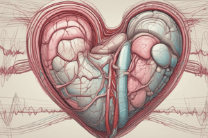
Cardiac Muscle
- Located in the middle layer of the heart, the myocardium
- Situated between the endocardium (inner layer) and the epicardium (outer layer)
- Muscle cells are called cardiomyocytes
- Cardiomyocytes are short, branched, and interconnected
- Interconnected via intercalated discs, facilitating electrical and mechanical communication
Cell Structure
- Cardiomyocytes are short, branched, and interconnected
- Connected by intercalated discs
- These discs provide electrical communication and mechanical connection between cells
Cardiac Muscle Layers
- Epicardium: The outermost layer of the heart, acting as a protective barrier and a passageway for coronary arteries and nerves
- Composed of thin connective tissue and mesothelium
- Myocardium: The middle and thickest layer; the main muscle layer responsible for pumping blood
- Contains striated muscle cells (cardiomyocytes) connected by intercalated disks
- Endocardium: The innermost layer; lines the heart chambers and valves, facilitating smooth blood flow
- Composed of thin connective tissue and endothelial cells
Cardiac Muscle Contraction Mechanism
- Contraction begins with Ca2+ ions entering the cell (calcium-induced calcium release).
- Intercalated discs transmit the action potential from the T tubule to the sarcoplasmic reticulum.
- This stimulates calcium release from the sarcoplasmic reticulum.
- Calcium released activates actin-myosin filaments, thereby initiating contraction.
- Cardiac muscle exhibits less regular and slower contraction dynamics compared to skeletal muscle, due to the difference in structure (DIAD vs. triad).
Cardiac Anatomy
- The heart is located in the middle mediastinum, at the level of thoracic vertebrae 15-18.
- It has four chambers (two atria and two ventricles)
- Right and left sides are separated by the interatrial and interventricular septa.
- The interventricular septum is thicker than the interatrial septum to generate more pressure during ventricular contraction.
- The right atrium receives blood from the superior and inferior vena cava.
Heart Valves
- Heart valves are one-way valves that allow blood flow in a single direction.
- Atrioventricular (AV) valves (tricuspid and bicuspid/mitral) control blood flow between atria and ventricles.
- Semilunar valves (aortic and pulmonary) control blood flow from the ventricles to the aorta and pulmonary arteries
- Their opening and closing depend passively on pressure gradients.
Heart Wall
- Three layers: endocardium, myocardium, and epicardium
- Myocardium is the heart muscle composed of cardiomyocytes with intercalated discs, facilitating rapid signal transmission.
- The rapid spread of this signal across the heart without damping is called a syncytium.
Heart Syncytia
- Two functional syncytia in the heart: atrial and ventricular
- Separated by a non-conductive fibrous tissue called the annulus fibrosus
- This separation allows the atria to contract before the ventricles
Heart Pacemaker Cells
- Sinoatrial (SA) node is the primary pacemaker (60-100 bpm) located near the entrance of the superior vena cava in the right atrium.
- Atrioventricular (AV) node is the secondary pacemaker (40-60 bpm) located in the interatrial septum behind the tricuspid valve.
Blood Pressure Regulation
- Maintaining blood pressure requires both short-term and long-term mechanisms.
- Short-term mechanisms involve neural control (e.g., baroreceptor reflex) adjusting blood vessel diameter (vasoconstriction/vasodilation).
- Long-term mechanisms involving hormonal pathways (e.g., Renin-angiotensin-aldosterone system) regulate blood volume and electrolyte balance.
Cardiac Output
- Cardiac output is the volume of blood pumped by the heart per minute (CO = SV x HR).
- Stroke Volume (SV): Volume of blood pumped per heartbeat.
- Heart Rate (HR): Number of heartbeats per minute.
- Normal resting adult human heart rate is 60-100 bpm.
Cardiac Action Potential
- Action potentials in cardiac cells have unique phases.
- Phase 4 is the resting potential (-90mV); only K+ channels are open
- Phase 0 is rapid depolarization caused by Na+ influx
- Phase 1 is early repolarization from K+ efflux
- Phase 2 is the plateau phase caused by Ca2+ influx and K+ efflux
- Phase 3 is repolarization from continued K+ efflux
SA Node Pacemaker Potential
- SA node cells are specialized cardiomyocytes with a resting membrane potential (-60 to -55mV).
- This is closer to the threshold potential, making them more excitable compared to other cardiomyocytes.
Funny Channels
- Funny channels (Na+ leak channels) in the SA node membrane contribute to maintaining a less negative resting membrane potential.
- They facilitate spontaneous depolarization, allowing the SA node to initiate the heart beat.
Venous Circulation
- Veins act as capacitance vessels, storing blood.
- Venous return is driven by the low pressure gradient towards the heart.
- Veins have valves to prevent backflow and skeletal muscle pumps aid venous return.
- Factors such as posture (standing) can affect venous pressure, leading to venous pooling or varicose veins.
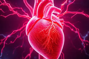
Coronary Circulation
- Coronary arteries supply oxygen and nutrients to the myocardium.
- The left coronary artery (LCA) branches into the left anterior descending (LAD) and circumflex arteries, supplying the left side of the heart.
- The right coronary artery (RCA) supplies the right side of the heart.
Abnormal Heart Rates
- Tachycardia: Heart rate above 100 bpm.
- Bradycardia: Heart rate below 60 bpm.
- Arrhythmia: Irregular heart beat patterns.
Studying That Suits You
Use AI to generate personalized quizzes and flashcards to suit your learning preferences.