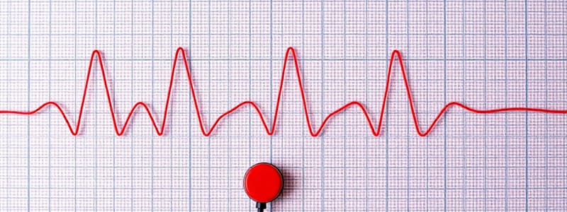
Podcast
Questions and Answers
What is represented by the P wave in an EKG?
What is represented by the P wave in an EKG?
- Ventricular depolarization
- Septal depolarization
- AV node delay
- Atrial depolarization (correct)
What happens during the PR interval of an EKG?
What happens during the PR interval of an EKG?
- Ventricular depolarization occurs
- Atrial depolarization occurs
- There is no net electrical vector movement (correct)
- The heart is in complete diastole
How does the Q wave appear on an EKG, and what does it represent?
How does the Q wave appear on an EKG, and what does it represent?
- Negative deflection, representing septal depolarization (correct)
- Positive deflection, representing atrial depolarization
- Positive deflection, representing ventricular repolarization
- Flat line, indicating AV node delay
Which lead is commonly used for EKG interpretation and has its positive electrode at the apex of the heart?
Which lead is commonly used for EKG interpretation and has its positive electrode at the apex of the heart?
What causes the R wave on an EKG?
What causes the R wave on an EKG?
What does a flat line on the EKG indicate?
What does a flat line on the EKG indicate?
What is the significance of pathological Q waves on an EKG?
What is the significance of pathological Q waves on an EKG?
Where does electrical activity travel after the AV node?
Where does electrical activity travel after the AV node?
What does the mean QRS vector primarily represent?
What does the mean QRS vector primarily represent?
Which waveform on the EKG specifically indicates depolarization of the ventricles?
Which waveform on the EKG specifically indicates depolarization of the ventricles?
What does the ST segment represent on an EKG?
What does the ST segment represent on an EKG?
Which EKG lead provides a view of the heart from the inferior perspective?
Which EKG lead provides a view of the heart from the inferior perspective?
What is the axis of Lead I on an EKG?
What is the axis of Lead I on an EKG?
What constitutes Einthoven's Law?
What constitutes Einthoven's Law?
What view does the augmented unipolar limb lead aVF provide?
What view does the augmented unipolar limb lead aVF provide?
Which leads are primarily used to assess the electrical activity of the interventricular septum?
Which leads are primarily used to assess the electrical activity of the interventricular septum?
What direction does the mean QRS vector typically point?
What direction does the mean QRS vector typically point?
During which phase does the T wave occur on the EKG?
During which phase does the T wave occur on the EKG?
Which of the following leads would show a significant indication of a posterior myocardial infarction (MI)?
Which of the following leads would show a significant indication of a posterior myocardial infarction (MI)?
What does the PR interval measure on the EKG?
What does the PR interval measure on the EKG?
Flashcards
Electrocardiogram (EKG)
Electrocardiogram (EKG)
A graphic representation of the electrical activity in the heart, recorded by electrodes placed on the body.
Electrodes and Electrical Activity
Electrodes and Electrical Activity
Electrodes placed on the body pick up electrical signals from the heart. If the electrical activity is moving towards the positive electrode, there will be a positive deflection on the EKG. If the electrical activity is moving away from the positive electrode, there will be a negative deflection.
Lead 2
Lead 2
A common lead used for EKG interpretation. The positive electrode is placed at the apex of the heart. The negative electrode is placed at the base of the heart.
Atrial Depolarization (P Wave)
Atrial Depolarization (P Wave)
Signup and view all the flashcards
AV Node Delay (PR Interval)
AV Node Delay (PR Interval)
Signup and view all the flashcards
Interventricular Septum Depolarization (Q Wave)
Interventricular Septum Depolarization (Q Wave)
Signup and view all the flashcards
Ventricular Depolarization (QRS Complex)
Ventricular Depolarization (QRS Complex)
Signup and view all the flashcards
Key EKG Wave Forms
Key EKG Wave Forms
Signup and view all the flashcards
Mean QRS Vector
Mean QRS Vector
Signup and view all the flashcards
ST Segment
ST Segment
Signup and view all the flashcards
Q wave
Q wave
Signup and view all the flashcards
R wave
R wave
Signup and view all the flashcards
S wave
S wave
Signup and view all the flashcards
T Wave
T Wave
Signup and view all the flashcards
P Wave
P Wave
Signup and view all the flashcards
Bipolar Limb Leads
Bipolar Limb Leads
Signup and view all the flashcards
Precordial Leads
Precordial Leads
Signup and view all the flashcards
Combined EKG Leads
Combined EKG Leads
Signup and view all the flashcards
PR Interval
PR Interval
Signup and view all the flashcards
Augmented Unipolar Limb Leads
Augmented Unipolar Limb Leads
Signup and view all the flashcards
aVR
aVR
Signup and view all the flashcards
aVL
aVL
Signup and view all the flashcards
aVF
aVF
Signup and view all the flashcards
Study Notes
Cardiac Conduction System and EKG Basics
-
Electrodes and Electrical Activity:
- EKG is a graphical representation of electrical activity in the heart.
- Electrodes placed on the body pick up electrical signals from the heart.
- Positive electrode picks up positive deflection when electrical activity moves towards it.
- Positive electrode picks up negative deflection when electrical activity moves away from it.
- A flat line indicates slow conduction or electrical activity moving perpendicular to the lead axis.
-
Lead 2:
- A common lead used for EKG interpretation.
- Positive electrode placed at the apex of the heart.
- Negative electrode placed at the base of the heart.
-
Atrial Depolarization (P Wave):
- SA node initiates electrical activity, moving towards the AV node.
- This movement is in the direction of the positive electrode in Lead 2.
- Produces a positive deflection on the EKG, known as the P wave.
-
AV Node Delay (PR Interval):
- AV node is a slow conductor, delaying transmission of electrical potentials for about 0.1 seconds.
- No net electrical vector movement during this time.
- Results in a flat line on the EKG, known as the PR interval.
- PR interval is crucial for diagnosing heart blocks based on its duration.
-
Interventricular Septum Depolarization (Q Wave):
- Electrical activity travels from the AV node to the bundle of His and then down the bundle branches.
- The left bundle branch depolarizes the interventricular septum, moving from left to right.
- This movement is opposite to the Lead 2 axis, creating a negative deflection on the EKG, called the Q wave.
-
Ventricular Depolarization (QRS Complex):
- After septum depolarization, electrical activity spreads through the Purkinje fibers, depolarizing the ventricles.
- Left ventricle has a larger muscle mass and generates a stronger electrical signal than the right ventricle.
- The net ventricular depolarization vector points downwards and slightly to the left, in the direction of the positive electrode in Lead 2.
- This produces a positive deflection on the EKG known as the R wave.
Key Points to Remember
- Q wave represents septal depolarization.
- R wave represents ventricular depolarization.
- P wave represents atrial depolarization.
- PR interval represents the delay in the AV node.
- Pathological Q waves indicate possible myocardial infarction, a topic for further study.
Mean QRS Vector
- The mean QRS vector represents the combined electrical activity of the left and right ventricles.
- The left ventricle has a thicker myocardium, generating more electrical activity and contributing to a larger vector magnitude.
- The mean QRS vector typically points downwards and slightly leftward, towards the positive electrode of lead II.
- This downward direction results in a large positive deflection in the EKG.
EKG Waveforms & Ventricular Depolarization
- P wave: Represents depolarization from the SA node to the AV node.
- PR interval: Represents the time it takes for the AV node to conduct the electrical impulse.
- Q wave: Represents depolarization of the septum, moving from left to right and slightly upwards.
- R wave: The largest positive deflection, indicates ventricular depolarization, particularly the apex of the ventricles.
- S wave: A negative deflection, indicates depolarization of the bases of the ventricles, as electrical activity moves upwards from the apex.
ST Segment
- Represents the period when the entire ventricular myocardium is depolarized but not yet repolarized.
- Characterized by a flat, isoelectric line on the EKG.
- There is no net electrical vector during the ST segment.
T Wave
- Represents the repolarization of the ventricles.
- The negative charge moving upwards within the ventricles results in a positive deflection on the EKG.
- As negative charges move away from the positive electrode, a positive deflection arises.
EKG Leads & Plane Projection
- Bipolar limb leads (Lead I, II, III): Measure electrical activity in the frontal plane, viewing the heart in an anterior-posterior direction.
- Augmented unipolar limb leads (aVR, aVL, aVF): Also measure frontal plane activity. Providing a frontal plane view.
- Precordial leads (V1-V6): Measure electrical activity in the horizontal plane (transverse plane), viewing the heart in a superior-inferior direction.
Bipolar Limb Leads: Placement & Axis
- Lead I: Negative electrode on the right arm, positive electrode on the left arm.
- Lead II: Negative electrode on the right arm, positive electrode on the left leg.
- Lead III: Negative electrode on the left arm, positive electrode on the left leg.
- Each lead has a specific axis that defines its direction of measurement.
The Basics of Electrocardiograms
- Electrocardiograms (ECGs/EKGs) are a valuable tool for assessing the electrical activity of the heart.
- Bipolar Limb Leads: Placed on the right arm, left arm, and left leg to visualize heart activity from various angles.
- Lead 1: Left side; positive on left arm, negative on right arm.
- Lead 2: Bottom; positive on left leg, negative on right arm.
- Lead 3: Bottom; positive on left leg, negative on left arm.
- Einthoven's Law: Lead 1 + Lead 3 = Lead 2.
- Augmented Unipolar Limb Leads: Generate negative charges at two points and a positive electrode at the third, creating a vector between the negative electrodes.
- AVR: Right side of the heart.
- AVL: Left and lateral view of the heart.
- AVF: Inferior view of the heart.
- Chest Leads (V1-V6): Unipolar leads with a single positive electrode placed at specific locations on the chest wall. Used to obtain horizontal plane views.
- V1: Right fourth intercostal space, parasternal side.
- V2: Left fourth intercostal space, parasternal side.
- V4: Fifth intercostal space, mid-clavicular line.
- V3: Between V2 and V4.
- V5: Fifth intercostal space, anterior axillary line.
- V6: Fifth intercostal space, mid-axillary line.
- Chest Leads Interpretation:
- V1 and V2: Primarily reflect activity from the interventricular septum.
- V3 and V4: Primarily reflect activity from the anterior wall of the ventricles.
- V5 and V6: Primarily reflect activity from the left lateral wall of the ventricles.
- V1 and V2 posterior MI: ST segment depression and T-wave inversion can suggest a posterior myocardial infarction, requiring additional posterior leads (V7, V8, V9) for confirmation.
The Different Viewing Planes used in EKGs
- Bipolar Limb Leads: Frontal plane view.
- Augmented Unipolar Limb Leads: Frontal plane view.
- Chest Leads: Horizontal plane (transverse plane) view.
- Combining all leads provides a comprehensive picture of heart's electrical activity.
Studying That Suits You
Use AI to generate personalized quizzes and flashcards to suit your learning preferences.


