Podcast
Questions and Answers
What is the main function of the myocardium?
What is the main function of the myocardium?
- It separates the heart chambers.
- It encloses the heart.
- It protects the heart from infection.
- It contracts to pump blood. (correct)
The tricuspid valve is located between the left atrium and the left ventricle.
The tricuspid valve is located between the left atrium and the left ventricle.
False (B)
What are the atrioventricular valves?
What are the atrioventricular valves?
The mitral valve and the tricuspid valve.
The outermost layer of the heart is called the ______.
The outermost layer of the heart is called the ______.
Match the following heart valves with their descriptions:
Match the following heart valves with their descriptions:
Which of the following layers is the thickest in the heart?
Which of the following layers is the thickest in the heart?
The pericardial sac has two layers, the parietal pericardium and the risk visceral pericardium.
The pericardial sac has two layers, the parietal pericardium and the risk visceral pericardium.
The amount of fluid in the pericardial space is about ______ milliliters.
The amount of fluid in the pericardial space is about ______ milliliters.
What is the primary function of the fluid in the pericardial space?
What is the primary function of the fluid in the pericardial space?
The order of the layers of the heart from inside to outside is the epicardium, myocardium, endocardium, and parietal pericardium.
The order of the layers of the heart from inside to outside is the epicardium, myocardium, endocardium, and parietal pericardium.
What are the blood vessels that supply oxygen and nutrients to the heart muscle called?
What are the blood vessels that supply oxygen and nutrients to the heart muscle called?
The space between the parietal pericardium and visceral pericardium contains approximately ______ milliliters of fluid.
The space between the parietal pericardium and visceral pericardium contains approximately ______ milliliters of fluid.
Match the following components of the heart with their functions:
Match the following components of the heart with their functions:
Where do the coronary arteries originate?
Where do the coronary arteries originate?
The left and right coronary arteries are located on the inside of the heart.
The left and right coronary arteries are located on the inside of the heart.
What is the term for the small branches that come off the coronary arteries?
What is the term for the small branches that come off the coronary arteries?
What is the role of the superior and inferior vena cava?
What is the role of the superior and inferior vena cava?
The left side of the heart contains deoxygenated blood.
The left side of the heart contains deoxygenated blood.
What are the two phases of the heart's cycle?
What are the two phases of the heart's cycle?
The blood travels from the right atrium through the _______ valve into the right ventricle.
The blood travels from the right atrium through the _______ valve into the right ventricle.
Match the following cardiac structures with their functions:
Match the following cardiac structures with their functions:
Which valve is also known as the bicuspid valve?
Which valve is also known as the bicuspid valve?
The pulmonary veins carry deoxygenated blood to the heart.
The pulmonary veins carry deoxygenated blood to the heart.
What color is the blood represented on the right side of the heart, and why?
What color is the blood represented on the right side of the heart, and why?
What is the purpose of the atrial kick?
What is the purpose of the atrial kick?
The tricuspid and mitral valves remain closed when the ventricles contract.
The tricuspid and mitral valves remain closed when the ventricles contract.
What are the two phases of the heart's systolic cycle?
What are the two phases of the heart's systolic cycle?
The blood from the right ventricle is forced into the ________ through the pulmonic valve.
The blood from the right ventricle is forced into the ________ through the pulmonic valve.
Match the following heart valves to their functions:
Match the following heart valves to their functions:
What percentage of blood in the ventricles comes from the atria during the diastolic phase?
What percentage of blood in the ventricles comes from the atria during the diastolic phase?
The left atrium receives blood from the pulmonary artery.
The left atrium receives blood from the pulmonary artery.
What happens when the atria contract?
What happens when the atria contract?
What causes the 'dub' sound in the heart?
What causes the 'dub' sound in the heart?
The normal stroke volume in adults is 50 milliliters.
The normal stroke volume in adults is 50 milliliters.
What is cardiac output?
What is cardiac output?
The normal heart rate in adults ranges from _____ beats per minute.
The normal heart rate in adults ranges from _____ beats per minute.
If the heart rate is 74 beats per minute and the stroke volume is 70 milliliters, what is the cardiac output?
If the heart rate is 74 beats per minute and the stroke volume is 70 milliliters, what is the cardiac output?
Preload is the amount of blood that fills the ventricles before they _____ .
Preload is the amount of blood that fills the ventricles before they _____ .
Cardiac output is unrelated to blood pressure.
Cardiac output is unrelated to blood pressure.
Match the following terms with their definitions:
Match the following terms with their definitions:
What is the main purpose of fluoroscopy in cardiac catheterization?
What is the main purpose of fluoroscopy in cardiac catheterization?
A total blockage in the coronary artery will show a visible pathway when contrast dye is injected.
A total blockage in the coronary artery will show a visible pathway when contrast dye is injected.
In balloon angioplasty, a _______ is inflated and deflated to break the plaque.
In balloon angioplasty, a _______ is inflated and deflated to break the plaque.
What is injected to visualize blockages during cardiac catheterization?
What is injected to visualize blockages during cardiac catheterization?
Only inpatient cardiac catheterizations require blood tests before the procedure.
Only inpatient cardiac catheterizations require blood tests before the procedure.
What should be monitored in patients undergoing cardiac catheterization due to the use of contrast dye?
What should be monitored in patients undergoing cardiac catheterization due to the use of contrast dye?
Flashcards
Heart Chambers
Heart Chambers
The heart has four chambers: right atrium, right ventricle, left atrium, and left ventricle.
Heart Valves
Heart Valves
The heart valves control blood flow through the chambers.
Tricuspid Valve
Tricuspid Valve
Located between the right atrium and right ventricle.
Mitral/Bicuspid Valve
Mitral/Bicuspid Valve
Signup and view all the flashcards
Semilunar Valves
Semilunar Valves
Signup and view all the flashcards
Myocardium
Myocardium
Signup and view all the flashcards
Heart Layers
Heart Layers
Signup and view all the flashcards
Percardial Sac
Percardial Sac
Signup and view all the flashcards
Pericardial fluid function
Pericardial fluid function
Signup and view all the flashcards
Heart layers (inside to outside)
Heart layers (inside to outside)
Signup and view all the flashcards
Coronary arteries
Coronary arteries
Signup and view all the flashcards
Coronary veins
Coronary veins
Signup and view all the flashcards
Coronary artery origin
Coronary artery origin
Signup and view all the flashcards
Blood flow pathway
Blood flow pathway
Signup and view all the flashcards
Heart Physiology
Heart Physiology
Signup and view all the flashcards
Coronary Artery Branches
Coronary Artery Branches
Signup and view all the flashcards
Superior Vena Cava
Superior Vena Cava
Signup and view all the flashcards
Inferior Vena Cava
Inferior Vena Cava
Signup and view all the flashcards
Right Atrium
Right Atrium
Signup and view all the flashcards
Pulmonary Artery
Pulmonary Artery
Signup and view all the flashcards
Pulmonary Veins
Pulmonary Veins
Signup and view all the flashcards
Left Atrium
Left Atrium
Signup and view all the flashcards
Diastole
Diastole
Signup and view all the flashcards
Systole
Systole
Signup and view all the flashcards
Fluoroscopy
Fluoroscopy
Signup and view all the flashcards
Cardiac Catheterization
Cardiac Catheterization
Signup and view all the flashcards
Blood flow through the heart
Blood flow through the heart
Signup and view all the flashcards
Atrial Systole
Atrial Systole
Signup and view all the flashcards
PCI (Percutaneous Coronary Intervention)
PCI (Percutaneous Coronary Intervention)
Signup and view all the flashcards
PTCA (Percutaneous Transluminal Coronary Angioplasty)
PTCA (Percutaneous Transluminal Coronary Angioplasty)
Signup and view all the flashcards
Ventricular Systole
Ventricular Systole
Signup and view all the flashcards
Atrial Kick
Atrial Kick
Signup and view all the flashcards
Contrast Dye
Contrast Dye
Signup and view all the flashcards
Pre-Procedure Nursing Interventions
Pre-Procedure Nursing Interventions
Signup and view all the flashcards
Tricuspid and Mitral/Bicuspid Valves
Tricuspid and Mitral/Bicuspid Valves
Signup and view all the flashcards
Post-Procedure Nursing Interventions
Post-Procedure Nursing Interventions
Signup and view all the flashcards
Pulmonary and Aortic Valves
Pulmonary and Aortic Valves
Signup and view all the flashcards
Heart Valves Importance
Heart Valves Importance
Signup and view all the flashcards
Kidney Function Tests
Kidney Function Tests
Signup and view all the flashcards
Heart Contraction Cycle
Heart Contraction Cycle
Signup and view all the flashcards
Stroke Volume
Stroke Volume
Signup and view all the flashcards
Cardiac Output
Cardiac Output
Signup and view all the flashcards
Heart Rate
Heart Rate
Signup and view all the flashcards
Preload
Preload
Signup and view all the flashcards
Afterload
Afterload
Signup and view all the flashcards
Tricuspid and Mitral Valves
Tricuspid and Mitral Valves
Signup and view all the flashcards
Aortic and Pulmonic Valves
Aortic and Pulmonic Valves
Signup and view all the flashcards
Contractility
Contractility
Signup and view all the flashcards
Study Notes
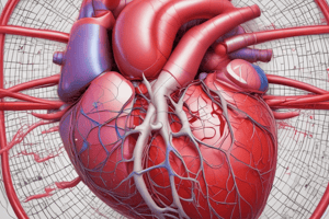
Cardiac Anatomy and Physiology
- The heart has four chambers: right atrium, right ventricle, left atrium, and left ventricle.
- Valves separate the atria and ventricles (tricuspid and mitral valves), and the ventricles from the arteries (pulmonary and aortic valves).
- The heart muscle has three layers: epicardium (outer), myocardium (middle, muscular), and endocardium (inner).
- The myocardium is the thickest layer, responsible for contraction.
- The pericardium surrounds the heart, containing fluid to cushion and lubricate.
- Coronary arteries supply oxygen and nutrients to the heart muscle and coronary veins return deoxygenated blood.
Heart Valves
- Tricuspid valve: between right atrium and right ventricle
- Mitral (bicuspid) valve: between left atrium and left ventricle
- Pulmonary valve: between right ventricle and pulmonary artery
- Aortic valve: between left ventricle and aorta
Blood Flow Through the Heart
- Blood enters the right atrium from the superior and inferior vena cava.
- Blood flows through the tricuspid valve to the right ventricle.
- Blood is pumped through the pulmonary valve into the pulmonary artery to the lungs.
- Blood returns from the lungs via the pulmonary veins to the left atrium.
- Blood flows through the mitral valve to the left ventricle.
- Blood is pumped through the aortic valve into the aorta and to the rest of the body.
- Atrial contraction contributes about 25% of the blood flow to the ventricles.
Cardiac Cycle
- Diastole: relaxation phase, filling of the chambers
- Systole: contraction phase, ejection of blood
- Atrial systole: atrial contraction, forces blood into ventricles
- Ventricular systole: ventricular contraction, forces blood into arteries
- Lub-dub sounds: closing of the heart valves
Cardiac Output
- Stroke volume: amount of blood pumped per heartbeat (typically 70 mL)
- Heart rate: number of heartbeats per minute (typically 60-100 bpm)
- Cardiac output: amount of blood pumped per minute (stroke volume x heart rate)
Studying That Suits You
Use AI to generate personalized quizzes and flashcards to suit your learning preferences.