Podcast
Questions and Answers
Which of the following is the primary function of the detrusor muscle arrangement in the bladder?
Which of the following is the primary function of the detrusor muscle arrangement in the bladder?
- To allow the bladder to change shape in response to external pressure.
- To provide structural integrity allowing the bladder to stretch in any direction. (correct)
- To facilitate voluntary control over bladder contractions.
- To enable the bladder to contract only in one direction.
The internal urethral sphincter is made of what type of muscle and controlled by which nervous system?
The internal urethral sphincter is made of what type of muscle and controlled by which nervous system?
- Smooth muscle, autonomic nervous system (correct)
- Skeletal muscle, autonomic nervous system
- Skeletal muscle, somatic nervous system
- Smooth muscle, somatic nervous system
What is the main function of the external urethral sphincter?
What is the main function of the external urethral sphincter?
- Voluntary control of urine flow (correct)
- Sensing bladder fullness
- Involuntary control of urine flow
- Initiating the micturition reflex
What is the collective function of the nervous system concerning the lower urinary tract?
What is the collective function of the nervous system concerning the lower urinary tract?
During the storage phase of the lower urinary tract function, which of the following events occurs?
During the storage phase of the lower urinary tract function, which of the following events occurs?
Which nerve is responsible for causing the detrusor muscle to contract, and what is the primary neurotransmitter involved in this process?
Which nerve is responsible for causing the detrusor muscle to contract, and what is the primary neurotransmitter involved in this process?
The stimulation of Beta 3 ($\beta_3$) receptors results in what?
The stimulation of Beta 3 ($\beta_3$) receptors results in what?
Which nerve is responsible for the voluntary contraction of the external urethral sphincter?
Which nerve is responsible for the voluntary contraction of the external urethral sphincter?
What is the primary purpose of the urethral sphincter in men?
What is the primary purpose of the urethral sphincter in men?
Which of the following events occurs during the normal voiding reflex?
Which of the following events occurs during the normal voiding reflex?
What leads to the urge to urinate?
What leads to the urge to urinate?
What is the role of the cerebral cortex in micturition?
What is the role of the cerebral cortex in micturition?
Which scenario is an example of the brain's continence centers at work?
Which scenario is an example of the brain's continence centers at work?
What is the composition of the detrusor muscle?
What is the composition of the detrusor muscle?
What is the effect of sympathetic nervous system activation on the internal urethral sphincter?
What is the effect of sympathetic nervous system activation on the internal urethral sphincter?
Where does the spinal motor outflow controlling the external urethral sphincter originate?
Where does the spinal motor outflow controlling the external urethral sphincter originate?
During bladder filling, what happens to intravesicular pressure, and why?
During bladder filling, what happens to intravesicular pressure, and why?
What is the combined effect of detrusor muscle contraction and external urethral sphincter relaxation?
What is the combined effect of detrusor muscle contraction and external urethral sphincter relaxation?
Through which type of receptors does acetylcholine (ACh) primarily cause the detrusor muscle to contract?
Through which type of receptors does acetylcholine (ACh) primarily cause the detrusor muscle to contract?
Which component of the nervous system must be reduced to allow for complete voiding?
Which component of the nervous system must be reduced to allow for complete voiding?
Flashcards
Internal Urethral Sphincter
Internal Urethral Sphincter
Primary muscle of continence; continuation of the Detrusor muscle made of smooth muscle at the bladder neck.
External Urethral Sphincter
External Urethral Sphincter
Anatomical sphincter consisting of localized circular muscle thickening derived from pelvic floor muscles, under somatic, voluntary control.
Detrusor Urinae Muscle
Detrusor Urinae Muscle
A meshwork of muscle fibres in roughly 3 layers: inner longitudinal, middle circular, outer longitudinal. Supplied by the autonomic nervous system.
Functions of Innervation
Functions of Innervation
Signup and view all the flashcards
Urinary Tract Activity Phases
Urinary Tract Activity Phases
Signup and view all the flashcards
Parasympathetic Stimulation in Voiding
Parasympathetic Stimulation in Voiding
Signup and view all the flashcards
Micturition Centres
Micturition Centres
Signup and view all the flashcards
Full Bladder Threshold
Full Bladder Threshold
Signup and view all the flashcards
Continence Centres
Continence Centres
Signup and view all the flashcards
Sympathetic Stimulation in Storage
Sympathetic Stimulation in Storage
Signup and view all the flashcards
Study Notes
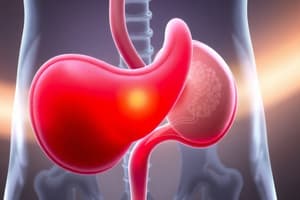
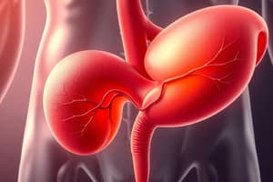
The Anatomy of the Bladder
- The body of the bladder temporarily stores urine
- The neck connects the bladder to the urethra
- The trigone consists of ureteric orifices and an internal urethral orifice at the angles of a triangle
- The detrusor muscle is made from a meshwork of muscle fibres in roughly 3 layers
- Fibres are arranged in inner longitudinal, middle circular, and outer longitudinal layers
- This arrangement provides bladder strength regardless of the direction it's stretched
- The detrusor muscle receives spinal nerve supply bilaterally, and is supplied by the autonomic nervous system, so is not under voluntary control
- The internal urethral sphincter is a continuation of the detrusor muscle, made of smooth muscle
- The internal urethral sphincter is a physiological sphincter at the bladder neck
- The internal urethral sphincter is a physiological sphincter with no muscle thickening - action is due to structure
- The internal urethral sphincter is the primary muscle of continence
- The external urethral sphincter is an anatomical sphincter of localised circular muscle thickening for action
- The external urethral sphincter derived from pelvic floor muscles
- The external urethral sphincter is skeletal muscle, under somatic, voluntary control
- The external urethral sphincter contracts to constrict the urethra and "hold in” urine
Innervation of the Bladder
- The nervous system provides sensations of bladder filling and pain
- The nervous system allows the bladder to relax and accommodate increasing volumes of urine
- The nervous system initiates and maintains voiding so the bladder empties completely, with minimal residual volume
- The nervous system provides integrated regulation of the smooth and skeletal muscle sphincters of the urethra.
- Lower urinary tract activity is divided into two phases: filling and voiding
- During filling, the bladder relaxes and accommodates increasing urine volumes, while urethral sphincters increase tone to maintain continence
- During voiding, the urethral sphincters relax, and the bladder contracts.
- The extent of bladder contraction is greater in men
- In men, the prostatic urethra functions to prevent retrograde ejaculation into the bladder.
- Pelvic nerves perform parasympathetic functions (S2-S4)
- Ach M3 receptors cause contraction
- Hypogastric nerves perform sympathetic functions (T10-L2)
- NA B3 receptors cause relaxation
- Hypogastric nerves (T10-L2): NA a₁ receptors cause contraction of the internal urethral sphincter
- The contraction of the internal urethral sphincter is a sympathetic function
- Pudendal nerves perform somatic functions (S2-S4)
- Spinal motor outflow comes from Onof's Nucleus of the ventral horn of the cord
- Ach Nicotinic receptors cause contraction of the external urethral sphincter
The Normal Voiding Reflex
- The threshold for feeling a full bladder is ~400ml
- When the bladder is full, an urge to urinate arises
- Brain and Spinal micturition centres control normal voiding reflex
- Parasympathetic neurons are involved in micturition
- Increased parasympathetic stimulation to the bladder via the Pelvic nerve causes the Detrusor to contract, and increase intravesicular pressure
- The Cerebral Cortex makes an executive decision to urinate, reducing somatic stimulation to the External Urethral Sphincter
- Detrusor contraction coupled with the relaxation of the External Urethral Sphincter results in the bladder emptying through the urethra.
- Ureters, urinary bladder, and internal and external urethral sphincters work together to pass and store urine in the urinary bladder
- Bladder walls have many folds, and distend when filling with urine, so intravesicular pressure hardly changes as the bladder fills.
- At around 400ml of bladder filling, afferent nerves from the bladder wall signal the need to void (pain/temperature sensation), with possible stretch receptors
- Active Brain and Spinal Continence Centres and Sympathetic Neurons increase sympathetic stimulation to the bladder via the hypogastric nerve
- That stimulation causes the Detrusor to relax, and the Internal Urethral Sphincter to contract
- The Cerebral Cortex decides not to urinate, and increases somatic stimulation to the External Urethral Sphincter
- Increasing stimulation causes the External Urethral Sphincter contracts, constricting the urethra
- The relaxation of the Detrusor, and the simultaneous contraction of the Internal and External Urethral sphincters reduces intravesicular pressure, constricting the urethra and preventing micturition.
Studying That Suits You
Use AI to generate personalized quizzes and flashcards to suit your learning preferences.