Podcast
Questions and Answers
Blood is best classified as which type of tissue?
Blood is best classified as which type of tissue?
- Nervous
- Connective (correct)
- Muscle
- Epithelial
Which of the following is NOT a primary function of blood?
Which of the following is NOT a primary function of blood?
- Distributing nutrients
- Producing hormones (correct)
- Stabilizing body temperature
- Transporting dissolved gases
The process of blood cell formation is called:
The process of blood cell formation is called:
- Histogenesis
- Homeostasis
- Hemostasis
- Hematopoiesis (correct)
What are the two major components of blood?
What are the two major components of blood?
Which of the following formed elements is responsible for transporting oxygen?
Which of the following formed elements is responsible for transporting oxygen?
The clotting reaction in blood primarily functions to:
The clotting reaction in blood primarily functions to:
Which blood type is known as the universal donor?
Which blood type is known as the universal donor?
Defense against toxins and pathogens is a function of blood primarily carried out by:
Defense against toxins and pathogens is a function of blood primarily carried out by:
Which of the following blood components is responsible for producing antibodies?
Which of the following blood components is responsible for producing antibodies?
Which formed element aids in the activation of the intrinsic pathway of coagulation?
Which formed element aids in the activation of the intrinsic pathway of coagulation?
Which of the following formed elements lacks a nucleus?
Which of the following formed elements lacks a nucleus?
Which of the following leukocytes releases histamine and heparin?
Which of the following leukocytes releases histamine and heparin?
In red bone marrow, myeloid stem cells differentiate to form all of the following cell types EXCEPT:
In red bone marrow, myeloid stem cells differentiate to form all of the following cell types EXCEPT:
Which type of leukocyte is most abundant in the blood?
Which type of leukocyte is most abundant in the blood?
What is the approximate life span of an erythrocyte?
What is the approximate life span of an erythrocyte?
Which of the following organs is NOT located in the mediastinum?
Which of the following organs is NOT located in the mediastinum?
Which heart chamber forms the right border of the heart?
Which heart chamber forms the right border of the heart?
What valve guards the opening between the left atrium and left ventricle?
What valve guards the opening between the left atrium and left ventricle?
The epicardium is also known as the:
The epicardium is also known as the:
Which of the following cardiac structures is responsible for delaying the impulse between the atria and ventricles?
Which of the following cardiac structures is responsible for delaying the impulse between the atria and ventricles?
Which sequence correctly describes the direction of blood flow in the pulmonary circuit?
Which sequence correctly describes the direction of blood flow in the pulmonary circuit?
Which of the following best describes the location of the heart?
Which of the following best describes the location of the heart?
Which of the following is NOT a function of the fibrous pericardium?
Which of the following is NOT a function of the fibrous pericardium?
The sternocostal surface of the heart is primarily formed by the:
The sternocostal surface of the heart is primarily formed by the:
Which vessels supply blood to the heart muscle itself?
Which vessels supply blood to the heart muscle itself?
During ventricular systole, which valves are open?
During ventricular systole, which valves are open?
Which event occurs during the early phase of ventricular diastole?
Which event occurs during the early phase of ventricular diastole?
Which of the following represents the correct sequence of structures in the cardiac conduction system?
Which of the following represents the correct sequence of structures in the cardiac conduction system?
Which of the following statements accurately describes the location and function of the SA node?
Which of the following statements accurately describes the location and function of the SA node?
Which cardiac vein typically drains directly into the coronary sinus?
Which cardiac vein typically drains directly into the coronary sinus?
During which phase of the cardiac cycle are all four heart valves typically closed?
During which phase of the cardiac cycle are all four heart valves typically closed?
If a patient has blood type AB, which antibodies are present in their plasma?
If a patient has blood type AB, which antibodies are present in their plasma?
A blood sample is spun in a centrifuge. Which of the following would NOT be found in the buffy coat layer?
A blood sample is spun in a centrifuge. Which of the following would NOT be found in the buffy coat layer?
Which of the following is the most accurate comparison of the intrinsic and extrinsic coagulation pathways:
Which of the following is the most accurate comparison of the intrinsic and extrinsic coagulation pathways:
A researcher is studying the effects of a drug that selectively inhibits the function of T cells. Which specific aspect of immune defense would be most directly compromised?
A researcher is studying the effects of a drug that selectively inhibits the function of T cells. Which specific aspect of immune defense would be most directly compromised?
A patient with a history of rheumatic fever develops a heart murmur. Further examination reveals damage to the chordae tendineae. Which valve is most likely affected by this damage?
A patient with a history of rheumatic fever develops a heart murmur. Further examination reveals damage to the chordae tendineae. Which valve is most likely affected by this damage?
During ventricular systole, what prevents the atrioventricular valves from everting into the atria?
During ventricular systole, what prevents the atrioventricular valves from everting into the atria?
A pathologist observes a blood smear and notes an unusually high number of cells with bilobed nuclei and bright orange-red cytoplasmic granules. Which type of leukocyte is most likely elevated?
A pathologist observes a blood smear and notes an unusually high number of cells with bilobed nuclei and bright orange-red cytoplasmic granules. Which type of leukocyte is most likely elevated?
Following a myocardial infarction, a patient exhibits signs of impaired cardiac contractility. Which layer of the heart is most likely damaged?
Following a myocardial infarction, a patient exhibits signs of impaired cardiac contractility. Which layer of the heart is most likely damaged?
A researcher is investigating new treatments to accelerate erythropoiesis. Which of the following would likely provide the MOST direct therapeutic benefit?
A researcher is investigating new treatments to accelerate erythropoiesis. Which of the following would likely provide the MOST direct therapeutic benefit?
A cardiologist notes that a patient's cardiac output decreases significantly during inhalation but returns to normal during exhalation. This pattern suggests a possible impairment in which structure?
A cardiologist notes that a patient's cardiac output decreases significantly during inhalation but returns to normal during exhalation. This pattern suggests a possible impairment in which structure?
Flashcards
What type of tissue is blood?
What type of tissue is blood?
Blood is a fluid connective tissue that circulates through the cardiovascular system.
Where are blood cells formed?
Where are blood cells formed?
Blood cells are formed in the red bone marrow, and the process is called hemopoiesis.
What are the major blood components?
What are the major blood components?
Plasma and Formed Elements (Erythrocytes, Leukocytes, Platelets).
How many chambers does the heart have?
How many chambers does the heart have?
Signup and view all the flashcards
What are the two circuits of the heart?
What are the two circuits of the heart?
Signup and view all the flashcards
Parietal vs Visceral Membranes
Parietal vs Visceral Membranes
Signup and view all the flashcards
Visceral vs Parietal Pericardium
Visceral vs Parietal Pericardium
Signup and view all the flashcards
What are the three layers of the heart wall?
What are the three layers of the heart wall?
Signup and view all the flashcards
Where the heart positioned?
Where the heart positioned?
Signup and view all the flashcards
When are AV valves open?
When are AV valves open?
Signup and view all the flashcards
When are semilunar valves open?
When are semilunar valves open?
Signup and view all the flashcards
What are the heart's conduction structures?
What are the heart's conduction structures?
Signup and view all the flashcards
What does an EKG measure?
What does an EKG measure?
Signup and view all the flashcards
Diastole vs systole
Diastole vs systole
Signup and view all the flashcards
The cardiac cycle
The cardiac cycle
Signup and view all the flashcards
What are the major coronary arteries?
What are the major coronary arteries?
Signup and view all the flashcards
Where do cardiac veins drain?
Where do cardiac veins drain?
Signup and view all the flashcards
What are the cardiac nerve supplies?
What are the cardiac nerve supplies?
Signup and view all the flashcards
What is transport function blood?
What is transport function blood?
Signup and view all the flashcards
What is distribute function blood?
What is distribute function blood?
Signup and view all the flashcards
What is transport metabolic waste function blood?
What is transport metabolic waste function blood?
Signup and view all the flashcards
What is deliver enzymes and hormones function blood?
What is deliver enzymes and hormones function blood?
Signup and view all the flashcards
What is stabilize function blood?
What is stabilize function blood?
Signup and view all the flashcards
What is prevent fluid losses blood function blood?
What is prevent fluid losses blood function blood?
Signup and view all the flashcards
What is defend function blood?
What is defend function blood?
Signup and view all the flashcards
What is stabilize body temperature function blood?
What is stabilize body temperature function blood?
Signup and view all the flashcards
What is Characteristics of Red blood cells (Erythrocytes)?
What is Characteristics of Red blood cells (Erythrocytes)?
Signup and view all the flashcards
What is function of Red blood cells (Erythrocytes)?
What is function of Red blood cells (Erythrocytes)?
Signup and view all the flashcards
What is characteristics of Neutrophils?
What is characteristics of Neutrophils?
Signup and view all the flashcards
What is function of Neutrophils?
What is function of Neutrophils?
Signup and view all the flashcards
What is characteristics of Eosinophils?
What is characteristics of Eosinophils?
Signup and view all the flashcards
What is function of Eosinophils?
What is function of Eosinophils?
Signup and view all the flashcards
What is characteristics of Basophils?
What is characteristics of Basophils?
Signup and view all the flashcards
What is function of Basophils?
What is function of Basophils?
Signup and view all the flashcards
What is characteristics of Monocytes?
What is characteristics of Monocytes?
Signup and view all the flashcards
What is function of Monocytes?
What is function of Monocytes?
Signup and view all the flashcards
What is characteristics of Lymphocytes?
What is characteristics of Lymphocytes?
Signup and view all the flashcards
What is function of Lymphocytes?
What is function of Lymphocytes?
Signup and view all the flashcards
What is characteristics of Platelets?
What is characteristics of Platelets?
Signup and view all the flashcards
What is function of Platelets?
What is function of Platelets?
Signup and view all the flashcards
RBC
RBC
Signup and view all the flashcards
Basophils release.
Basophils release.
Signup and view all the flashcards
Platelets are.
Platelets are.
Signup and view all the flashcards
Blood through.
Blood through.
Signup and view all the flashcards
Study Notes
- Human Anatomy is Bio 2032.
- Contact Cristina V. Dieni, PhD at 942-421, ext 2248 or [email protected].
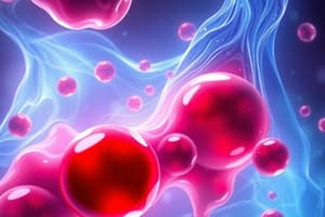
Blood Tissue Type
- Blood is a fluid connective tissue.
- Circulates through the cardiovascular system.
Blood Functions
- Transports dissolved gases, nutrients, and metabolic wastes.
- Delivers enzymes and hormones.
- Stabilizes pH and electrolyte composition.
- Prevents fluid losses through damaged vessels via clotting.
- Defends against toxins and pathogens using white blood cells and antibodies.
- Stabilizes body temperature by absorbing and redistributing heat.
Blood Cell Formation (Hemopoiesis)
- Blood cells form in red bone marrow.
- Hemopoiesis is the process where hematopoietic stem cells give rise to all blood cells.
- Pluripotential stem cells differentiate into two cell lines.
- Myeloid stem cells differentiate into erythrocytes, platelets, basophils, eosinophils, neutrophils, and monocytes.
- Lymphoid stem cells migrate to the thymus and differentiate into T cells.
Major Blood Components
- Plasma is a major blood component.
- Formed elements like erythrocytes (red blood cells), leukocytes (white blood cells), and platelets are major components.
Red Blood Cells (Erythrocytes)
- Red blood cells lack a nucleus and organelles.
- The lifespan of a red blood cell is about 120 days.
- Red blood cells are flexible, which allows them to travel through blood vessels easily.
- Red blood cells allow more room for hemoglobin.
- Without mitochondria, oxygen can be transported to tissues instead of being "used" by the mitochondria.
Blood Types & Rh Factor
- Red blood cells have surface antigens that determine blood type.
- Type A blood has RBCs with surface antigen A only.
- Type B blood has RBCs with surface antigen B only.
- Type AB blood has RBCs with both A and B surface antigens.
- Type O blood lacks both A and B surface antigens.
- Plasma contains antibodies that attack foreign surface antigens.
- Universal donors have type O blood.
- Universal recipients have type AB blood.
Granulocytes
- Neutrophils contain chemicals to kill bacteria.
- Eosinophils granules release chemicals that reduce inflammation.
- Basophils granules release histamine and heparin.
- Histamine dilates blood vessels.
- Heparin prevents abnormal blood clotting.
Agranulocytes
- Monocytes are large, phagocytic.
- Lymphocytes are responsible for specific immunity.
- T cells, B cells, and NK cells can differentiate.
Platelets
- Platelets derive from megakaryocytes.
- Megakaryocytes fragment into membrane-enclosed packets.
- Platelet thromboplastin factor is the main chemical.
- Thrombocytes were formerly called platelets.
- Platelets are involved in blood clotting (hemostasis).
- They release chemicals to initiate the clotting process.
- Platelets clump together to form a platelet plug.
- They contain actin and myosin which contract the clot.
Cardiovascular System: Heart Size and Chambers
- The heart is about the size of one's loosely clenched fist.
- The heart consists of four chambers: two atria and two ventricles.
- It pumps blood in two circuits: pulmonary and systemic.
Membranes
- Parietal membranes line cavity walls.
- Visceral membranes adhere to organ surfaces.
Pericardium
- The pericardial membrane forms two layers.
- Visceral pericardium (epicardium) adheres to the heart surface.
- The parietal pericardium consists of a fibrous outer layer called the pericardial sac and a serous pericardium that lines the inside.
- The serous pericardium produces a small amount of serous fluid.
Structure of the Heart Wall
- The heart wall consists of three layers.
- The epicardium (visceral pericardium) is the external surface.
- The myocardium consists of cardiac muscle.
- The endocardium is the internal surface and is continuous with the lining of blood vessels.
Heart Orientation
- The heart lies slightly to the left of midline in the mediastinum.
- The superior portion of the heart is the base.
- The apex is the inferior portion.
- The heart sits at an oblique angle.
- The right border is formed by the right atrium.
- The inferior border is formed by the right ventricle.
- The heart is rotated slightly toward the left.
- The sternocostal surface is formed by the right atrium and right ventricle.
- The posterior surface is formed by the left atrium.
Valves
- Atrioventricular valves bicuspid & tricuspid are open.
- Semilunar valves are pulmonary and aortic.
Blood Flow Through the Heart
- The right side of the heart is filled with deoxygenated blood.
Specialized Muscle Fibers in the Heart
- Major structures include the SA Node, AV Node, AV Bundle, Bundle Branches, and Purkinje Fibers.
Cardiac Cycle
- Step 1: SA node activity and atrial activation begin (Time = 0 msec).
- Step 2: Stimulus reaches the AV node (Elapsed time = 50 msec).
- Step 3: 100 msec delay at the AV node; atrial contraction begins (Elapsed time = 150 msec).
- Step 4: Impulse travels along the interventricular septum via the AV bundle and bundle branches to the Purkinje fibers (Elapsed time = 175 msec).
- Step 5: Impulse is distributed by Purkinje fibers throughout the ventricular myocardium; atrial contraction is completed, ventricular contraction begins (Elapsed time = 225 msec).
- Diastole records relaxation.
- Systole records contraction.
- The EKG (electrocardiogram) records electrical activity of heart
Cardiac Cycle Contraction and Relaxation
- The cardiac cycle has phases of contraction (systole) and relaxation (diastole).
Coronary Blood Vessels
- Coronary blood vessels supply blood to the heart muscles.
- They branch off the ascending aorta.
- Major coronary vessels include the Right coronary artery (RCA) and Left coronary artery (LCA).
- Cardiac veins drain into the coronary sinus.
Coronary Arteries
- Branches from the RCA: Marginal artery and the Posterior interventricular artery.
- Branches from the LCA: Anterior interventricular artery, Great Cardiac vein and Circumflex artery.
Cardiac Nerve Supply
- Parasympathetic vagus nerve.
- Sympathetic nerve.
Studying That Suits You
Use AI to generate personalized quizzes and flashcards to suit your learning preferences.