Podcast
Questions and Answers
Which part of the nervous system do autonomic neurons belong to?
Which part of the nervous system do autonomic neurons belong to?
- Both Central and Somatic Nervous Systems
- Peripheral Nervous System (correct)
- Somatic Nervous System
- Central Nervous System
Somatic motor neurons have two neurons traveling from the spinal cord to the effector.
Somatic motor neurons have two neurons traveling from the spinal cord to the effector.
False (B)
Preganglionic neurons originate in the midbrain or hindbrain or from the thoracic, ______, or sacral spinal cord.
Preganglionic neurons originate in the midbrain or hindbrain or from the thoracic, ______, or sacral spinal cord.
lumbar
Where are autonomic ganglia located?
Where are autonomic ganglia located?
Visceral effector organs will atrophy if a nerve is cut.
Visceral effector organs will atrophy if a nerve is cut.
What is the term given to the target becoming even more sensitive to stimulation?
What is the term given to the target becoming even more sensitive to stimulation?
What neurotransmitter do somatic motor neurons release?
What neurotransmitter do somatic motor neurons release?
Match the preganglionic neurons with their origin:
Match the preganglionic neurons with their origin:
The sympathetic division is also called the craniosacral division.
The sympathetic division is also called the craniosacral division.
Which of the following is secreted by the adrenal medulla when stimulated by the sympathetic nervous system?
Which of the following is secreted by the adrenal medulla when stimulated by the sympathetic nervous system?
Flashcards
Autonomic Neuron Function
Autonomic Neuron Function
Input to the brain and spinal cord that directs activity of motor neurons, innervating muscles and glands.
Preganglionic Neurons
Preganglionic Neurons
Originate in midbrain/hindbrain, or thoracic, lumbar, sacral spinal cord, influencing postganglionic neurons in ganglia.
Autonomic Ganglia Location
Autonomic Ganglia Location
Located in head, neck, abdomen, forming chains along either side of spinal cord; where preganglionic neurons synapse.
Muscle Contraction Rhythms
Muscle Contraction Rhythms
Signup and view all the flashcards
Neurotransmitter Release
Neurotransmitter Release
Signup and view all the flashcards
Sympathetic Function
Sympathetic Function
Signup and view all the flashcards
Parasympathetic Function
Parasympathetic Function
Signup and view all the flashcards
Adrenergic Receptor Sensitivity
Adrenergic Receptor Sensitivity
Signup and view all the flashcards
Complementary Effects
Complementary Effects
Signup and view all the flashcards
Cooperative Effects
Cooperative Effects
Signup and view all the flashcards
Study Notes
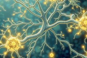
Neural Control of Involuntary Effectors: Autonomic Neurons
- Autonomic neurons consists of the brain and spinal cord
- Sensory neurons provide input and directs motor neuron activity to innervate muscles/glands
- Association neurons integrate sensory data, directing responses to maintain homeostasis and adapt to the environment
Somatic vs Autonomic Differences
- Somatic motor neurons feature cell bodies located in the spinal cord, with a single neuron extending from the spinal cord to the effector
- The autonomic motor system uses a two-neuron chain in the PNS
- The first neuron has its cell body in the brain/spinal cord and synapses in an autonomic ganglion
- The second neuron has its cell body in the ganglion and synapses on the effector
Autonomic Neuron Location
- Preganglionic neurons originate in the midbrain/hindbrain, or the thoracic, lumbar, or sacral spinal cord
- Postganglionic neurons originate in a ganglion
- Autonomic ganglia are located in the head, neck, and abdomen, and alongside the spinal cord
Visceral Effector Organs
- Visceral effector organs are somewhat independent of innervation, and wont atrophy if a nerve is cut
- Denervation hypersensitivity: targets become more sensitive to stimulation
Muscle Contraction Without Stimulation
- Cardiac and some smooth muscles contract rhythmically without nerve stimulation
- Autonomic innervation can alter intrinsic contraction speed
- Autonomic motor neurons stimulate or inhibit, depending on the organ and receptors present
Neurotransmitters Released
- Somatic motor neurons release acetylcholine, which is always excitatory
- Autonomic neurons release acetylcholine and norepinephrine, which may be excitatory or inhibitory
Somatic vs Autonomic Table
- Somatic efferent organs are skeletal muscles and autonomic are cardiac muscle, smooth muscle, and glands
- Somatic has no ganglia, autonomic has cell bodies of postganglionic autonomic fibers located in paravertebral, prevertebral (collateral), and terminal ganglia
- Somatic has one neuron from the CNS to the effector, autonomic has 2
- Somatic neuromuscular junction is a specialized motor end plate, autonomic junction has no specialization of postsynaptic membrane, and all areas of smooth muscle cells contain receptor proteins for neurotransmitters
- Somatic nerve impulse effect on muscle is excitatory only, autonomic is either excitatory or inhibitory
- Somatic fibers are fast-conducting, thick (9-13 um), and myeinated, autonomic fibers are slow-conducting; preganglionic fibers lightly myelinated but thin (3 um); postganglionic fibers unmyeinated and very thin (about 1.0 um)
- Effect of somatic denervation is flaccid paralysis and atrophy, effect of autonomic denervation is muscle tone and function persist; target cells show denervation hypersensitivity
Divisions of the Autonomic Nervous System: Sympathetic Division
- Preganglionic neurons originate from the thoracic and lumbar regions of the spinal cord
- This is also called the thoracolumbar division
- Preganglionic neurons synapse in sympathetic ganglia parallel to the spinal cord
- Called paravertebral ganglia
- These ganglia form a sympathetic chain
Convergence and Divergence
- Preganglionic neurons can branch and synapse in ganglia at any level, allowing for:
- Divergence: One preganglionic neuron synapses on several postganglionic neurons at different levels
- Convergence: Several preganglionic neurons at different levels synapse on one postganglionic neuron
- This arrangement allows the sympathetic division to act as a single unit through mass activation and be tonically active
Collateral Ganglia
- Many sympathetic neurons exiting the spinal cord below the diaphragm dont synapse in the sympathetic chain, but form splanchnic nerves synapsing in collateral ganglia instead
- Collateral ganglia include celiac, superior mesenteric, and inferior mesenteric ganglia
- Postganglionic neurons here innervate the digestive, urinary, and reproductive systems
Adrenal Glands
- The adrenal medulla secretes epinephrine/norepinephrine during sympathetic nervous system stimulation (part of mass activation)
- Embryologically, the adrenal medulla is a modified ganglion innervated directly by preganglionic sympathetic neurons
Summary of the Sympathetic Division
- Eye spinal origin of preganglionic fibers is C8 and T1, origin of postganglionic fibers is cervical ganglia
- Head and neck spinal origin of preganglionic fibers is T1 to T4, origin of postganglionic fibers is cervical ganglia
- Heart and lungs spinal origin of preganglionic fibers is T1 to T5, origin of postganglionic fibers is upper thoracic (paravertebral) ganglia
- Upper extremities spinal origin of preganglionic fibers is T2 to T9, origin of postganglionic fibers is lower cervical and upper thoracic (paravertebral) ganglia
- Upper abdominal viscera adrenal spinal origin of preganglionic fibers is T4 to T9, origin of postganglionic fibers is celiac and superior mesenteric (collateral) ganglia
- Adrenal spinal origin of preganglionic fibers is T10 and T11, there is not applicable origin of postganglionic fibers
- Urinary and reproductive systems spinal origin of preganglionic fibers is T12 to L2, origin of postganglionic fibers is celiac and interior mesenteric (collateral) ganglia
- Lower extremities spinal origin of preganglionic fibers is T9 to L2, origin of postganglionic fibers is lumbar and upper sacral (paravertebral) ganglia
Parasympathetic Division
- Preganglionic neurons originate from the brain or sacral region of the spinal cord, which is also called the craniosacral division
- Preganglionic neurons synapse on ganglia located near/in effector organs, called terminal ganglia
- Preganglionic neurons do NOT travel with somatic neurons and terminal ganglia supply very short postganglionic neurons to the effectors
Cranial Nerves and the Parasympathetic Division
- The oculomotor, facial, glosso-pharyngeal, and vagus nerves carry parasympathetic preganglionic neurons
- Oculomotor (III) nerve: preganglionic fibers exit midbrain and synapse on the ciliary ganglion, then postganglionic fibers innervate the ciliary muscle of the eye
- Facial (VII) nerve: preganglionic fibers exit the pons and synapse in the pterygopalatine ganglion where postganglionic fibers synapse on nasal mucosa, pharynx, palate, and lacrimal glands. Also preganglionic fibers exit the pons and synapse in the Submandibular ganglion where postganglionic fibers synapse on salivary glands.
- Glossopharyngeal: Preganglionic fibers synapse on otic ganglion, then Postganglionic fibers innervate salivary gland.
- Vagus nerve (X): preganglionic fibers exit medulla, branch into several plexi/nerves, and travel to ganglia within effector organs (heart, lungs, esophagus, stomach, pancreas, liver, intestines)
Sacral Nerves of Parasympathetic Division
- Preganglionic nerves from the sacral region of the spinal cord innervate the lower part of the large intestine, rectum, urinary and reproductive organs
- Terminal ganglia are located here
Summary of Parasympathetic Division: Table
- Oculomotor (third cranial) nerve fibers originate in midbrain (cranial) - Location of Terminal Ganglia Ciliary ganglion, effector organs eye (smooth muscle in iris and ciliary body)
- Facial (seventh cranial) nerve fibers originate in pons (cranial) - Location of Terminal Ganglia Pterygopalatine and submandibular ganglia, effector organs are lacrimal, mucous, and salivary glands
- Glossopharyngeal (ninth cranial) nerve fibers originate in medulla oblongata (cranial) - Location of Terminal Ganglia Otic ganglion, effector organ is the parotid gland
- Vegus (tenth cranial) nerve fibers originate in medulla oblongata (cranial) - Location of Terminal Ganglia Terminal ganglia in or near organ, effector organs are the heart, lungs, gastrointestinal tract, liver, pancreas
- Pelvic spinal nerves spinal fibers originate in S2-S4 (sacral) - Location of Terminal Ganglia Terminal ganglia near organs, effector organs are the lower half of large intestine, rectum, urinary bladder, and reproductive organs
Functions of the Autonomic Nervous System: Sympathetic division
- Sympathetic division activates via "fight or flight" mode by the release of norepinephrine from postganglionic neurons and the secretion of epinephrine from the adrenal medulla
- Intense physical activity readiness is increased in emergencies by increased heart rate and blood glucose levels. The body then diverts blood to skeletal muscles
- Tonically regulates heart, blood vessels, and organs
Parasympathetic division Functions
- Antagonistic to the sympathetic division
- “Rest and digest” is activated through ACh release from postganglionic neurons
- The heart rate slows while digestive activities increase
Adrenergic and Cholinergic Synaptic Transmission
- Cholinergic synaptic transmission is where acetylcholine (Ach) is used by all preganglionic neurons (sympathetic and parasympathetic)
- It is also the neurotransmitter released from most parasympathetic postganglionic neurons
- Some sympathetic postganglionic neurons release Ach which innervate sweat glands and skeletal muscle blood vessels and are called cholinergic symapses
- Adrenergic synaptic transmission is where norepinephrine is the neurotransmitter released by most sympathetic postganglionic neurons
- These synapses are called adrenergic
- Varicosities: axons of postganglionic neurons have swellings called varicosities that release neurotransmitter along the axon
- Forms "synapses en passant" which is classified as 'in passing'
- Sympathetic and parasympathetic neurons innervate the same tissues but release different neurotransmitters
Response to Adrenergic Stimulation
- Can involve epinephrine in the blood and norepinephrine from sympathetic nerves
- Stimulation: heart, dilatory muscles of the iris, smooth muscles of many blood vessels (causes vessel constriction)
- Inhibition: Bronchioles in lungs, other blood vessels that cause dilation
- alpha (a) and beta adrenergic receptors uses G-proteins and second messenger systems
- Beta (B) receptors (b) use cAMP
- Alpha (a) receptors (a) use Ca2+ second messenger system
- Aplha receptors are more sensitive to norepinephrine and beta receptors are more sensitive to blood epinephrine
- Alpha2 Receptors Located on presynaptic axons
- When stimulated, Alpha 2 Receptors result in inhibition of norepinephrine release in the synapse
- May be a negative-feedback system
- Some drugs to lower blood pressure act here to inhibit presynaptic neurons in the brain, inhibiting the whole sympathoadrenal system.
- Subtypes that will give different responses
- Drugs mimic adrenergic responses
- Agonists promote the process stimulated by the NT
- Antagonists- drugs that block the action of the NT
Response to Cholinergic Stimulation
- ACh released from preganglionic neurons of both the sympathetic/parasympathetic division is stimulatory
- ACh from postganglionic neurons of the parasympathetic division is usually stimulatory but some are inhibitory
- Sympathetic and parasympathetic effects are opposite in general
Cholinergic Receptors
- Nicotinic cholinergic receptors are found in autonomic ganglia
- Stimulated by Ach from preganglionic neurons
- These serve as ligand-gated ion channels for Na+ & K+ and are blocked by curare
- Muscarinic Cholinergic receptors are found in visceral muscle organs.
- Stimulated by release of Ach from post ganglionic neurons
- Five types have been identified and can be stimulatory or inhibitory depending which chemical opening for either (K+ or Ca2+= channels)
- Use G-proteins and second messenger system + blocked by atropine.
Other Autonomic Neurotransmitters
- Some postganglionic autonomic neurons don't release ACh or norepinephrine, they are called “nonadrenergic, noncholinergic fibers"
- Proposed include ATP, vasoactive intestinal peptide (VIP), and nitric oxide (NO)
- Nonadrenergic, Noncholinergic Fibers is important for erection of the penis and innervate blood vessels, causing relaxation and vasodilation using NO
- NO can also produce smooth muscle relaxation in the stomach, intestines, urinary bladder, and the brain.
Organs with Dual Innervation
- Innervated by both sympathetic and parasympathetic neurons
- Most of the time these are antagonists
- Heart rate – sym increases, para decreases
- Digestive functions – sym decreases, para increases
- Pupil diameter – sym dilates, para constricts
Complementary Effects
- When both divisions produce equivalent effects on the same target
- Example - Salivary gland secretion: Parasympathetic division stimulates secretion of watery saliva then Sympathetic division constricts blood vessels so the secretion is thicker
- Cooperative Effects: When both divisions produce different effects that work together to promote a single action
- Example - Erection and ejaculation: parasympathetic division causes vasodilation + erection and sympathetic causes the ejaculation
- Example - Urination: Parasympathetic division aids in urinary bladder contraction; sympathetic helps with bladder muscle tone to control urination
Organs Without Dual Innervation
- The adrenal medulla, arrector pili muscles in skin, sweat glands in skin, and most blood vessels are innervated by the sympathetic division only
- Regulation through an increase and decrease in sympathetic nerve activity
- Essential for body temperature regulation through blood vessels and sweat glands
Control of ANS by Higher Brain Centers
- Many visceral functions are regulated by autonomic reflexes, such that sensory input is sent to brain centers by the vagus nerve
- Sensory input integrates the information and modify the activity of preganglionic neurons
- The medulla oblongata controls cardiovascular, pulmonary, urinary, reproductive, and digestive functions
- In the medulla, the hypothalamus is the major regulatory center of the autonomic nervous system for body temperature, hunger, thirst, and pituitary gland
- Limbic system: The regulation center of emotion based autonomic responses during emotional states (blushing, pallar, fainting, cold sweating and racing heart rate)
- Cerebellum: The regulation center of motion sickness resulting in nausea, sweating and changing cardiovascular
- Frontal and temporal lobes for different emotions and personalities
Other Functions of Higher Brain Centers
- Autonomic dysreflexia: stroke, pulmonary edema, and myocardial infarction in people with spinal cord injuries at or above the sixth thoracic level (T6) of the spinal cord can be caused by Autonomic dysreflexia.
- At first there is a loss of spinal reflexes below the level of the injury, but after some time the reflexes reappear in an exaggerated state
- Aging is connected with with enhanced sympathetic activity causing increased sympathetic tone, and an increased risk of hypertension, together with cardiovascular diseases.
Studying That Suits You
Use AI to generate personalized quizzes and flashcards to suit your learning preferences.