Podcast
Questions and Answers
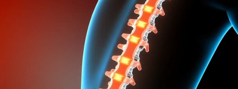
What is a primary characteristic of ankylosing spondylitis as seen on spinal X-rays?
What is a primary characteristic of ankylosing spondylitis as seen on spinal X-rays?
- Thickening of the vertebral discs
- Increased curvature of the lumbar spine
- Presence of osteophytes at the C7-T1 junction
- Loss of anterior concavity of vertebral bodies (correct)
Which demographic is most affected by ankylosing spondylitis?
Which demographic is most affected by ankylosing spondylitis?
- Women over 50 years old
- Children under 10 years old
- Elderly individuals above 70 years old
- Men between 20 and 40 years old (correct)
What percentage of patients with ankylosing spondylitis present with axial symptoms?
What percentage of patients with ankylosing spondylitis present with axial symptoms?
- 60%
- 75%
- 80% (correct)
- 90%
What is the significance of the HLA-B27 gene in relation to ankylosing spondylitis?
What is the significance of the HLA-B27 gene in relation to ankylosing spondylitis?
Which of the following conditions is classified under the spondyloarthropathies related to inflammatory low back pain?
Which of the following conditions is classified under the spondyloarthropathies related to inflammatory low back pain?
What is a common cause of patellar tendinopathy in athletes?
What is a common cause of patellar tendinopathy in athletes?
Which symptom is typically associated with patellar tendinopathy?
Which symptom is typically associated with patellar tendinopathy?
What does functional impotence usually lead to in cases of patellar tendinopathy?
What does functional impotence usually lead to in cases of patellar tendinopathy?
Which condition is NOT related to patellar tendinopathy?
Which condition is NOT related to patellar tendinopathy?
What is a common incident rate of iliotibial band syndrome among runners?
What is a common incident rate of iliotibial band syndrome among runners?
Which muscle contributes fibers to the iliotibial band?
Which muscle contributes fibers to the iliotibial band?
Which of the following activities is likely to trigger pain in individuals with iliotibial band syndrome?
Which of the following activities is likely to trigger pain in individuals with iliotibial band syndrome?
What is the primary factor involved in the development of patellofemoral pain syndrome in adolescents and young adults?
What is the primary factor involved in the development of patellofemoral pain syndrome in adolescents and young adults?
Which anatomical factor does NOT contribute to the alteration of patellar tracking?
Which anatomical factor does NOT contribute to the alteration of patellar tracking?
What symptom is NOT commonly associated with patellofemoral pain syndrome?
What symptom is NOT commonly associated with patellofemoral pain syndrome?
In individuals over 40 years of age, what percentage is likely to present with osteoarthritis when experiencing patellofemoral pain?
In individuals over 40 years of age, what percentage is likely to present with osteoarthritis when experiencing patellofemoral pain?
What clinical action is NOT typically required for the diagnosis of patellofemoral pain syndrome?
What clinical action is NOT typically required for the diagnosis of patellofemoral pain syndrome?
Which of the following factors does NOT increase the likelihood of developing patellofemoral pain syndrome?
Which of the following factors does NOT increase the likelihood of developing patellofemoral pain syndrome?
Which of the following describes the pain behavior pattern associated with patellofemoral pain syndrome?
Which of the following describes the pain behavior pattern associated with patellofemoral pain syndrome?
Which of the following conditions is patellofemoral pain syndrome often confused with?
Which of the following conditions is patellofemoral pain syndrome often confused with?
What biomechanical factor is associated with the alteration of patellar tracking?
What biomechanical factor is associated with the alteration of patellar tracking?
What are the possible factors contributing to iliotibial band syndrome?
What are the possible factors contributing to iliotibial band syndrome?
Which anatomical location is the insertion point for the iliotibial band?
Which anatomical location is the insertion point for the iliotibial band?
Which of the following symptoms is NOT commonly associated with iliotibial band syndrome?
Which of the following symptoms is NOT commonly associated with iliotibial band syndrome?
What is a common event that triggers the pain of iliotibial band syndrome?
What is a common event that triggers the pain of iliotibial band syndrome?
Which condition can be confused with iliotibial band syndrome?
Which condition can be confused with iliotibial band syndrome?
What biomechanical factor may lead to iliotibial band syndrome?
What biomechanical factor may lead to iliotibial band syndrome?
In the context of patellofemoral pain syndrome, the Q angle is associated with which type of force?
In the context of patellofemoral pain syndrome, the Q angle is associated with which type of force?
What kind of modifications are listed as potential changes that can improve iliotibial band syndrome symptoms?
What kind of modifications are listed as potential changes that can improve iliotibial band syndrome symptoms?
Which anatomical location is implicated in the friction causing iliotibial band syndrome?
Which anatomical location is implicated in the friction causing iliotibial band syndrome?
Flashcards
Ankylosing Spondylitis (AS)
Ankylosing Spondylitis (AS)
A chronic inflammatory disease affecting the spine and the joints. 80% of patients have axial symptoms, meaning their spine is primarily affected.
Bamboo Spine
Bamboo Spine
The fusion of vertebrae due to AS, which results in a rigid, 'bamboo-like' shape of the spine.
Syndesmophytes
Syndesmophytes
Small bone growths that form on the vertebrae in AS. They bridge the gaps between vertebrae, eventually leading to fusion.
HLA-B27
HLA-B27
Signup and view all the flashcards
Coccygodynia
Coccygodynia
Signup and view all the flashcards
Patellar Tendinopathy
Patellar Tendinopathy
Signup and view all the flashcards
Iliotibial Band Syndrome
Iliotibial Band Syndrome
Signup and view all the flashcards
Functional Impotence
Functional Impotence
Signup and view all the flashcards
Pain on Tensioning of Patellar Tendon
Pain on Tensioning of Patellar Tendon
Signup and view all the flashcards
Pain on Palpation of Patellar Tendon
Pain on Palpation of Patellar Tendon
Signup and view all the flashcards
Pain on Contraction of Patellar Tendon
Pain on Contraction of Patellar Tendon
Signup and view all the flashcards
Iliotibial Band (ITB)
Iliotibial Band (ITB)
Signup and view all the flashcards
What is the iliotibial band (ITB)?
What is the iliotibial band (ITB)?
Signup and view all the flashcards
Where does the ITB originate?
Where does the ITB originate?
Signup and view all the flashcards
Where does ITB insert?
Where does ITB insert?
Signup and view all the flashcards
What is ITB syndrome?
What is ITB syndrome?
Signup and view all the flashcards
When is ITB syndrome pain worse?
When is ITB syndrome pain worse?
Signup and view all the flashcards
What causes ITB syndrome?
What causes ITB syndrome?
Signup and view all the flashcards
What other factors can contribute to ITB syndrome?
What other factors can contribute to ITB syndrome?
Signup and view all the flashcards
What is the Q angle?
What is the Q angle?
Signup and view all the flashcards
What is patellofemoral pain syndrome?
What is patellofemoral pain syndrome?
Signup and view all the flashcards
Patellofemoral Joint Contact Changes
Patellofemoral Joint Contact Changes
Signup and view all the flashcards
Why does Patellofemoral Tracking Go Wrong?
Why does Patellofemoral Tracking Go Wrong?
Signup and view all the flashcards
How is PFPS Diagnosed?
How is PFPS Diagnosed?
Signup and view all the flashcards
How Common is PFPS?
How Common is PFPS?
Signup and view all the flashcards
What Factors Can Cause PFPS?
What Factors Can Cause PFPS?
Signup and view all the flashcards
Where and When Does PFPS Pain Occur?
Where and When Does PFPS Pain Occur?
Signup and view all the flashcards
How Does PFPS Pain Behave?
How Does PFPS Pain Behave?
Signup and view all the flashcards
What Other Conditions Can Mimic PFPS?
What Other Conditions Can Mimic PFPS?
Signup and view all the flashcards
Study Notes
Pelvis
- Conditions related to the pelvis include ankylosing spondylitis, coccygodynia, meralgia paresthetica, and pubalgia.
C/O-Medical records
- This section outlines the crucial steps in medical record management, from patient initial complaint (C/O) through to final plan (Rx1).
Ankylosing spondylitis
- Characterised as a chronic form of arthritis, often affecting the spine (spondyloarthropathy).
- Bamboo-shaped spine is a common feature, often with inflammation, leading to spinal fusion, and a stooped posture.
- 80% of patients experience axial symptoms.
- Typically affecting the lumbopelvic spine.
Spondyloarthropathies classification
- Seronegative arthritis / peripheral spondyloarthritis is associated with Crohn's disease or ulcerative colitis, characterized by: peripheral, symmetrical, and bilateral involvement of joints like knees, ankles, elbows, and wrists.
- Enteropathic arthritis/spondyloarthritis progress independently of the primary intestinal disease. Predisposition is shown to HLA-B27 but less than in ankylosing spondylitis.
- Differentiated spondyloarthritis : Includes peripheral arthritis, sacroiliitis, and enthesitis with inflammatory low back pain. Diagnostic classification is often unclear or cannot be definitively established with imaging tests alone.
- Reactive arthritis / Polyarticular. Asymmetric, commonly affecting feet and peripheral joints, mostly characterized by pain in the hindfoot.
AS incidence
- Lumbopelvic joint is frequently affected, as well as the lumbar vertebrae.
- Areas where ligaments and tendons attach to bones, especially in the spine.
- Cartilage between the sternum and ribs can also be impacted.
- Hip and shoulder joints can also be affected.
Coccygodynia
- Defined as pain in or around the tailbone (coccyx).
- Often with undefined spreading of pain.
- Higher frequency in women (5:1).
Coccygodynia: Involved factors
- Traumatic origin – local trauma.
- Prolonged sitting, especially on hard surfaces.
- Other factors: arthritis, childbirth.
- Obesity listed as a risk factor.
TP pelvic floor
- This section describes tenderness spots in the pelvis and their locations.
Coccygodynia: 24-hour behavior
- Pain while sitting worsens with leaning backward.
- Pain during bowel movements or sexual intercourse.
- Pain can be reduced with particular cushions.
Coccygodynia: Current and past history
- To reduce symptoms: leaning forward when sitting, and using pressure-reducing cushions (sometimes wedge-shaped).
- Applying heat or ice.
- Medication.
- Symptoms can resolve in weeks with rest.
- Prolonged sitting should be avoided.
Coccygodynia: Special questions
- Trauma in area (direct or indirect) should be investigated.
- Bowel or urination problems, or any sexual difficulties should be explored..
- If using cushions to reduce symptoms, continuing that practise is recommended.
- Upright sitting should be maintained to avoid recurrence of symptoms.
Meralgia paresthetica
- Mononeuropathy (sensory-only) of the femorocutaneous nerve.
- Often caused by injury or entrapment, mainly at the inguinal level.
- The condition is more common in people aged between 30 and 40, without a gender bias.
- Incidence rate of 4.3 per 10,000 people in a given region.
Meralgia paresthetica: Factors Involved
- Idiopathic.
- Mechanical causes – conditions that increase abdominal pressure, situations where the nerve is compressed.
- Metabolic factors –type 2 diabetes, alcoholism.
- Iatrogenic causes– during hip, lumbar spine, abdominal, obstetric, gynaecological surgery.
Meralgia paresthetica: Past and Present Background
- Obesity.
- Pregnancy.
- Tight fitting clothing.
- Surgery
Meralgia paresthetica: Special questions
- Differential diagnosis includes: metastasis at the iliac crest, lumbar disc herniation, avulsion fracture, chronic appendicitis, spinal surgery and diabetes.
- Differentiate from trochanteritis.
Meralgia paresthetica: Prognosis
- 62% of cases resolve spontaneously.
- Conservative measures are effective for 90% of cases.
- 73% of infiltration cases improve completely; relapse rate is 7%.
- Cases caused by compression, posture adjustment, or tissue retraction have quicker recovery times than direct injury.
Pubalgia
- This is not a specific diagnosis. It is a generic term used to describe groin pain.
- Different etiologies are involved, including sports hernia, sportsman’s hernia, sportsman’s groin, Gilmore’s groin, athletic pubalgia, and core muscle injury.
- Different aspects of groin pain should be considered as a differential diagnosis.
Pubalgia: Factors involved
- Young males, and particularly males involved in sports.
- About 58% of cases are related to soccer.
- Includes sports-related overload.
- Repetitive movements.
- Postural imbalances.
- Muscular imbalances.
- Dysplasia or micro-traumatisms.
- About 70% of cases involve adductor muscles.
Pubalgia: Body chart
- Localised to the pubic region.
Pubalgia: 24h behavior
- Localized pain; worse with palpation; Possible irradiation.
- Symptoms increase after exercise.
- Symptoms do not worsen with increased intra-abdominal pressure.
- Improves with ice, rest and NSAIDs.
- Symptoms may recur.
Pubalgia: History
- Insidious onset.
- Pain improvement with rest and non-steroidal anti-inflammatory drugs (NSAIDs).
- Possible fissure, or gracile disinsertion.
- Chronic phase: constant pain and limitations.
- Joint overuse may cause micro-traumas.
- Swelling caused by oedema and muscular contractions.
Pubalgia: Special Questions
- Lumbar: radiculopathy, canal stenosis, disc herniation.
- Hip pathology.
- Inguinal hernia.
- Muscle rupture.
- Urinary tract infections.
- Adductor tendinopathy.
Knee: Clinical processes
- Osteoarthritis.
- Patellar tendinopathy.
- Iliotibial band syndrome.
- Patellofemoral pain syndrome.
Knee Osteoarthritis
- A degenerative joint condition characterized by progressive loss of cartilage and marginal bone hypertrophy.
- Changes within the synovial membrane occur.
- Worldwide incidence is estimated to be 240 per 100,000 persons per year.
- OA frequently affects the knee joint and is the most common cause of disability in individuals over 65 years of age (up to 10% of affected individuals).
- It does not affect the entire knee, but typically one or two of the knee compartments.
Knee OA diagnostic criteria
- Criteria are based on clinical records, laboratory testing, and imaging evaluation.
- Clinical records include data such as: pain (gonalgia), age (>50), stiffness (<50 minutes), crepitus, tenderness of the bone; no temperature increases, but bone volume increase is seen.
- Laboratory testing includes: VHS (<40 mm/h), rheumatoid factor (<1:40).
- Imaging evaluation includes osteophytes (bony outgrowths)
Knee OA Classification
- Primary (or idiopathic): 70% of cases in the world.
- Secondary – related to past injuries, inflammations, etc.
Knee OA: factors involved
- Age (around 50 years old).
- Sex (female).
- Genetic component is less prominent than other factors.
- Weight alterations.
- Systemic factors (metabolic syndrome).
- Physical activity – is not considered a direct risk factor for developing this condition..
- Symptoms do not always correlate with radiological alterations in 50% of individuals.
Knee OA: 24-hour behavior
- Typical initial symptoms include mild pain following strenuous activity.
- Pain which worsens when walking up or down stairs or ramps, or during squatting.
- Over time in advanced cases there can be stiffness, instability and swelling (due to effusion) resulting in limited mobility and even deformity.
Knee OS: Actual and past history
- Previous joint injuries (especially those occurring 10-15 years ago)
- Meniscus problems, ACL tears.
- Weight alterations, overuse, local trauma, infections, metabolic disorders
Knee OA: Special questions
- Consider differential diagnoses of other rheumatic or musculoskeletal conditions, including: osteoarthritis (osteoarthrosis, rheumatoid arthritis, fibromyalgia, gout, lupus, psoriatic arthritis, Reiter’s disease, Sjogren's disease).
Patellar tendinopathy
- Overuse injury within the patella tendon, usually accompanied by pain, thickening, and reduced functioning.
- Pain is frequently insidious at onset and is triggered by increased physical activity.
- Pain becomes more persistent with increasing frequency and intensity of exercise.
- Tendinosis is usually more common in impact athletes, with an incidence of 30-45% in this population.
- However, non athletes can also be affected between 8% and 50% depending on working activity
- Several classification methods are used to assess the severity of the injury.
Patellar tendinopathy: Factors involved
- Men aged 35 or older.
- Biomechanical risk factors (including excessive foot pronation, high hip anteversion, increased Q-angle).
- Previous tendon or knee injuries.
- Genetic factors affecting tendon development and healing.
- Incorrect training methods.
- Environmental factors can contribute.
Iliotibial band syndrome
- Runners' knee syndrome.
- Acute external knee pain, generally associated with repetitive knee flexion and extension.
- Iliotibial band (ITB) friction against lateral femoral condyle a common source of pain.
- An increased frequency exists in runners (1.6–12%) and cyclists (15–24%).
- ITB is a source of up to 22% of lower limb related injuries.
Iliotibial band syndrome: Factors involved
- Narrow or wide iliotibial bands which results in friction with the lateral femoral condyle.
- Iliotibial band compression on underlying adipose tissue.
- Weak hip abductors (gluteus medius).
- Poor foot biomechanics (resulting in dysmetria), excessive running and athletic endurance training.
- Clothing can also be contributing factor.
Patellofemoral pain syndrome
- A common cause of dull pain around and front of the knee in the anterior aspect of the knee.
- Patella tracking dysfunction can result in pain.
- Patella never fully makes contact with the femur in all positions.
- Cartilage degeneration and inflammation can result in secondary osteoarthritis.
- There can be a biomechanical component which is dependent on issues with femoral anteversion, Q-angle, genu valgus/recurvatum or tibial varus/pronation of foot.
- Possible muscular weakness in hip external abductors or knee extensors causes muscular problems that can create instability.
- Occurs frequently in females between 40 and 50 years old.
- The incidence is about 18% in men and sports men, and 33% in sportswomen.
- 70% of those with patellofemoral pain over 40 years old have comorbid osteoarthritis.
Patellofemoral pain syndrome: Factors involved
- Age, particularly young adults and adolescents.
- Gender, with females being more likely to be affected.
- Overweight and obesity.
- Reduced flexibility.
- Lack of a good pre-exercise warm-up routine that prepares body for sports activity.
- High level of sporting activity and/or training.
Hip osteoarthritis (OA)
- A degenerative joint disease of the hip joint where cartilage and subchondral bone degrades.
- The hip and subchondral bone thicken, with development of osteophytes.
- Chronic synovitis is commonly present.
- The causes are often multifactorial: wear and tear arthritis, infection, crystal deposition, and/ or autoimmune disorders..
- The condition is often found in individuals over 50 years old, with higher incidence in women and significant association with obesity.
Hip OA: Factors involved
- Advanced age – the likelihood of developing this condition increases in individuals 50 years or older.
- Increasing prevalence with age into 75+ age category.
- Female gender.
- Obesity and other weight-related issues.
- Genetic predisposition.
- Previous joint injury/inflammation.
- Repeated stress applied to the hip joint .
- Muscle weakness.
Hip abductor-rotator tendinosis
- Pain in hip abductor-rotator tendons. Pain often felt around greater trochanter.
- Pain may be felt during or after exercise, or even with prolonged periods of standing or sitting.
Tendinosis or tendonitis?
- Tendinosis – is a degenerative condition resulting from imbalance between tendon tissue damage and self-repair.
- Resulting in compression, friction, and degeneration of the tendon structure.
- Tendonitis – is a common overuse injury that involves inflammatory damage.
- Both can occur simultaneously.
Hip abductor-rotator tendinosis: Special questions
- Localised trauma to the region
- Sports practices, footwear.
- Type of job performed.
- Metabolic dysfunctions.
- Physical conditions including past injuries are relevant..
Studying That Suits You
Use AI to generate personalized quizzes and flashcards to suit your learning preferences.
Related Documents
Description
Test your knowledge on the key characteristics and demographics of ankylosing spondylitis. This quiz covers spinal X-ray findings, the significance of the HLA-B27 gene, and axial symptoms related to this condition. Perfect for students and healthcare professionals alike!


