Podcast
Questions and Answers
Which branch of the lateral circumflex femoral artery is not mentioned in the provided information?
Which branch of the lateral circumflex femoral artery is not mentioned in the provided information?
- Obturator branch (correct)
- Perforating branch
- Profunda femoris branch
- Descending branch
Which of the following femoral arteries does not have a transverse branch?
Which of the following femoral arteries does not have a transverse branch?
- Profunda femoris artery
- Lateral circumflex femoral artery
- Medial circumflex femoral artery (correct)
- Femoral artery
What is the common characteristic of the branches of the lateral circumflex femoral artery mentioned in the provided information?
What is the common characteristic of the branches of the lateral circumflex femoral artery mentioned in the provided information?
- All are directed towards the gluteal region
- All have a descriptive name
- All are named based on their direction (correct)
- All have a similar course
What can be inferred about the medial circumflex femoral artery from the provided information?
What can be inferred about the medial circumflex femoral artery from the provided information?
What is the relationship between the lateral and medial circumflex femoral arteries?
What is the relationship between the lateral and medial circumflex femoral arteries?
Match the following anatomical structures with their locations:
Match the following anatomical structures with their locations:
Match the following ankle structures with their functions:
Match the following ankle structures with their functions:
Match the following ankle structures with their anatomical relationships:
Match the following ankle structures with their anatomical relationships:
Match the following ankle structures with their primary components:
Match the following ankle structures with their primary components:
Match the following ankle structures with their functional significance:
Match the following ankle structures with their functional significance:
Flashcards are hidden until you start studying
Study Notes
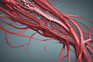
Profunda Femoris (Deep Femoral) Artery
- Originates from the femoral artery, which is the largest branch.
- Terminates as the 4th perforating artery.
Branches of Profunda Femoris Artery
- Muscular branch: Supplies blood to the adductor muscles.
- Articular (acetabular) branch: Supplies blood to the head of the femur.
Femoral Artery
- Main artery of the lower limb.
Branches of Femoral Artery
- Lateral circumflex femoral artery:
- Divides into three branches: ascending, transverse, and descending.
- Medial circumflex femoral artery:
- Divides into two branches: ascending and one unnamed branch.
Tributaries of the Saphenous Vein
- The Great (Long) Saphenous Vein drains the lateral side of the foot, ankle, and back of the leg.
- The Small Saphenous Vein is another tributary of the Saphenous Vein system.
Formation of the Popliteal Vein
- The Popliteal Vein is formed by the union of venae commitants of both the anterior and posterior tibial arteries.
- The Popliteal Vein is located near the Popliteal fascia.
Great (Long) Saphenous Vein
- The Great (Long) Saphenous Vein begins at the medial end of the dorsal venous arch of the foot.
- It is also located near the tibial n. retinaculum and inferior ext. retinaculum.
Peroneal Retinaculae
- Definition: Thickened bands of deep fascia at the lateral aspect of the ankle joint.
- The Peroneal Retinaculae are located near the Dorsalis pedis vessels.
Studying That Suits You
Use AI to generate personalized quizzes and flashcards to suit your learning preferences.