Podcast
Questions and Answers
What is the primary function of the stomach in relation to food?
What is the primary function of the stomach in relation to food?
- To absorb nutrients from food
- To store and mix food with gastric secretions (correct)
- To control the rate of digestion
- To eliminate waste from the body
Which surface of the stomach lies under the cover of the lower ribs and costal cartilages?
Which surface of the stomach lies under the cover of the lower ribs and costal cartilages?
- Both anterior and posterior surfaces (correct)
- Neither anterior nor posterior surface
- Posterior surface
- Anterior surface
What is the function of the pyloric orifice?
What is the function of the pyloric orifice?
- To store food
- To communicate with the duodenum (correct)
- To absorb nutrients from food
- To control the rate of digestion
What is the name of the indentation located in the lower part of the lesser curvature?
What is the name of the indentation located in the lower part of the lesser curvature?
What is the name of the muscle layer that forms the pyloric sphincter?
What is the name of the muscle layer that forms the pyloric sphincter?
Which part of the stomach is usually full of gas?
Which part of the stomach is usually full of gas?
What is the name of the region of the stomach that extends from the level of the cardial notch to the level of the angular notch?
What is the name of the region of the stomach that extends from the level of the cardial notch to the level of the angular notch?
What is the name of the area immediately adjacent to the cardial orifice?
What is the name of the area immediately adjacent to the cardial orifice?
Where does the common bile duct lie initially?
Where does the common bile duct lie initially?
What is the function of the gallbladder?
What is the function of the gallbladder?
What is the name of the short, dilated chamber embedded in the duodenal wall?
What is the name of the short, dilated chamber embedded in the duodenal wall?
What is the name of the sphincter surrounding the hepatopancreatic ampulla?
What is the name of the sphincter surrounding the hepatopancreatic ampulla?
What is the part of the gallbladder that projects below the inferior border of the liver?
What is the part of the gallbladder that projects below the inferior border of the liver?
Where does the common bile duct open into the duodenum?
Where does the common bile duct open into the duodenum?
What is the name of the duct that joins the common hepatic duct to form the common bile duct?
What is the name of the duct that joins the common hepatic duct to form the common bile duct?
What can compress the common bile duct, causing jaundice?
What can compress the common bile duct, causing jaundice?
What is the name of the fossa that forms the anterior part of the right limb of the 'H' shaped arrangement of grooves and fossae on the liver?
What is the name of the fossa that forms the anterior part of the right limb of the 'H' shaped arrangement of grooves and fossae on the liver?
Which of the following vessels carries oxygenated blood from the placenta to the liver during intrauterine life?
Which of the following vessels carries oxygenated blood from the placenta to the liver during intrauterine life?
What is the name of the ligament that forms the posterior boundary of the falciform ligament?
What is the name of the ligament that forms the posterior boundary of the falciform ligament?
What is the name of the part of the liver located between the fossa for the gallbladder and the fissure for the round ligament of liver?
What is the name of the part of the liver located between the fossa for the gallbladder and the fissure for the round ligament of liver?
Which of the following structures is NOT part of the porta hepatis?
Which of the following structures is NOT part of the porta hepatis?
What is the name of the structure that forms the posterior part of the left limb of the 'H' shaped arrangement of grooves and fossae on the liver?
What is the name of the structure that forms the posterior part of the left limb of the 'H' shaped arrangement of grooves and fossae on the liver?
What is the name of the duct that allows a large proportion of oxygenated blood from the placenta to bypass the liver and go directly to the IVC during intrauterine life?
What is the name of the duct that allows a large proportion of oxygenated blood from the placenta to bypass the liver and go directly to the IVC during intrauterine life?
What is the name of the part of the liver located between the groove for the IVC and the fissure for the ligamentum venosum?
What is the name of the part of the liver located between the groove for the IVC and the fissure for the ligamentum venosum?
What is the main function of the spleen in regards to blood cells?
What is the main function of the spleen in regards to blood cells?
Which of the following structures is NOT related to the visceral surface of the spleen?
Which of the following structures is NOT related to the visceral surface of the spleen?
What is the usual orientation of the long axis of the spleen?
What is the usual orientation of the long axis of the spleen?
What is the function of the gastrosplenic ligament?
What is the function of the gastrosplenic ligament?
What is the location of the spleen in relation to the diaphragm?
What is the location of the spleen in relation to the diaphragm?
What is the name of the ligament that connects the spleen to the left kidney?
What is the name of the ligament that connects the spleen to the left kidney?
What is the shape of the diaphragmatic surface of the spleen?
What is the shape of the diaphragmatic surface of the spleen?
What is the normal position of the anterior pole of the spleen in adults?
What is the normal position of the anterior pole of the spleen in adults?
What is the name of the wider part of the pyloric orifice?
What is the name of the wider part of the pyloric orifice?
Which structure lies posterior to the stomach?
Which structure lies posterior to the stomach?
What is the branching pattern of the left gastroepiploic artery?
What is the branching pattern of the left gastroepiploic artery?
Where does the venous blood from the stomach ultimately drain into?
Where does the venous blood from the stomach ultimately drain into?
What is the surface of the liver that faces inferiorly, posteriorly, and to the left?
What is the surface of the liver that faces inferiorly, posteriorly, and to the left?
What is the name of the border that separates the diaphragmatic and visceral surfaces of the liver?
What is the name of the border that separates the diaphragmatic and visceral surfaces of the liver?
Which of the following is NOT a posterior relation of the stomach?
Which of the following is NOT a posterior relation of the stomach?
What is the name of the artery that supplies the stomach and is a branch of the celiac trunk?
What is the name of the artery that supplies the stomach and is a branch of the celiac trunk?
Where does the portal vein terminate?
Where does the portal vein terminate?
What is the tributary that drains the territory supplied by the inferior mesenteric artery?
What is the tributary that drains the territory supplied by the inferior mesenteric artery?
What is the name of the vein that drains blood from the lesser curvature of the stomach?
What is the name of the vein that drains blood from the lesser curvature of the stomach?
What is the consequence of portal hypertension?
What is the consequence of portal hypertension?
What is the site of portal-systemic anastomosis in the anterior abdominal wall?
What is the site of portal-systemic anastomosis in the anterior abdominal wall?
What is the name of the vein that drains the gallbladder?
What is the name of the vein that drains the gallbladder?
What is the consequence of enlargement of esophageal veins in portal hypertension?
What is the consequence of enlargement of esophageal veins in portal hypertension?
What is the site of portal-systemic anastomosis in the rectum and anal canal?
What is the site of portal-systemic anastomosis in the rectum and anal canal?
What is the name of the ligament that connects the liver to the anterior abdominal wall?
What is the name of the ligament that connects the liver to the anterior abdominal wall?
What is the name of the vein that forms by the union of the superior mesenteric and splenic veins?
What is the name of the vein that forms by the union of the superior mesenteric and splenic veins?
What is the location of the major duodenal papilla in relation to the duodenum?
What is the location of the major duodenal papilla in relation to the duodenum?
What is the name of the ligament that connects the spleen to the diaphragm?
What is the name of the ligament that connects the spleen to the diaphragm?
Which of the following structures is NOT a part of the small intestine?
Which of the following structures is NOT a part of the small intestine?
What is the location of the root of the mesentery of the small intestine?
What is the location of the root of the mesentery of the small intestine?
What is the function of the teniae coli in the large intestine?
What is the function of the teniae coli in the large intestine?
What is the name of the artery that supplies the pancreas?
What is the name of the artery that supplies the pancreas?
What is the location of the pancreas in relation to the duodenum?
What is the location of the pancreas in relation to the duodenum?
What is the name of the ligament that connects the pancreas to the spleen?
What is the name of the ligament that connects the pancreas to the spleen?
What is the function of the marginal artery in the large intestine?
What is the function of the marginal artery in the large intestine?
What is the location of the McBurney's point in relation to the abdomen?
What is the location of the McBurney's point in relation to the abdomen?
Flashcards are hidden until you start studying
Study Notes
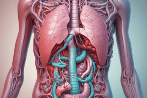
Stomach
- Located in the left hypochondriac, epigastric, and umbilical regions, mostly under the cover of lower ribs and costal cartilages.
- Functions: stores food, mixes food with gastric secretions to form a semi-fluid mass (chyme), and controls the rate of delivery of chyme to the small intestine.
- Has 2 surfaces: anterior and posterior.
- Has 2 borders: lesser curvature (shorter and on the right) and greater curvature (longer and on the left).
- Has an incisura angularis (angular notch) located in the lower part of the lesser curvature.
- Has 2 orifices: cardial and pyloric orifices.
- The cardial orifice communicates with the esophagus and has a cardial notch located between the left border of the esophagus and the fundus of the stomach.
- The pyloric orifice communicates with the duodenum and is surrounded by a sphincter (pyloric sphincter), formed by a thickening of the circular muscle layer of the stomach.
Parts of the Stomach
- Cardia (cardial part): area immediately adjacent to the cardial orifice.
- Fundus: dome-shaped, upper part of the stomach, limited inferiorly by a horizontal line drawn across the stomach starting at the cardial notch, usually full of gas.
- Body: extends from the level of the cardial notch to the level of the angular notch.
- Pyloric part: distal, funnel-shaped region of the stomach, extends from the angular notch to the pyloric orifice, its proximal, wider part is known as the pyloric antrum, and its distal, narrower part is known as the pyloric canal.
Anatomical Relations of the Stomach
- Anteriorly: anterior abdominal wall, left costal margin, diaphragm, left lobe of liver.
- Posteriorly: lesser sac, diaphragm, spleen, upper part of the left kidney, left suprarenal gland, splenic artery, pancreas, transverse mesocolon.
Blood Supply of the Stomach
- Arteries: left gastric artery (branch of the celiac trunk), right gastric artery (branch of the proper hepatic artery), left gastroepiploic artery (branch of the splenic artery), right gastroepiploic artery (branch of the gastroduodenal artery), and short gastric arteries (branches of the splenic artery).
- All arteries are accompanied by veins of the same name, and all venous blood from the stomach drains (directly or indirectly) into the portal vein.
Liver
- Located in the upper part of the abdominal cavity, just beneath the diaphragm, in the right hypochondriac, epigastric, and left hypochondriac regions, mostly under the cover of lower ribs and costal cartilages.
- Surfaces: diaphragmatic and visceral.
- Diaphragmatic surface: convex, smooth, molded to the undersurface of the diaphragm.
- Visceral surface: faces inferiorly, posteriorly, and to the left, molded to adjacent organs (gallbladder, esophagus, stomach, duodenum, right kidney, right suprarenal gland, right colic flexure).
- Diaphragmatic and visceral surfaces are separated anteriorly by a sharp border (inferior border of the liver), with no clear demarcation between them posteriorly.
Visceral Surface of the Liver
- Has a series of grooves and fossae arranged like a letter "H".
- The crossbar of the "H" is the porta hepatis (hilum of the liver), which contains the right and left hepatic ducts, branches of the proper hepatic artery, and the portal vein.
- The right limb of the "H" (right sagittal fissure) is formed anteriorly by the fossa for the gallbladder and posteriorly by the groove for the IVC.
- The left limb of the "H" (left sagittal fissure) is formed anteriorly by the fissure for the round ligament of the liver and posteriorly by the fissure for the ligamentum venosum.
Ligaments of the Liver
- Falciform ligament: a two-layered peritoneal fold that connects the diaphragmatic surface of the liver to the anterior abdominal wall (above the umbilicus), its liver attachment forms the boundary between the right and left lobes.
- Round ligament of the liver: a fibrous cord that connects the liver to the umbilicus.
Gallbladder
- Pear-shaped sac located on the visceral surface of the liver, stores bile, and concentrates it by absorbing water.
- Parts: fundus, body, and neck.
- Fundus: anterior, rounded end that projects below the inferior border of the liver, in contact with the anterior abdominal wall at the level of the tip of the right 9th costal cartilage.
- Body: middle part of the gallbladder, between the fundus and neck, contacts the visceral surface of the liver (superiorly) and the transverse colon and duodenum (inferiorly).
- Neck: narrow, tapering end, opposite the fundus, and directed towards the porta hepatis, usually makes an S-shaped bend and becomes continuous with the cystic duct.
Spleen
- Largest lymphoid organ of the body, located in the left hypochondriac region, between the stomach and diaphragm, entirely sheltered by lower ribs (9th to 11th).
- Functions: elimination of old and/or damaged blood cells, and filters antigens from blood and contributes to the immune response against such antigens.
- Surfaces: diaphragmatic (smooth and convex) and visceral (irregular and concave).
- Borders: superior (usually notched) and inferior.
- Poles: anterior and posterior.
- Long axis of the spleen lies along the 10th rib.
- Normal spleen cannot be palpated in the adult (anterior pole reaches normally as far forward as the midaxillary line).
- Intraperitoneal organ, its hilum is connected to the stomach and left kidney by gastrosplenic and splenorenal ligaments, respectively.
Small Intestine
- The longest part of the alimentary canal, extending from the pyloric orifice to the ileocecal junction.
- Most food digestion and absorption take place in the small intestine.
- Divided into three parts: duodenum, jejunum, and ileum.
Duodenum
- A C-shaped tube, approximately 10 inches long, that curves around the head of the pancreas.
- Located in the epigastric and umbilical regions.
- Divided into four parts: 1st (superior), 2nd (descending), 3rd (inferior, horizontal or transverse), and 4th (ascending).
- Important anatomical relations:
- Gastroduodenal artery passes posterior to the 1st part of duodenum.
- Gallbladder is anterior to the 1st part and upper portion of the 2nd part of duodenum.
- Superior mesenteric artery and vein pass anterior to the 3rd part of duodenum.
- Internal structure:
- Duodenal mucosa is smooth in the first 2cm.
- Mucosal surface has numerous folds (circular folds or plicae circulares) in the remaining duodenum.
- Major duodenal papilla is a small elevation located approximately halfway down the posteromedial wall of the 2nd part of duodenum.
- Minor duodenal papilla (inconstant) is located in the 2nd part of duodenum, about 2cm above the major duodenal papilla.
Jejunum and Ileum
- Jejunum begins at the duodenojejunal flexure, and ileum ends at the ileocecal junction.
- Both are intraperitoneal and freely movable, attached to the posterior abdominal wall by the mesentery of the small intestine.
- Root of the mesentery of the small intestine extends inferiorly and to the right from the left side of L2 to the right sacroiliac joint.
Differences between Jejunum and Ileum
- Coils of jejunum lie in the upper part of the infracolic compartment, while coils of ileum are in the lower part of the infracolic compartment and pelvic cavity.
- Jejunum has a larger diameter and thicker walls than ileum.
- Circular folds are larger and more numerous in jejunum than in ileum.
- Jejunal arteries form fewer arcades than ileal arteries.
- Aggregated lymphoid follicles/nodules (Peyer’s patches) are present in ileum along its antimesenteric border.
- Ileum has more mesenteric fat than jejunum.
Large Intestine
- Extends from the ileocecal junction to the anus.
- Mainly concerned with absorbing water and electrolytes and storing undigested materials until they can be eliminated from the body as feces.
- Parts: cecum with vermiform appendix, ascending colon, transverse colon, descending colon, sigmoid colon, rectum, and anal canal.
Differences between Large and Small Intestines
- Teniae coli are three bands of longitudinally arranged smooth muscle fibers, approximately equally spaced around the circumference of the large intestine.
- Fatty appendices (epiploic/omental appendices) are present in the large intestine but not in the small intestine.
- Wall of the small intestine is smooth, while the wall of the large intestine is sacculated (haustra).
Vermiform Appendix
- A narrow muscular tube containing a large amount of lymphoid tissue.
- Intraperitoneal, with a small mesentery (mesoappendix) that contains appendicular vessels.
- McBurney’s point is the area of greatest tenderness in appendicitis.
- Three teniae coli converge at the base of the appendix (useful in locating the appendix during surgery).
- Position of the appendix is variable, and sensory fibers carrying pain from the appendix terminate in the spinal cord at the level of T10.
Pancreas
- An exocrine and endocrine gland.
- Exocrine part produces enzymes involved in the digestion of proteins, fats, and carbohydrates.
- Endocrine part (pancreatic islets [of Langerhans]) produces hormones (mainly insulin and glucagon).
- Elongated organ that lies in the epigastric and left hypochondriac regions, deeply located on the posterior abdominal wall, behind the peritoneal sac (most of it is secondary retroperitoneal).
- Parts: head, neck, body, and tail.
Marginal Artery
- A continuous arterial channel that skirts the inner margin of the colon from the ileocecal junction to the rectosigmoid junction.
- Formed by anastomoses between branches of the ileocolic, right colic, middle colic, left colic, and sigmoid arteries.
- Can serve as a source of collateral circulation to a part of the colon after its chief arterial supply has been obstructed or ligated.
Portal Vein
- Drains blood from most of the alimentary canal (from the lower ⅓ of the esophagus to the upper ½ of the anal canal), spleen, and pancreas, and terminates in the liver.
- Begins posterior to the neck of the pancreas by the union of the superior mesenteric and splenic veins.
- Runs superiorly and to the right, passing posterior to the 1st part of the duodenum, and runs within the hepatoduodenal ligament (posterior to the proper hepatic artery and common bile duct).
- Divides into right and left terminal branches at the porta hepatis.
Portal-Systemic Anastomoses
- In normal conditions, portal venous blood passes through the liver and drains into the IVC, which then carries it to the heart (direct route).
- Other routes of communication exist between the portal and systemic (SVC, IVC) circulations, which become important if the direct route is blocked, causing portal hypertension.
- Sites of portal-systemic anastomoses include:
- Lower part of the esophagus
- Walls of the rectum and anal canal
- Anterior abdominal wall
- Wherever non-peritoneal areas of intestines, liver, and pancreas are in contact with the body wall
Studying That Suits You
Use AI to generate personalized quizzes and flashcards to suit your learning preferences.