Podcast
Questions and Answers
Which of the following is a primary function of the nasal cavity?
Which of the following is a primary function of the nasal cavity?
- Producing hormones that regulate body temperature.
- Synthesizing vitamin D through exposure to sunlight.
- Filtering, warming, and moistening air before it reaches the lungs. (correct)
- Secreting digestive enzymes to aid in breaking down food particles.
What is the main function of the soft palate?
What is the main function of the soft palate?
- To form the floor of the nasal cavity.
- To house the openings to the Eustachian tubes.
- To provide structural support for the tongue.
- To divide the nasopharynx from the lower pharynx. (correct)
Which section of the pharynx is located directly behind the nasal cavity?
Which section of the pharynx is located directly behind the nasal cavity?
- Laryngopharynx
- Hypopharynx
- Oropharynx
- Nasopharynx (correct)
What is the primary function of the pharynx?
What is the primary function of the pharynx?
Which structure connects the nasopharynx to the ears?
Which structure connects the nasopharynx to the ears?
What is the primary role of the oropharynx?
What is the primary role of the oropharynx?
Which type of epithelium lines the laryngopharynx?
Which type of epithelium lines the laryngopharynx?
What critical function does the larynx perform during swallowing?
What critical function does the larynx perform during swallowing?
Why are the right and left hilum difficult to visualize on chest X-rays?
Why are the right and left hilum difficult to visualize on chest X-rays?
During inhalation, what role do the innermost intercostal muscles play?
During inhalation, what role do the innermost intercostal muscles play?
How do the floating ribs differ from the first seven ribs in terms of their attachment?
How do the floating ribs differ from the first seven ribs in terms of their attachment?
Why is the pleural cavity considered a potential space?
Why is the pleural cavity considered a potential space?
How many lobes does the right lung have, and what separates them?
How many lobes does the right lung have, and what separates them?
Which of the following muscles is NOT a primary muscle involved in respiration, but rather an accessory muscle that assists in elevating the rib cage?
Which of the following muscles is NOT a primary muscle involved in respiration, but rather an accessory muscle that assists in elevating the rib cage?
What structural component separates pulmonary lobules?
What structural component separates pulmonary lobules?
What is the combined number of ribs in the human thoracic cage?
What is the combined number of ribs in the human thoracic cage?
Which of the following statements accurately describes the function of the pulmonary artery?
Which of the following statements accurately describes the function of the pulmonary artery?
During exhalation, which action helps decrease the size of the chest cavity?
During exhalation, which action helps decrease the size of the chest cavity?
Which of the following statements correctly describes the position of the left hilum compared to the right hilum?
Which of the following statements correctly describes the position of the left hilum compared to the right hilum?
Which of the following occurs at the respiratory membrane?
Which of the following occurs at the respiratory membrane?
If a surgeon needs to access a specific bronchopulmonary segment, what anatomical characteristic makes this possible?
If a surgeon needs to access a specific bronchopulmonary segment, what anatomical characteristic makes this possible?
Where are the intercostal muscles located and what is their primary function?
Where are the intercostal muscles located and what is their primary function?
What is the hilum of the lung?
What is the hilum of the lung?
How does the autonomic nervous system influence lung function?
How does the autonomic nervous system influence lung function?
Which of the following structural changes occurs in the bronchi as they branch into smaller tubes?
Which of the following structural changes occurs in the bronchi as they branch into smaller tubes?
What is the primary function of the cilia lining the bronchioles?
What is the primary function of the cilia lining the bronchioles?
Which of the following accurately describes the primary function of the respiratory system?
Which of the following accurately describes the primary function of the respiratory system?
In what way does oxygen reach the bloodstream from the alveoli?
In what way does oxygen reach the bloodstream from the alveoli?
What is the pleural cavity?
What is the pleural cavity?
During respiration, where does the diffusion of oxygen from the pulmonary alveoli into the blood primarily occur?
During respiration, where does the diffusion of oxygen from the pulmonary alveoli into the blood primarily occur?
Which structure separates the nasal cavity from the oral cavity?
Which structure separates the nasal cavity from the oral cavity?
Which of the following is NOT a function of the larynx?
Which of the following is NOT a function of the larynx?
What distinguishes the respiratory bronchioles from the terminal bronchioles?
What distinguishes the respiratory bronchioles from the terminal bronchioles?
Why are nosebleeds common, and what characteristic of the nose contributes to this?
Why are nosebleeds common, and what characteristic of the nose contributes to this?
Which type of cartilage is the epiglottis composed of, and what is its function?
Which type of cartilage is the epiglottis composed of, and what is its function?
How does the respiratory system contribute to maintaining homeostasis in the body?
How does the respiratory system contribute to maintaining homeostasis in the body?
What is meant by 'dead space' in the context of the bronchioles?
What is meant by 'dead space' in the context of the bronchioles?
Which best describes the role of mucus in the nasal cavity?
Which best describes the role of mucus in the nasal cavity?
Damage to the nasal septum would directly affect which of the following functions?
Damage to the nasal septum would directly affect which of the following functions?
Flashcards
Respiratory System
Respiratory System
The system responsible for the exchange of oxygen and carbon dioxide between the body and the atmosphere.
Respiration
Respiration
Inhaling air into the lungs and exhaling air out of the lungs.
Upper Respiratory System
Upper Respiratory System
Nasal cavity, pharynx, larynx and trachea.
Lower Respiratory System
Lower Respiratory System
Signup and view all the flashcards
Nose
Nose
Signup and view all the flashcards
Nasal cavity
Nasal cavity
Signup and view all the flashcards
Nose's Function
Nose's Function
Signup and view all the flashcards
Nasal Hairs and Mucus
Nasal Hairs and Mucus
Signup and view all the flashcards
Nasal Cavity Functions
Nasal Cavity Functions
Signup and view all the flashcards
Hard Palate
Hard Palate
Signup and view all the flashcards
Soft Palate
Soft Palate
Signup and view all the flashcards
Pharynx Function
Pharynx Function
Signup and view all the flashcards
Nasopharynx
Nasopharynx
Signup and view all the flashcards
Oropharynx
Oropharynx
Signup and view all the flashcards
Laryngopharynx Function
Laryngopharynx Function
Signup and view all the flashcards
Larynx Function
Larynx Function
Signup and view all the flashcards
Larynx
Larynx
Signup and view all the flashcards
Thyroid Cartilage
Thyroid Cartilage
Signup and view all the flashcards
Cricoid Cartilage
Cricoid Cartilage
Signup and view all the flashcards
Epiglottis
Epiglottis
Signup and view all the flashcards
Bronchi
Bronchi
Signup and view all the flashcards
Bronchioles
Bronchioles
Signup and view all the flashcards
Cilia
Cilia
Signup and view all the flashcards
Alveoli
Alveoli
Signup and view all the flashcards
What is the Hilum?
What is the Hilum?
Signup and view all the flashcards
What are Intercostal Muscles?
What are Intercostal Muscles?
Signup and view all the flashcards
What are External Intercostals?
What are External Intercostals?
Signup and view all the flashcards
What are Internal Intercostals?
What are Internal Intercostals?
Signup and view all the flashcards
What are Innermost Intercostals?
What are Innermost Intercostals?
Signup and view all the flashcards
What is the Thoracic Cage?
What is the Thoracic Cage?
Signup and view all the flashcards
What happens during Inhalation?
What happens during Inhalation?
Signup and view all the flashcards
What happens during Exhalation?
What happens during Exhalation?
Signup and view all the flashcards
Pleural Cavity
Pleural Cavity
Signup and view all the flashcards
Lobes of the Right Lung
Lobes of the Right Lung
Signup and view all the flashcards
Lobes of the Left Lung
Lobes of the Left Lung
Signup and view all the flashcards
Bronchopulmonary Segment
Bronchopulmonary Segment
Signup and view all the flashcards
Cardiac Notch
Cardiac Notch
Signup and view all the flashcards
Pulmonary Artery Function
Pulmonary Artery Function
Signup and view all the flashcards
Respiratory Membrane
Respiratory Membrane
Signup and view all the flashcards
Hilum
Hilum
Signup and view all the flashcards
Study Notes
- The respiratory system is our breathing equipment.
- The respiratory system facilitates the inhalation and exhalation of gases that circulate around body tissues and cells, excreting excess and waste.
- Oxygen is our "food" and carbon dioxide is our "waste".
- Respiration involves the exchange of oxygen and carbon dioxide between the atmosphere and body cells.
- This includes inspiration, expiration, diffusion of oxygen from the pulmonary alveoli to blood, carbon dioxide from blood to the alveoli, and the transport of oxygen to and carbon dioxide from body cells.
- The respiratory system consists of an upper and lower section.
Upper Respiratory System
- The upper respiratory system is comprised of the nasal cavity, pharynx, larynx, and trachea.
Lower Respiratory System
- The lower respiratory system is comprised of the Bronchi/Bronchioles, Lungs, Pleura, Diaphragm and Ribs and Intercostal muscules
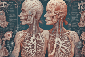
The Nose and Nasal Cavity
- It is the only part of the respiratory system visible externally.
- It serves as the route for air entry into the respiratory system.
- Air is drawn in via the nostrils and enters the large nasal cavity.
- The nasal cavity is divided into two by the nasal septum, which consists of hyaline cartilage.
- The nasal cavity is partitioned from the oral cavity by the palate.
- Dense vascularity in the nose can lead to nosebleeds with large blood loss.
- The nose and nasal hairs trap particles and dust to adhere to mucus; mucus prevents drying.
- Air travels over moist mucosa and becomes saturated with water for humidification.
- The Inervation of the nose responsible for sense of smell via the olfactory nerve.
- As air passes through the nasal cavity it warms or cools to within 1 degree of body temperature.
- The entire mucosa of the nasal fossae is covered by mucus, secreted by ciliated columnar epithelium which contains secreting goblet cells.
- The nose houses our sense of smell and contributes greatly to taste sensation through its posterior communication with the mouth.
Palates
- The hard palate forms the floor of the nasal cavity and separates the nasal and oral cavities
- The soft palate extends posterior to the hard palate.
- It divides the superior nasopharynx from lower pharynx
The Pharynx
- It makes up the part of the throat that is situated immediately behind the nasal cavity.
- It sits behind the mouth and above the oesophagus and larynx.
- It is conventionally divided into three sections: nasopharynx, oropharynx, and laryngopharynx and is important in vocalization.
- The function of the pharynx is to transfer food from the mouth to the oesophagus and to warm, moisten and filter air before it moves into the trachea.
- The pharynx is a part of the digestive and respiratory systems.
Nasopharynx
- It is 2 to 3 cm wide and 3 to 4 cm long.
- The nasopharynx is the space above the soft palate at the back of the nose and connects the nose to the mouth, allowing a person to breathe through the nose.
- The soft palate separates the nasopharynx from the oropharynx.
- It contains adenoid tissue, which fights infection, and the openings to the Eustachian tubes, which lead to the ears.
- It provides a major drainage path for lymphatic fluids and generally drains into the throat, nose, or ears.
Oropharynx
- Accepts air from the nasopharynx and passes it to the laryngeal pharynx.
- It also accepts food from the mouth and passes it to the oesophagus.
- It is important that the oropharynx and associated structures prevent food or liquids from entering the lungs, as choking may ensue, with potentially fatal consequences.
Laryngopharynx
- Lined by stratified squamous epithelium.
- The function is to pass food and air.
- It works as a passage for food and air.
- During swallowing, the entry of air temporarily stops, allowing food to pass safely to the oesophagus.
- This prevents food from entering our respiratory tract and from choking the trachea (windpipe).
Larynx
- Allows air to pass through it while keeping food and drink from blocking the airway.
- It is also the body’s "voice box" as it contains the vocal folds that produce the sounds of speech and singing.
- It consists of the the Thyroid cartilage (Adam's apple) with hyaline cartilage, the Cricoid cartilage with hyaline cartilage and the Epiglottis with elastic cartilage.
Lower Respiratory Tract: Bronchi (sing. Bronchus)
- Conducts air into the lungs.
- The right and left Bronchus branch into smaller secondary and tertiary branches which then branch into smaller tubes known as bronchioles.
- No gas exchange takes place in the Bronchi.
- It contains progressively less cartilage and more smooth muscle.
Bronchioles
- The bronchioles have no cartilage but smooth muscle dominates
- Progressing down the bronchioles, the muscle and connective tissue begin to disappear, leaving a single layer of epithelial cells resulting in Alveoli.
- The function of the Bronchioles is to deliver air to the network of millions of alveoli
- There are 3 types of bronchioles: Lobular, Terminal and Respiratory.
- Lobular Bronchioles are the passages that first enter the lungs
- Terminal Bronchioles are the smaller passages in each lung
- Respiratory Bronchioles are the leads on from terminal branches that lead to alveolar ducts
- The lobular and terminal branches are often referred to as the dead space because no air exchange takes place in these passages.
- The walls of the Bronchioles are lined with finger like projections known as cilia.
- The function of the cilia is to remove debris and microbes
Alveoli
- There are millions of alveoli.
- If Alveoli were stretched end to end they covers a tennis court!
- Air exchange takes place in the alveoli.
- Once O₂ reaches the alveoli it diffuses through a single cell in an alveolus, followed by a single cell in a capillary to enter the bloodstream
- At the same time, CO₂ is released from the capillary to the alveoli and then exhaled.
- Flow of air from extenal evironment happen because lungs pressure is changing.
- Alveolar gas exchange is affected by surface area, partial pressure gradients of gases and the matching of ventilation and profusion
Pleural Cavities and Membranes
- The Pleural cavity is a fluid-filled space between the 2 pulmonary pleurae
- The outer Pleura (parietal) is attached to the chest (thoracic) wall.
- The inner Pleura (visceral) covers the lungs and adjoining structures including the blood vessels, bronchi and nerves.
- The Pleural Cavity is considered a potential space because the 2 Pleurae adhere to each other through a serous thin film (in normal conditions)
The Anatomy of the Lungs
- Lungs consist of lobes that are separated from each other by fissures.
- The right lung consists of superior, middle and inferior lobes.
- The left lung consists of superior and inferior lobes.
- The bronchopulmonary segment is a division of a lobe, and each lobe houses bronchopulmonary segments.
- Each segment receives air from its own tertiary bronchus and is supplied with blood by its own artery.
- A pulmonary lobule is a subdivision formed as the bronchi branch into bronchioles.
- Each lobule receives its own bronchiole that has multiple branches.
- An interlobular septum is a wall composed of connective tissue, separating the lobules from each other.
- The cardiac notch is the indentation seen on the left lung, and allows space for the heart.
- The blood supply to the lungs plays an important role in the gas exchange and serves as a transport system for gases throughout the body.
- Innervation by the parasympathetic and sympathetic nervous system provides a level of control through dilation and constriction of the airway.
- The major function of the lungs is to perform gaseous exchange.
- This requires blood from the pulmonary circulation.
- This blood supply contains deoxygenated blood, which travels to the lungs where erythrocytes pick up the O2 to transport to body tissues.
- The pulmonary artery carries deoxygenated blood to the alveoli, and branches multiple times as it follows the bronchi, becoming smaller and smaller in diameter.
- One arteriole and one venule supply and drain one pulmonary lobule.
- As they near the alveoli, the pulmonary arteries become the pulmonary capillary network.
- The capillary network consists of tiny vessels with very thin walls.
- The pulmonary capillary network branches and follows the bronchioles and the structure of the alveoli.
- At this point the capillary wall meets the alveolar wall creating the respiratory membrane.
- Once the blood is oxygenated, it drains from the alveoli, by way of multiple pulmonary veins, which exit the lungs through the hilum.
- The Hilum are where the bronchi, arteries, veins and nerves enter and exit the lungs.
- Both right and left Hilum are similar in size, but the left hilum usually is slightly higher in the chest than the right.
- it can It is difficult to visualize them on chest X rays, further tests e.g CT scan are required to determine if a problem exists in the area.
The Intercoastal muscles
- Intercoastal muscules are a group of muscles that are situated in the ribs.
- There are 3 layers that assist in the breathing process.
- The 3 layers of the intercoastal mucles are, external , internal and innermost.
- The external muscles sit outside the ribs
- The internal muscles sit between the ribs
- The innermost muscles sit inside the ribs
- They are innervated by the intercostal nerves, and blood supply is from the intercostal artery and vein.
Thoracic Cage
- Surrounds and protects the heart and lungs
- It consists of 12 Thoracic vertebrae, 24 ribs plus the sternum
- First 7 ribs are attached to the Sternum by costal cartilage
- Of the remaining 5, 3 have costal cartilage connected to the cartilage above and the last 2 floating ribs are connected by their cartilage to muscle in the abdominal wall.
During Inhalation
- The Diaphragm relaxes, lungs expand
- The innermost intercostal muscles relax while the external intercostal muscles contract causing the chest cavity to expand.
- This expansion allows the lungs to fill with air due to the negative pressure created by the extra space
- As air fills the lungs, gases are exchanged and it is time to exhale
During Exhalation
- Air needs to be forced out.
- The chest cavity must become smaller.
- The diaphragm and the external intercostal muscles contract applying force to the base and sides of the lungs.
- The innermost intercostal muscles contract while the external intercostal muscles relax.
- The cavity contracts and air is forced out
Accessory Muscles of Respiration
- Accessory muscles do not actively play a part in breathing but are considered accessory as they help in elevating the rib cage.
- They are the sternocleidomastoid and the scalene
- Some other neck muscles are also considered as accessory muscles of respiration.
Studying That Suits You
Use AI to generate personalized quizzes and flashcards to suit your learning preferences.
Related Documents
Description
Test your knowledge of the upper and lower respiratory tracts. Questions cover nasal cavity, pharynx, larynx, ribs and lungs. This quiz assesses understanding of respiratory system functions.