Podcast
Questions and Answers
What is the function of the fibrous pericardium?
What is the function of the fibrous pericardium?
- To prevent the heart from expanding too much (correct)
- To produce pericardial fluid
- To attach the heart to the posterior surface of the sternum
- To facilitate the movement of the heart and great vessels
Which of the following is NOT a characteristic of the fibrous pericardium?
Which of the following is NOT a characteristic of the fibrous pericardium?
- It is attached to the central tendon of the diaphragm
- It is the outermost layer of the pericardium
- It is a single-walled membrane (correct)
- It is made of connective tissue
What is the relationship between the fibrous pericardium and the tunica adventitia of the great vessels?
What is the relationship between the fibrous pericardium and the tunica adventitia of the great vessels?
- The tunica adventitia is a part of the fibrous pericardium
- They are separate structures
- They are fused together (correct)
- The fibrous pericardium is a part of the tunica adventitia
What is the pericardial cavity?
What is the pericardial cavity?
Which layer of the serous pericardium directly covers the heart and the roots of the great vessels?
Which layer of the serous pericardium directly covers the heart and the roots of the great vessels?
What is the function of the pericardial fluid?
What is the function of the pericardial fluid?
What is the attachment of the fibrous pericardium to the posterior surface of the sternum called?
What is the attachment of the fibrous pericardium to the posterior surface of the sternum called?
What is the purpose of the parietal layer of the serous pericardium?
What is the purpose of the parietal layer of the serous pericardium?
What is the function of the thin film of serous fluid in the pericardium?
What is the function of the thin film of serous fluid in the pericardium?
What is the characteristic sound heard during auscultation in cases of pericarditis?
What is the characteristic sound heard during auscultation in cases of pericarditis?
Which layer of the heart wall is formed by the visceral layer of serous pericardium?
Which layer of the heart wall is formed by the visceral layer of serous pericardium?
What is the function of the ventricular septum?
What is the function of the ventricular septum?
Which of the following blood vessels empties into the right atrium?
Which of the following blood vessels empties into the right atrium?
What is the characteristic feature of the posterior part of the right atrium?
What is the characteristic feature of the posterior part of the right atrium?
What is the function of the pectinate muscles in the right atrium?
What is the function of the pectinate muscles in the right atrium?
What is the term for the ear-like pouch that projects from the right atrium?
What is the term for the ear-like pouch that projects from the right atrium?
What is the main difference between the left atrium and the right atrium?
What is the main difference between the left atrium and the right atrium?
What is the purpose of the interatrial septum in the left atrium?
What is the purpose of the interatrial septum in the left atrium?
What is the function of the pectinate muscles in the left atrium?
What is the function of the pectinate muscles in the left atrium?
Why is the left ventricle thicker than the right ventricle?
Why is the left ventricle thicker than the right ventricle?
What is the function of the trabeculae carneae in the left ventricle?
What is the function of the trabeculae carneae in the left ventricle?
What is the purpose of the aortic vestibule in the left ventricle?
What is the purpose of the aortic vestibule in the left ventricle?
What is the relationship between the aortic orifice and the aortic valve?
What is the relationship between the aortic orifice and the aortic valve?
Why does the left ventricle perform more work than the right ventricle?
Why does the left ventricle perform more work than the right ventricle?
What prevents blood backflow into the left atrium?
What prevents blood backflow into the left atrium?
At which level is the pulmonary valve located?
At which level is the pulmonary valve located?
What is the function of the pulmonary sinuses?
What is the function of the pulmonary sinuses?
Which of the following arises from the posterior aortic sinus?
Which of the following arises from the posterior aortic sinus?
What occurs in the capillaries of systemic circulation?
What occurs in the capillaries of systemic circulation?
What is the direction of blood flow in the systemic veins?
What is the direction of blood flow in the systemic veins?
Where do the superior and inferior vena cavae drain blood into?
Where do the superior and inferior vena cavae drain blood into?
What is the direction of blood flow from the left ventricle?
What is the direction of blood flow from the left ventricle?
What is the primary function of the fibrous heart skeleton?
What is the primary function of the fibrous heart skeleton?
What type of valve is also known as the tricuspid valve?
What type of valve is also known as the tricuspid valve?
Which of the following is NOT a function of the fibrous heart skeleton?
Which of the following is NOT a function of the fibrous heart skeleton?
What is the name of the valve that separates the left atrium from the left ventricle?
What is the name of the valve that separates the left atrium from the left ventricle?
What is the purpose of the chordae tendineae in the atrioventricular valves?
What is the purpose of the chordae tendineae in the atrioventricular valves?
What is the main difference between the atrioventricular valves and the semilunar valves?
What is the main difference between the atrioventricular valves and the semilunar valves?
What is the function of the semilunar valves in the heart?
What is the function of the semilunar valves in the heart?
What is the term for the type of connective tissue that forms the fibrous heart skeleton?
What is the term for the type of connective tissue that forms the fibrous heart skeleton?
Flashcards are hidden until you start studying
Study Notes
Pericardium
- The pericardium is a double-walled fibrous membrane that encloses the heart and the roots of great vessels.
- It lies posterior to the body of the sternum and the second to sixth costal cartilages.
- The fibrous pericardium is the tough, outermost layer that prevents the heart from expanding too much.
- The internal surface of the fibrous pericardium is lined with serous pericardium.
- The fibrous pericardium is fused with the tunica adventitia of great vessels, attached to the posterior surface of the sternum by sternopericardial ligaments, and fused with the central tendon of the diaphragm.
Serous Pericardium
- The serous pericardium is the inner layer of the pericardium, made up of two layers: parietal and visceral layers.
- The parietal layer is the outer layer, firmly attached to the fibrous pericardium.
- The visceral layer is the innermost layer, directly covering the heart and roots of great vessels.
- The pericardial cavity is the space between the two layers of the serous pericardium, holding the pericardial fluid.
Pericarditis and Pericardial Effusion
- Normally, the layers of serous pericardium make no detectable sound during auscultation.
- Pericarditis makes the surfaces rough, resulting in a pericardial friction rub, which sounds like the rustle of silk when listening with a stethoscope.
- Certain inflammatory diseases may produce pericardial effusion.
Heart Wall
- The wall of the heart consists of three layers: epicardium, myocardium, and endocardium.
- The epicardium is a thin external layer formed by the visceral layer of serous pericardium.
- The myocardium is a thick middle layer composed of cardiac muscle.
- The endocardium is a thin internal layer, or lining membrane of the heart, that also covers its valves.
Heart Chambers
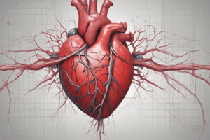
- The heart is divided into four chambers: right atrium, left atrium, right ventricle, and left ventricle.
- The atria are separated by the atrial septum, and the ventricles are separated by the ventricular septum.
- The ventricles have thicker walls and pump blood to the lungs and body.
Right Atrium
- The right atrium forms the right border of the heart and receives venous blood from the superior vena cava, inferior vena cava, and coronary sinus.
- The ear-like right auricle is a small, conical muscular pouch that projects from the right atrium and overlaps the ascending aorta.
Left Atrium
- The left atrium forms most of the base of the heart, where the pairs of valveless right and left pulmonary veins enter.
- The interior of the left atrium has a smooth-walled part, a smaller muscular auricle containing pectinate muscles, four pulmonary veins entering its posterior wall, a slightly thicker wall than that of the right atrium, and an interatrial septum that slopes posteriorly and to the right.
Left Ventricle
- The left ventricle forms the apex of the heart, nearly all of its left (pulmonary) surface and border, and most of the diaphragmatic surface.
- Because arterial pressure is much higher in the systemic than in the pulmonary circulation, the left ventricle performs more work than the right ventricle.
- The interior of the left ventricle has a double-leaflet mitral valve, walls that are two to three times as thick as those of the right ventricle, a conical cavity, and walls covered with thick muscular ridges, trabeculae carneae.
Fibrous Skeleton of the Heart
- The fibrous heart skeleton is located between the atria and ventricles and is formed from dense irregular connective tissue.
- It separates the atria and ventricles, anchors heart valves by forming supportive rings at their attachment points, provides electrical insulation between atria and ventricles, and provides a rigid framework for the attachment of cardiac muscle tissue.
Valves of the Heart
- There are two types of valves in the heart: atrioventricular valves and semilunar valves.
- Atrioventricular valves separate the atria from the ventricles, and semilunar valves separate the ventricles from the output large vessels (aorta and pulmonary arteries).
Right Atrioventricular Valve
- The right atrioventricular valve, also called the tricuspid valve, separates the right atrium from the right ventricle and has three triangular flaps.
- Venous blood flows from the right atrium, through the valve into the right ventricle, and is forced closed when the right ventricle begins to contract, preventing blood backflow into the right atrium.
Left Atrioventricular Valve
- The left atrioventricular valve, also called the bicuspid valve or mitral valve, separates the left atrium from the left ventricle and has chordae tendineae.
- Oxygenated blood flows from the left atrium into the left ventricle, and is forced closed when the left ventricle begins to contract, preventing blood backflow into the left atrium.
Semilunar Valves
- The pulmonary valve, located at the apex of the conus arteriosus, is at the level of the left third costal cartilage.
- The aortic valve, obliquely placed, is located between the left ventricle and the root of the aorta.
Pulmonary and Systemic Circulation
- Systemic circulation: oxygenated blood from the left ventricle is pumped into the aorta and then into smaller systemic arteries; gas exchange in tissues occurs from capillaries; systemic veins then carry deoxygenated blood and waste products.
- Pulmonary circulation: deoxygenated blood from the right ventricle is pumped into the pulmonary artery and then into smaller pulmonary arteries; gas exchange in lungs occurs from capillaries; pulmonary veins then carry oxygenated blood and return it to the left atrium.
Studying That Suits You
Use AI to generate personalized quizzes and flashcards to suit your learning preferences.


