Podcast
Questions and Answers
Which bony landmark is suspended in the neck by muscles between the floor of the mouth and the larynx?
Which bony landmark is suspended in the neck by muscles between the floor of the mouth and the larynx?
- Clavicle
- Hyoid bone (correct)
- Mastoid
- Manubrium of sternum
Which anatomical region in the body serves functions including supporting the head, movement of the head/neck, swallowing, breathing, and phonation?
Which anatomical region in the body serves functions including supporting the head, movement of the head/neck, swallowing, breathing, and phonation?
- Neck (correct)
- Pelvis
- Abdomen
- Thorax
What are the functions of the neck highlighted in the text?
What are the functions of the neck highlighted in the text?
- Supporting the head, movement of the head/neck, swallowing, breathing, and phonation (correct)
- Supporting the arms
- Supporting the torso
- Supporting the legs
Which of the following is NOT included in the superficial muscles of the neck, supra/infrahyoid muscles, and prevertebral muscles covered in the session?
Which of the following is NOT included in the superficial muscles of the neck, supra/infrahyoid muscles, and prevertebral muscles covered in the session?
Which bony landmark is located at the floor of the mouth and the larynx?
Which bony landmark is located at the floor of the mouth and the larynx?
What makes the neck one of the more complicated anatomical regions in the body?
What makes the neck one of the more complicated anatomical regions in the body?
Which bony landmark is located at the base of the skull?
Which bony landmark is located at the base of the skull?
What is the function of the supra/infrahyoid muscles highlighted in the session?
What is the function of the supra/infrahyoid muscles highlighted in the session?
Which triangle is formed by starting at the middle of the hyoid bone and drawing a straight line down the midline of the neck to the manubrium?
Which triangle is formed by starting at the middle of the hyoid bone and drawing a straight line down the midline of the neck to the manubrium?
Which structure can typically be seen crossing superficial to the omoclavicular (subclavian) triangle?
Which structure can typically be seen crossing superficial to the omoclavicular (subclavian) triangle?
Which triangle is formed superior to the inferior belly of the omohyoid and contains the occipital artery and spinal accessory nerve (CN XI)?
Which triangle is formed superior to the inferior belly of the omohyoid and contains the occipital artery and spinal accessory nerve (CN XI)?
Which triangle is formed caudal to the inferior belly of the omohyoid, within the posterior triangle, and contains the subclavian artery?
Which triangle is formed caudal to the inferior belly of the omohyoid, within the posterior triangle, and contains the subclavian artery?
Which nerve passes through the occipital triangle within the posterior triangle?
Which nerve passes through the occipital triangle within the posterior triangle?
Which triangle is formed by starting at the superior nuchal line on the occipital bone where the SCM and trapezius muscles meet and then following the anterior border of the trapezius down to the clavicle?
Which triangle is formed by starting at the superior nuchal line on the occipital bone where the SCM and trapezius muscles meet and then following the anterior border of the trapezius down to the clavicle?
Which triangle is subdivided into 2 smaller triangles using the inferior belly of the omohyoid?
Which triangle is subdivided into 2 smaller triangles using the inferior belly of the omohyoid?
Which structure is formed by starting at the middle of the hyoid bone and drawing a straight line down the midline of the neck to the manubrium?
Which structure is formed by starting at the middle of the hyoid bone and drawing a straight line down the midline of the neck to the manubrium?
Which vessel passes through the occipital triangle within the posterior triangle?
Which vessel passes through the occipital triangle within the posterior triangle?
Which muscle forms the boundary of the muscular triangle?
Which muscle forms the boundary of the muscular triangle?
Which muscle primarily facilitates movement of the scapulae?
Which muscle primarily facilitates movement of the scapulae?
Which muscle is not a part of the infrahyoid muscles of the neck?
Which muscle is not a part of the infrahyoid muscles of the neck?
Which muscle is part of the prevertebral muscles of the neck?
Which muscle is part of the prevertebral muscles of the neck?
Which muscle is part of the superficial ventral muscles of the neck?
Which muscle is part of the superficial ventral muscles of the neck?
Which triangle of the neck contains the submandibular gland and hypoglossal nerve?
Which triangle of the neck contains the submandibular gland and hypoglossal nerve?
Which muscle is located in the interscalene space?
Which muscle is located in the interscalene space?
Which muscle divides the neck into anterior and posterior triangles?
Which muscle divides the neck into anterior and posterior triangles?
Which triangle of the neck extends from the midline of the chin to the midline of the hyoid bone?
Which triangle of the neck extends from the midline of the chin to the midline of the hyoid bone?
Which structure traverses the interscalene space?
Which structure traverses the interscalene space?
Which muscle forms the carotid triangle of the neck?
Which muscle forms the carotid triangle of the neck?
Flashcards are hidden until you start studying
Study Notes
Anatomy of the Neck: Muscles, Landmarks, and Triangles
- The neck landmarks include the hyoid bone, upper border of thyroid cartilage, and bifurcation of carotid arteries at specific cervical levels.
- The ventral muscles of the neck include the superficial muscles such as platysma and sternocleidomastoid, with detailed information on their attachments and actions.
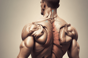
- The trapezius muscle primarily facilitates movement of the scapulae.
- The suprahyoid and infrahyoid muscles of the neck include muscles like digastric, stylohyoid, mylohyoid, geniohyoid, sternohyoid, sternothyroid, thyrohyoid, and omohyoid.
- The prevertebral muscles of the neck consist of longus colli, longus capitis, rectus capitis anterior, and rectus capitis lateralis, each with specific actions and innervation.
- The scalene muscles, including anterior, middle, and posterior scalene, have distinct functions and attachments.
- The interscalene space is a gap between the anterior and middle scalene muscles, traversed by structures like the subclavian artery and brachial plexus.
- The neck is divided into two major anatomic triangles by the sternocleidomastoid muscle: the anterior and posterior triangles.
- The anterior triangle is further divided into four subdivisions: submental, submandibular, and carotid triangles, each with specific contents and boundaries.
- The submental triangle extends from the midline of the chin to the midline of the hyoid bone and contains submental lymph nodes that drain the tongue and floor of the mouth.
- The submandibular triangle is the area between the inferior border of the mandible and the anterior and posterior bellies of the digastric muscle, containing structures like the submandibular gland and hypoglossal nerve.
- The carotid triangle is formed by the superior belly of the omohyoid, the inferior belly of the digastric, and the anterior border of the sternocleidomastoid muscle.
Studying That Suits You
Use AI to generate personalized quizzes and flashcards to suit your learning preferences.


