Podcast
Questions and Answers
Which muscle pulls the neck of the mandible forward along with the articular disc?
Which muscle pulls the neck of the mandible forward along with the articular disc?
- Temporalis
- Masseter
- Lateral pterygoid (correct)
- Medial pterygoid
What is the primary function of the posterior fibers of the Temporalis muscle?
What is the primary function of the posterior fibers of the Temporalis muscle?
- Protrusion of the mandible
- Elevation of the mandible
- Rotation of the mandible
- Retraction of the mandible (correct)
At what age do teeth begin to erupt?
At what age do teeth begin to erupt?
- At 6 years of age
- At 2 years of age
- Around 6 months after birth (correct)
- At birth
How many incisors are present in each jaw?
How many incisors are present in each jaw?
What covers the tongue?
What covers the tongue?
What divides the tongue into right and left halves?
What divides the tongue into right and left halves?
How is the upper surface of the tongue divided?
How is the upper surface of the tongue divided?
What is the frenulum of the tongue?
What is the frenulum of the tongue?
What is the origin of the sternocleidomastoid muscle?
What is the origin of the sternocleidomastoid muscle?
Which nerve innervates the thyrohyoid muscle?
Which nerve innervates the thyrohyoid muscle?
What is the action of the scalene muscles when acting unilaterally?
What is the action of the scalene muscles when acting unilaterally?
What are the muscles included in the infrahyoid muscles?
What are the muscles included in the infrahyoid muscles?
Which muscle is responsible for depressing the hyoid bone and larynx?
Which muscle is responsible for depressing the hyoid bone and larynx?
What is the insertion point of the sternocleidomastoid muscle?
What is the insertion point of the sternocleidomastoid muscle?
What are the actions of the sternocleidomastoid muscle when contracting bilaterally?
What are the actions of the sternocleidomastoid muscle when contracting bilaterally?
What is the origin of the scalene muscles?
What is the origin of the scalene muscles?
What is the origin of the Rectus Capitis Lateralis muscle?
What is the origin of the Rectus Capitis Lateralis muscle?
What is the insertion point of the Rectus Capitis Lateralis muscle?
What is the insertion point of the Rectus Capitis Lateralis muscle?
Which nerves innervate the Rectus Capitis Lateralis muscle?
Which nerves innervate the Rectus Capitis Lateralis muscle?
What action does the Rectus Capitis Lateralis muscle perform unilaterally?
What action does the Rectus Capitis Lateralis muscle perform unilaterally?
What is the origin of the Rectus Capitis Anterior muscle?
What is the origin of the Rectus Capitis Anterior muscle?
What is the bilateral action of the Longus Capitis muscle?
What is the bilateral action of the Longus Capitis muscle?
Where does the Superior Oblique of Longus Colli originate?
Where does the Superior Oblique of Longus Colli originate?
What is the origin of the Vertical portion of Longus Colli?
What is the origin of the Vertical portion of Longus Colli?
Flashcards are hidden until you start studying
Study Notes
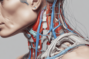
Neck Muscles
- The thyrohyoid muscle is innervated by C1 fibers via the hypoglossal nerve (CN XII).
Scalene Muscles
- The scalene muscles have three parts: anterior, medius/middle, and posterior.
- They originate from the transverse processes of the 3rd to 6th cervical vertebrae.
- They insert into the 1st and 2nd ribs.
- They are innervated by C4, 5, and 6 (anterior rami of cervical nerves).
- When acting unilaterally, they elevate the 1st rib/2nd rib and laterally flex and rotate the cervical part of the vertebral column to the same side.
- When acting bilaterally, they cause cervical flexion.
Sternocleidomastoid Muscle
- The sternocleidomastoid muscle originates from the manubrium sterni and the medial 1/3 of the clavicle.
- It inserts into the mastoid process of the temporal bone and the occipital bone.
- It is innervated by the spinal part of the accessory nerve (CN 11) and C2 and 3.
- When contracting bilaterally, it causes cervical flexion and head extension.
- When contracting unilaterally, it causes ipsilateral flexion and contralateral rotation.
Suprahyoid and Infrahyoid Muscles
- The suprahyoid muscles include the digastrics, mylohyoid, stylohyoid, and geniohyoid (DMSG).
- The infrahyoid muscles include the thyrohyoid, omohyoid, sternothyroid, and sternohyoid (TOSS).
- The sternothyroid muscle is responsible for depressing the hyoid bone and larynx.
- The digastrics muscle elevates the hyoid bone and depresses the mandible.
- The mylohyoid muscle elevates the hyoid bone and floor of the mouth during swallowing.
Rectus Capitis Lateralis Muscle
- The rectus capitis lateralis muscle originates from the transverse process of the atlas (C1).
- It inserts into the jugular process of the occipital bone.
- It is innervated by the anterior rami of the 1st and 2nd cervical nerves (C1-C2).
- When acting bilaterally, it flexes the head on the neck.
- When acting unilaterally, it laterally flexes the head.
Rectus Capitis Anterior Muscle
- The rectus capitis anterior muscle originates from the lateral mass and transverse process of the atlas (C1).
- It inserts into the basilar part of the occipital bone.
- It is innervated by the anterior rami of the 1st and 2nd cervical nerves (C1-C2).
- It flexes the head.
Longus Capitis Muscle
- The longus capitis muscle originates from the anterior tubercles of the transverse processes of C3-C6.
- It inserts into the basilar part of the occipital bone.
- It is innervated by the anterior rami of the 1st to 3rd cervical nerves (C1-C3).
- It causes head flexion bilaterally and ipsilateral head rotation unilaterally.
Longus Colli Muscle
- The longus colli muscle has three segments: superior oblique, vertical portion, and inferior oblique.
- The superior oblique originates from the anterior tubercles of the transverse processes of C3-C5 vertebrae.
- The vertical portion originates from the anterior surfaces of the bodies of the lower three cervical and superior three thoracic vertebrae (C5-T3).
- The inferior oblique originates from the posterior fibers of the temporalis.
Mastigation
- The lateral pterygoid muscle pulls the neck of the mandible forward along with the articular disc.
- The temporalis, masseter, and medial pterygoid muscles are involved in the elevation of the mandible.
- The lateral pterygoid muscle is involved in the protrusion of the mandible, drawing the lower teeth forward over the upper teeth.
- The posterior fibers of the temporalis muscle are involved in the retraction of the mandible.
Teeth Development
- Deciduous teeth begin to erupt about 6 months after birth.
- All deciduous teeth have erupted by the end of 2 years.
- The lower jaw teeth usually appear first.
- There are 4 incisors, 2 canines, and 4 molars in each jaw.
- Teeth begin to erupt at 6 years of age, and the last tooth, the 3rd molar, typically erupts between 17 and 30 years of age.
Tongue
- The tongue is covered in a mucous membrane.
- The tongue is divided into right and left halves by a median fibrous septum.
- The upper surface of the tongue is divided into anterior and posterior parts by a V-shaped sulcus called the sulcus terminalis.
- The frenulum of the tongue is a fold of mucous membrane that connects the undersurface of the tongue to the floor of the mouth.
- The intrinsic muscles of the tongue are confined to the tongue and are not attached to bone.
Studying That Suits You
Use AI to generate personalized quizzes and flashcards to suit your learning preferences.


