Podcast
Questions and Answers
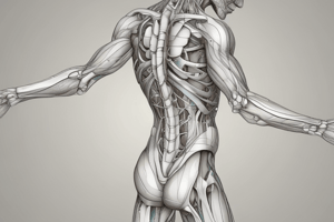
What is the anatomical position of a human?
What is the anatomical position of a human?
- Lying down with arms extended
- Sitting with legs crossed and arms raised
- Standing upright with palms facing backward
- Standing erect with face and eyes looking forward (correct)
Which plane divides the body into right and left parts?
Which plane divides the body into right and left parts?
- Coronal plane
- Transverse plane
- Horizontal plane
- Sagittal plane (correct)
What type of anatomy involves studying organs using imaging techniques?
What type of anatomy involves studying organs using imaging techniques?
- Microscopic anatomy
- Radiographic anatomy (correct)
- Developmental anatomy
- Applied anatomy
Which term describes movement toward the median plane?
Which term describes movement toward the median plane?
In which direction does the term 'superior' refer?
In which direction does the term 'superior' refer?
What describes the movement of a joint bending to decrease the angle between two bones?
What describes the movement of a joint bending to decrease the angle between two bones?
Which of the following directional terms means 'closer to the trunk'?
Which of the following directional terms means 'closer to the trunk'?
What type of study focuses on the normal structure of cells under a microscope?
What type of study focuses on the normal structure of cells under a microscope?
What is the main function of the tibial tuberosity?
What is the main function of the tibial tuberosity?
Which of the following muscles originates from the lateral surface of the tibia?
Which of the following muscles originates from the lateral surface of the tibia?
Which joint connects the lower end of the fibula with the fibular notch of the tibia?
Which joint connects the lower end of the fibula with the fibular notch of the tibia?
What is a characteristic of the medial longitudinal arch?
What is a characteristic of the medial longitudinal arch?
Which of the following bones does NOT contribute to the medial longitudinal arch?
Which of the following bones does NOT contribute to the medial longitudinal arch?
What is true about the fibular notch on the tibia?
What is true about the fibular notch on the tibia?
Which bone is known as the largest and strongest tarsal bone?
Which bone is known as the largest and strongest tarsal bone?
What distinguishes the hallux from other toes in terms of phalangeal structure?
What distinguishes the hallux from other toes in terms of phalangeal structure?
What is the purpose of the obturator canal in relation to the pelvic bone?
What is the purpose of the obturator canal in relation to the pelvic bone?
Which part of the femur serves as the attachment site for the iliopsoas muscle?
Which part of the femur serves as the attachment site for the iliopsoas muscle?
What is the angle that the neck of the femur makes with the line of the shaft?
What is the angle that the neck of the femur makes with the line of the shaft?
Which feature of the femur is primarily responsible for articulating with the tibia?
Which feature of the femur is primarily responsible for articulating with the tibia?
What is the function of the patellar ligament?
What is the function of the patellar ligament?
What shape is the patella and where does its apex face?
What shape is the patella and where does its apex face?
What prominent feature is located on the back of the upper third of the femur?
What prominent feature is located on the back of the upper third of the femur?
What forms the flat surface known as the tibial plateau?
What forms the flat surface known as the tibial plateau?
Which muscles are primarily responsible for flexion at the hip joint?
Which muscles are primarily responsible for flexion at the hip joint?
Which ligament provides anterior support to the knee joint?
Which ligament provides anterior support to the knee joint?
What movement occurs at the knee joint during full extension?
What movement occurs at the knee joint during full extension?
Which muscles contribute to lateral rotation of the leg at the knee joint?
Which muscles contribute to lateral rotation of the leg at the knee joint?
Which two menisci are found in the knee joint?
Which two menisci are found in the knee joint?
Which ligament connects the lateral epicondyle of the femur to the head of the fibula?
Which ligament connects the lateral epicondyle of the femur to the head of the fibula?
What type of joint is the ankle classified as?
What type of joint is the ankle classified as?
Which of the following actions is performed by the Popliteus muscle?
Which of the following actions is performed by the Popliteus muscle?
What type of joint is the superior tibiofibular joint?
What type of joint is the superior tibiofibular joint?
Which muscle is primarily responsible for dorsiflexion of the foot?
Which muscle is primarily responsible for dorsiflexion of the foot?
What is the primary action of the quadriceps femoris?
What is the primary action of the quadriceps femoris?
What ligament is associated with the inferior tibiofibular joint?
What ligament is associated with the inferior tibiofibular joint?
Where does the sartorius muscle insert?
Where does the sartorius muscle insert?
What type of movement is not allowed at the inferior tibiofibular joint?
What type of movement is not allowed at the inferior tibiofibular joint?
Which muscle assists with both flexion and lateral rotation of the thigh?
Which muscle assists with both flexion and lateral rotation of the thigh?
The inter-osseous membrane is located between which two bones?
The inter-osseous membrane is located between which two bones?
What is the primary action of the gluteus maximus muscle?
What is the primary action of the gluteus maximus muscle?
Which nerve supplies the gluteus medius and minimus muscles?
Which nerve supplies the gluteus medius and minimus muscles?
What condition is characterized by drooping of the pelvis to the contralateral side while walking?
What condition is characterized by drooping of the pelvis to the contralateral side while walking?
Which muscle originates from the anterior surface of the middle 3 sacral segments?
Which muscle originates from the anterior surface of the middle 3 sacral segments?
What is the insertion point of the tensor fasciae latae muscle?
What is the insertion point of the tensor fasciae latae muscle?
Which of the following muscles performs medial rotation of the thigh?
Which of the following muscles performs medial rotation of the thigh?
What action is primarily associated with the posterior femoral muscles?
What action is primarily associated with the posterior femoral muscles?
Which muscle is NOT part of the gluteal region?
Which muscle is NOT part of the gluteal region?
Flashcards
Anatomical Position
Anatomical Position
A standard reference position for the human body, used to describe locations and directions of body parts.
Sagittal Plane
Sagittal Plane
A vertical plane that divides the body into right and left parts.
Coronal Plane
Coronal Plane
A vertical plane that divides the body into front (anterior) and back (posterior) parts.
Transverse Plane
Transverse Plane
Signup and view all the flashcards
Anterior
Anterior
Signup and view all the flashcards
Posterior
Posterior
Signup and view all the flashcards
Superior
Superior
Signup and view all the flashcards
Inferior
Inferior
Signup and view all the flashcards
Femur: Structure
Femur: Structure
Signup and view all the flashcards
Femur: Head Function
Femur: Head Function
Signup and view all the flashcards
Femur: Trochanters
Femur: Trochanters
Signup and view all the flashcards
Tibia: Weight-bearing bone
Tibia: Weight-bearing bone
Signup and view all the flashcards
Tibia: Condyles
Tibia: Condyles
Signup and view all the flashcards
Patella: Function
Patella: Function
Signup and view all the flashcards
Obturator Canal
Obturator Canal
Signup and view all the flashcards
Femur: Shaft Features
Femur: Shaft Features
Signup and view all the flashcards
Tibial Tuberosity
Tibial Tuberosity
Signup and view all the flashcards
Medial Malleolus
Medial Malleolus
Signup and view all the flashcards
Fibular Notch
Fibular Notch
Signup and view all the flashcards
Soleal Line
Soleal Line
Signup and view all the flashcards
Fibula's Role
Fibula's Role
Signup and view all the flashcards
Head of Fibula
Head of Fibula
Signup and view all the flashcards
Lateral Malleolus
Lateral Malleolus
Signup and view all the flashcards
Tarsal Bones
Tarsal Bones
Signup and view all the flashcards
Hip Flexion Muscles
Hip Flexion Muscles
Signup and view all the flashcards
Hip Extension Muscles
Hip Extension Muscles
Signup and view all the flashcards
Hip Abduction Muscles
Hip Abduction Muscles
Signup and view all the flashcards
Hip Adduction Muscles
Hip Adduction Muscles
Signup and view all the flashcards
Knee Joint Type
Knee Joint Type
Signup and view all the flashcards
Knee Menisci
Knee Menisci
Signup and view all the flashcards
Knee Ligaments
Knee Ligaments
Signup and view all the flashcards
Knee Flexion Muscles
Knee Flexion Muscles
Signup and view all the flashcards
Lateral Ankle Ligament
Lateral Ankle Ligament
Signup and view all the flashcards
Dorsiflexion
Dorsiflexion
Signup and view all the flashcards
Plantarflexion
Plantarflexion
Signup and view all the flashcards
Superior Tibiofibular Joint
Superior Tibiofibular Joint
Signup and view all the flashcards
Inferior Tibiofibular Joint
Inferior Tibiofibular Joint
Signup and view all the flashcards
Interosseous Membrane
Interosseous Membrane
Signup and view all the flashcards
Sartorius Muscle
Sartorius Muscle
Signup and view all the flashcards
Quadriceps Femoris Muscle
Quadriceps Femoris Muscle
Signup and view all the flashcards
Vastus lateralis Origin
Vastus lateralis Origin
Signup and view all the flashcards
Vastus intermedius Origin
Vastus intermedius Origin
Signup and view all the flashcards
Posterior Femoral Muscles: Primary Actions
Posterior Femoral Muscles: Primary Actions
Signup and view all the flashcards
Gluteus Maximus: Function
Gluteus Maximus: Function
Signup and view all the flashcards
Gluteus Medius & Minimus: Function
Gluteus Medius & Minimus: Function
Signup and view all the flashcards
Tensor Fasciae Latae: Function
Tensor Fasciae Latae: Function
Signup and view all the flashcards
Trendelenburg Gait
Trendelenburg Gait
Signup and view all the flashcards
Lateral Rotators of the Thigh
Lateral Rotators of the Thigh
Signup and view all the flashcards
Study Notes
Anatomy of the Lower Limb
- Contents:
- Overview of lower limb bones (page 2)
- Joints of the lower limb (page 2)
- Muscles of the lower limb (page 2)
- Nerves of the lower limb (page 2)
- Blood supply (page 2)
- Important anatomical notes (page 2)
Introduction to Anatomy
-
Types of study:
- Macroscopic anatomy: Studying organ structure by naked eye
- Microscopic anatomy (Histology): Studying cellular structure under a microscope
- Developmental anatomy: Studying intrauterine life
- Applied anatomy: Application of anatomical facts in medicine and surgery
- Surface anatomy: Identifying organ borders on the skin
- Radiographic anatomy: Studying anatomy using imaging techniques (CT, MRI, X-ray)
-
Anatomical position: Standing erect, facing forward, arms hanging, palms forward
-
Anatomical planes:
- Sagittal: Divides body into right and left parts (median plane if equal halves)
- Coronal (Frontal): Divides body into front and back parts
- Transverse (Horizontal): Divides body into upper and lower parts
Anatomical Terms
-
Terms of relationship:
- Anterior (front)
- Posterior (back)
- Superior (above)
- Inferior (below)
- Medial (closer to the median plane)
- Lateral (farther from the median plane)
-
Terms of comparison:
- Cranial (closer to the head)
- Caudal (closer to the tail)
- Proximal (closer to the trunk)
- Distal (farther from the trunk)
- Ventral (closer to the anterior abdominal wall)
- Dorsal (closer to the backbone)
-
Terms of movements:
- Flexion: To bend
- Extension: To straighten
- Abduction: Movement away from the midline
- Adduction: Movement towards the midline
- Circumduction: Multi-axial movement (flexion, abduction, extension, adduction).
- Protraction: Movement forward
- Retraction: Movement backward
- Dorsiflexion: Move foot upward
- Plantarflexion: Move foot downward
- Rotation: Turning movement around a single axis
- Supination: Palm forward
- Pronation: Palm backward
Body Cavities
-
Ventral body cavities:
- Thoracic cavity: Contains lungs and heart
- Abdominal cavity: Contains digestive organs
- Pelvic cavity: Contains reproductive organs
- Lined by serous membranes
-
Dorsal body cavities:
- Cranial cavity: Contains the brain
- Vertebral canal: Contains the spinal cord
- Lined by meninges
Superficial Fascia
- Definition: Loose areolar or adipose tissue connecting skin to underlying bones/deep fascia
- Site: Well-developed in trunk wall, abdominal wall, buttocks, and face; absent in ear pinna, eyelids, penis, and scrotum
- Functions: Facilitates skin movement, forms a soft bed for blood vessels and nerves, retains body warmth, gives a smooth contour
Deep Fascia
- Definition: Dense connective tissue that forms sheets around muscles and tendons beneath superficial fascia
- Site: Absent in face, scalp, and anterior abdominal wall
- Functions: Invests muscles, creates tension for muscle action, invests delicate structures, thickens in the palms and soles for protection
Bones
- Bone tissue is a hard form of connective tissue
- Forms 18% of body weight
- Contains 206 bones
- Cells: Osteocytes (mature), Osteoblasts (young), Osteoclasts (remodeling)
- Bone matrix: Organic (30% collagen) and inorganic (70% calcium salts)
- Functions: Gives body shape, forms joints, protects organs, stores calcium and phosphorus
Types of Bones
-
Histological Classification: Two types: Compact (shaft of long bones) and Spongy (ends of long bones)
-
Morphological Classification: Long bones (limbs), shaft and two articular ends.
- Develop by intra-cartilaginous ossification
- Primary centers of ossification appear in the shaft (diaphysis) during week 8-12 gestation
- Secondary centers of ossification appear at the ends (epiphyses) around birth
- Epiphyseal cartilage plates remain to allow continued bone growth until the ages of 19-25
-
Blood Supply: Nutrient artery and vein, periosteal twigs, articular vessels (ends)
Types of Joints
-
Fibrous joints: Bones connected by fibrous tissue (sutures of skull)
-
Cartilaginous joints: Bones connected by cartilage (primary-no movement; secondary-limited movement)
-
Synovial joints: Cavity with synovial fluid, enclosed by a fibrous capsule.
- Features: Articular cartilage, fibrous capsule, synovial membrane, synovial fluid, ligaments
-
Structures inside the synovial joint: Articular disc (fibrocartilage), intra-articular ligaments, muscle tendons
-
Classification of synovial joints:
- Plane: (e.g. intercarpal)
- Uni-axial: (e.g. hinge- elbow)
- Bi-axial: (e.g. ellipsoid- radiocarpal, saddle- thumb carpometacarpal)
- Multi-axial: (e.g. ball and socket- hip and shoulder)
Pelvic Bone
- Structure: Formed by ilium, ischium, pubis, which fuse together by the age of 15.
- Acetabulum: Deep socket for articulation with the femur's head that forms the hip joint.
Femur
- Structure: Longest, heaviest, and strongest bone in the body
Tibia
- Structure: Larger, medial weight-bearing bone in the leg. Medial and lateral tibial condyles form the tibial plateau and the tibial tuberosity
Fibula
- Structure: Smaller, non-weight-bearing bone in the leg, with the prominent lateral malleolus.
Patella
- Structure: Largest sesamoid bone, roughly triangular and located anterior to the knee joint.
Arches of the Foot
- Function: Weight distribution, elasticity during movement, absorption of shocks, adaptation to surfaces
- Types: medial longitudinal, lateral longitudinal, trans verse
Muscles of the Lower Limb
- Muscles of the thigh: Sartorius, Rectus femoris, (four muscles of the quadriceps (vastus lateralis, medialis, intermedius, rectus femoris)
- Muscles of the gluteal region: Gluteus maximus, medius, minimus, tensor fasciae latae
Nerves of the Lower Limb
- Lateral rotators of the thigh: Innervated by the branches of L5, S1 and S2 nerves to their respective muscles on the lateral side of the thigh
- Gluteus muscles: Superior Gluteal nerve or inferior gluteal nerve depending
Studying That Suits You
Use AI to generate personalized quizzes and flashcards to suit your learning preferences.