Podcast
Questions and Answers
The pelvic bone is made up of the ilium, pubis, and ischium.
The pelvic bone is made up of the ilium, pubis, and ischium.
True (A)
The femur has a fovea for the ligament of the head on its shaft.
The femur has a fovea for the ligament of the head on its shaft.
False (B)
The greater trochanter is located on the medial side of the femur.
The greater trochanter is located on the medial side of the femur.
False (B)
The sacrum has a sacral canal that runs through it.
The sacrum has a sacral canal that runs through it.
Signup and view all the answers
The tarsal tunnel is located on the lateral side of the ankle.
The tarsal tunnel is located on the lateral side of the ankle.
Signup and view all the answers
The intertrochanteric crest is found between the greater and lesser trochanters on the femur.
The intertrochanteric crest is found between the greater and lesser trochanters on the femur.
Signup and view all the answers
The iliac crest is part of the ischium.
The iliac crest is part of the ischium.
Signup and view all the answers
The deep fascia of the thigh is very weak and thin.
The deep fascia of the thigh is very weak and thin.
Signup and view all the answers
The head of the femur articulates with the acetabulum of the pelvis.
The head of the femur articulates with the acetabulum of the pelvis.
Signup and view all the answers
The lateral condyle is located on the medial side of the femur.
The lateral condyle is located on the medial side of the femur.
Signup and view all the answers
The iliotibial tract runs from the tuberculum of the iliac crest to just below the ankle.
The iliotibial tract runs from the tuberculum of the iliac crest to just below the ankle.
Signup and view all the answers
The great saphenous vein passes anterior to the medial malleolus.
The great saphenous vein passes anterior to the medial malleolus.
Signup and view all the answers
Varicose veins can occur when valves in veins are functioning properly.
Varicose veins can occur when valves in veins are functioning properly.
Signup and view all the answers
The small saphenous vein ascends in the anterior leg close to the sural nerve.
The small saphenous vein ascends in the anterior leg close to the sural nerve.
Signup and view all the answers
Fascia lata helps in the efficient contraction of muscles that compress veins.
Fascia lata helps in the efficient contraction of muscles that compress veins.
Signup and view all the answers
The gluteus maximus and tensor fascia lata both attach to the iliotibial tract.
The gluteus maximus and tensor fascia lata both attach to the iliotibial tract.
Signup and view all the answers
The dorsal venous arch is part of the deep venous system in the leg.
The dorsal venous arch is part of the deep venous system in the leg.
Signup and view all the answers
Blood flow from healthy veins is assisted by valves that prevent backflow.
Blood flow from healthy veins is assisted by valves that prevent backflow.
Signup and view all the answers
The lateral boundary of the femoral triangle is the medial margin of the adductor longus.
The lateral boundary of the femoral triangle is the medial margin of the adductor longus.
Signup and view all the answers
The apex of the femoral triangle opens into the popliteal fossa.
The apex of the femoral triangle opens into the popliteal fossa.
Signup and view all the answers
The great saphenous vein drains into the femoral artery.
The great saphenous vein drains into the femoral artery.
Signup and view all the answers
The femoral sheath contains the femoral nerve, artery, and vein.
The femoral sheath contains the femoral nerve, artery, and vein.
Signup and view all the answers
The medial boundary of the femoral triangle is formed by the pectineus muscle.
The medial boundary of the femoral triangle is formed by the pectineus muscle.
Signup and view all the answers
The femoral canal contains loose connective tissue, fat, and lymphatics.
The femoral canal contains loose connective tissue, fat, and lymphatics.
Signup and view all the answers
Femoral hernias occur through the femoral ring.
Femoral hernias occur through the femoral ring.
Signup and view all the answers
The floor of the femoral triangle consists of only the pectineus muscle.
The floor of the femoral triangle consists of only the pectineus muscle.
Signup and view all the answers
Abdominal content protrudes into the canal more commonly in males.
Abdominal content protrudes into the canal more commonly in males.
Signup and view all the answers
The obturator nerve is responsible for the motor function of the adductor longus muscle.
The obturator nerve is responsible for the motor function of the adductor longus muscle.
Signup and view all the answers
The femoral nerve supplies sensory innervation to the skin of the medial thigh.
The femoral nerve supplies sensory innervation to the skin of the medial thigh.
Signup and view all the answers
A 25-year-old man presented with pain, swelling, and bruising in his knee after a car accident.
A 25-year-old man presented with pain, swelling, and bruising in his knee after a car accident.
Signup and view all the answers
The saphenous nerve is a branch of the obturator nerve.
The saphenous nerve is a branch of the obturator nerve.
Signup and view all the answers
Pain and swelling around the knee can occur after injuring it during a rugby game.
Pain and swelling around the knee can occur after injuring it during a rugby game.
Signup and view all the answers
The quadriceps muscle is innervated by the obturator nerve.
The quadriceps muscle is innervated by the obturator nerve.
Signup and view all the answers
Lumps near the pubic tubercle can indicate abdominal content protrusion.
Lumps near the pubic tubercle can indicate abdominal content protrusion.
Signup and view all the answers
A patellar fracture is usually caused by a hard blow to the front of the knee.
A patellar fracture is usually caused by a hard blow to the front of the knee.
Signup and view all the answers
Patellar dislocation occurs when the patella slips out of its normal position.
Patellar dislocation occurs when the patella slips out of its normal position.
Signup and view all the answers
Rehabilitation after a patellar fracture typically lasts only 2 weeks.
Rehabilitation after a patellar fracture typically lasts only 2 weeks.
Signup and view all the answers
Manual repositioning of a dislocated patella can involve extending the leg.
Manual repositioning of a dislocated patella can involve extending the leg.
Signup and view all the answers
Patellar fractures can be categorized into displaced and non-displaced fractures.
Patellar fractures can be categorized into displaced and non-displaced fractures.
Signup and view all the answers
The femoral artery is a continuation of the internal iliac artery.
The femoral artery is a continuation of the internal iliac artery.
Signup and view all the answers
After a patellar dislocation, swelling and impaired mobility are common symptoms.
After a patellar dislocation, swelling and impaired mobility are common symptoms.
Signup and view all the answers
The femoral artery becomes the popliteal artery at the popliteal fossa.
The femoral artery becomes the popliteal artery at the popliteal fossa.
Signup and view all the answers
Patellar dislocations do not require any rehabilitation.
Patellar dislocations do not require any rehabilitation.
Signup and view all the answers
The profunda femoris artery is a branch of the femoral artery.
The profunda femoris artery is a branch of the femoral artery.
Signup and view all the answers
Fascia lata is thickened medially and forms the iliotibial tract.
Fascia lata is thickened medially and forms the iliotibial tract.
Signup and view all the answers
The return of venous blood from the lower limbs to the heart is aided by gravity.
The return of venous blood from the lower limbs to the heart is aided by gravity.
Signup and view all the answers
The small saphenous vein drains into the femoral vein.
The small saphenous vein drains into the femoral vein.
Signup and view all the answers
Varicose veins are a result of properly functioning valves in the veins.
Varicose veins are a result of properly functioning valves in the veins.
Signup and view all the answers
Incompetent valves in healthy veins prevent blood from flowing backwards.
Incompetent valves in healthy veins prevent blood from flowing backwards.
Signup and view all the answers
The great saphenous vein passes posterior to the medial malleolus.
The great saphenous vein passes posterior to the medial malleolus.
Signup and view all the answers
Blood can accumulate in superficial veins when valves are not functioning properly.
Blood can accumulate in superficial veins when valves are not functioning properly.
Signup and view all the answers
The iliotibial tract assists in holding the leg in flexion once the leg is extended.
The iliotibial tract assists in holding the leg in flexion once the leg is extended.
Signup and view all the answers
The pelvic bone is made up of the ilium, pubis, and ischium.
The pelvic bone is made up of the ilium, pubis, and ischium.
Signup and view all the answers
The deep fascia of the thigh is very weak and thin.
The deep fascia of the thigh is very weak and thin.
Signup and view all the answers
The medial condyle of the femur is located on the lateral side of the bone.
The medial condyle of the femur is located on the lateral side of the bone.
Signup and view all the answers
The greater sciatic notch is found in the pelvic bone.
The greater sciatic notch is found in the pelvic bone.
Signup and view all the answers
The neck of the femur is located above the greater trochanter.
The neck of the femur is located above the greater trochanter.
Signup and view all the answers
The sacral canal runs through the sacrum.
The sacral canal runs through the sacrum.
Signup and view all the answers
The iliac crest is part of the ilium.
The iliac crest is part of the ilium.
Signup and view all the answers
The ischial spine is located on the pubis.
The ischial spine is located on the pubis.
Signup and view all the answers
The intertrochanteric crest is located between the medial and lateral epicondyles.
The intertrochanteric crest is located between the medial and lateral epicondyles.
Signup and view all the answers
The greater saphenous vein passes posterior to the medial malleolus.
The greater saphenous vein passes posterior to the medial malleolus.
Signup and view all the answers
Abdominal content protrudes into the canal more commonly in females.
Abdominal content protrudes into the canal more commonly in females.
Signup and view all the answers
The obturator nerve supplies sensory innervation to the skin of the anterior thigh.
The obturator nerve supplies sensory innervation to the skin of the anterior thigh.
Signup and view all the answers
Pain and swelling around the knee can occur due to a patellar dislocation.
Pain and swelling around the knee can occur due to a patellar dislocation.
Signup and view all the answers
The saphenous nerve is responsible for motor functions in the thigh.
The saphenous nerve is responsible for motor functions in the thigh.
Signup and view all the answers
The femoral artery becomes the popliteal artery at the hip joint.
The femoral artery becomes the popliteal artery at the hip joint.
Signup and view all the answers
Lumps near the pubic tubercle can indicate a femoral hernia.
Lumps near the pubic tubercle can indicate a femoral hernia.
Signup and view all the answers
A 25-year-old man was admitted with a sprained ankle after a car accident.
A 25-year-old man was admitted with a sprained ankle after a car accident.
Signup and view all the answers
Patellar fractures typically require rehabilitation that lasts about two months.
Patellar fractures typically require rehabilitation that lasts about two months.
Signup and view all the answers
Varicose veins are only a cosmetic issue and do not cause any pain.
Varicose veins are only a cosmetic issue and do not cause any pain.
Signup and view all the answers
The hamstring group primarily extends the thigh and flexes the knee.
The hamstring group primarily extends the thigh and flexes the knee.
Signup and view all the answers
The iliopsoas is considered one of the most powerful hip flexors.
The iliopsoas is considered one of the most powerful hip flexors.
Signup and view all the answers
The sartorius muscle flexes the thigh at the hip and also flexes the leg at the knee.
The sartorius muscle flexes the thigh at the hip and also flexes the leg at the knee.
Signup and view all the answers
The vastus intermedius is part of the biceps femoris muscle group.
The vastus intermedius is part of the biceps femoris muscle group.
Signup and view all the answers
All muscles pass two joints in the body.
All muscles pass two joints in the body.
Signup and view all the answers
The femoral nerve innervates the quadriceps muscle group.
The femoral nerve innervates the quadriceps muscle group.
Signup and view all the answers
Support stockings are a common treatment for varicose veins.
Support stockings are a common treatment for varicose veins.
Signup and view all the answers
The pectineus muscle is innervated by the obturator nerve.
The pectineus muscle is innervated by the obturator nerve.
Signup and view all the answers
Adductor muscles primarily function by flexing the thigh.
Adductor muscles primarily function by flexing the thigh.
Signup and view all the answers
The quadriceps femoris group includes four muscles.
The quadriceps femoris group includes four muscles.
Signup and view all the answers
Elevating legs is an effective treatment for swelling caused by varicose veins.
Elevating legs is an effective treatment for swelling caused by varicose veins.
Signup and view all the answers
Sciatic nerve primarily innervates the quadriceps muscles.
Sciatic nerve primarily innervates the quadriceps muscles.
Signup and view all the answers
The intermuscular septum attaches to the linea aspera of the femur.
The intermuscular septum attaches to the linea aspera of the femur.
Signup and view all the answers
The lateral circumflex artery is primarily responsible for supplying the medial compartment of the thigh.
The lateral circumflex artery is primarily responsible for supplying the medial compartment of the thigh.
Signup and view all the answers
Deep venous thrombosis commonly occurs in the upper extremities.
Deep venous thrombosis commonly occurs in the upper extremities.
Signup and view all the answers
The deep femoral artery gives off perforating branches that penetrate the adductor magnus.
The deep femoral artery gives off perforating branches that penetrate the adductor magnus.
Signup and view all the answers
If thrombi break loose from veins, they become lymph nodes.
If thrombi break loose from veins, they become lymph nodes.
Signup and view all the answers
The great saphenous vein is located primarily in the lateral aspect of the leg.
The great saphenous vein is located primarily in the lateral aspect of the leg.
Signup and view all the answers
Presence of a clot in deep veins can lead to swelling in peripheral tissues.
Presence of a clot in deep veins can lead to swelling in peripheral tissues.
Signup and view all the answers
The obturator artery is a branch of the internal iliac artery.
The obturator artery is a branch of the internal iliac artery.
Signup and view all the answers
The superficial epigastric artery branches off the femoral artery.
The superficial epigastric artery branches off the femoral artery.
Signup and view all the answers
Study Notes
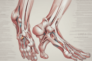
Anatomy of the Leg
- The leg supports body weight and allows for locomotion and balance.
- The pelvic bone is comprised of the ilium, pubis, and ischium.
- The femur is the largest bone in the body, connecting the hip and knee.
- The deep fascia of the thigh, fascia lata, limits outward extension of contracting muscles.
- The iliotibial tract extends from the iliac crest to below the knee, and is key in keeping the leg extended.
- The superficial veins of the leg include the great saphenous, small saphenous, and short saphenous vein.
- The great saphenous vein runs up the medial leg, while the small saphenous vein runs up the lateral leg.
- Varicose veins are dilated, elongated, and tortuous superficial veins with incompetent or absent valves.
- The femoral triangle is an area of muscle in the upper thigh, located between the inguinal ligament, sartorius, and adductor longus muscles.
- The femoral triangle contains the femoral nerve, artery, vein and lymph nodes.
- The femoral sheath surrounds the femoral artery and vein, and is continuous with transversals fascia superiorly.
- Femoral hernias occur when abdominal content protrudes through the femoral ring.
- The femoral canal is a weak area where femoral hernias can occur, specifically in females.
- The obturator nerve (L2-L4) innervates the adductor muscles and sensory nerves in the medial thigh.
- The femoral nerve (L2-L4) innervates the sartorius, pectineus, quadriceps muscles, and provides sensory innervation to the anterior thigh and medial leg.
- The saphenous nerve is a branch of the femoral nerve and provides sensory innervation to the medial leg.
Blood Supply of the Leg
- The external iliac artery becomes the femoral artery, palpable at the mid-inguinal point.
- The femoral artery passes through the adductor canal and hiatus and becomes the popliteal artery.
- The medial circumflex artery branches from the femoral artery, supplying the medial thigh muscles.
- The lateral circumflex artery branches from the femoral artery, supplying the lateral thigh muscles.
- The deep femoral artery (profunda femoris), a major branch of the femoral artery, supplies the posterior thigh muscles.
- The popliteal artery branches into the anterior tibial artery, posterior tibial artery, and fibular artery.
Common Leg Injuries
- Patellar fracture: Commonly caused by a hard blow to the knee, can be displaced or non-displaced.
- Patellar dislocation: Usually due to a direct blow or sudden twist of the leg, causing the patella to slip out of its groove.
Cases
- Case 1: A 25-year-old man with pain, swelling, and bruising to the front of the knee and two lumps, likely indicates a patellar fracture and dislocation following a car accident.
- Case 2: A 20-year-old man with knee pain, swelling, and bruising after a rugby game, could indicate a patellar fracture or dislocation.
Studying That Suits You
Use AI to generate personalized quizzes and flashcards to suit your learning preferences.
Description
This quiz explores the anatomy of the leg, including key components such as bones, muscles, and veins. You'll learn about the structure of the pelvic bone, the significance of the femur, and the role of various veins in the leg. Test your knowledge on these important aspects of human anatomy.