Podcast
Questions and Answers
Which muscle elevates the hyoid bone?
Which muscle elevates the hyoid bone?
- Pretracheal
- Sternothyroid
- Infrahyoid
- Suprahyoid (correct)
What is the function of the sternothyroid muscle?
What is the function of the sternothyroid muscle?
- Elevates the larynx
- Elevates the hyoid bone
- Depresses the hyoid bone
- Depresses the larynx (correct)
Which fascial layer encloses the platysma?
Which fascial layer encloses the platysma?
- Carotid sheath
- Superficial cervical fascia (correct)
- Pretracheal fascia
- Prevertebral fascia
What is the innervation of the muscles of facial expression in the platysma?
What is the innervation of the muscles of facial expression in the platysma?
Which of the following fascial layers is also known as the investing or superficial layer?
Which of the following fascial layers is also known as the investing or superficial layer?
What is the name of the fascial layer that encloses the thyroid gland?
What is the name of the fascial layer that encloses the thyroid gland?
Which fascial layer forms the fascial floor of the posterior triangle?
Which fascial layer forms the fascial floor of the posterior triangle?
What is the relationship between the alar fascia and the prevertebral fascia?
What is the relationship between the alar fascia and the prevertebral fascia?
What is the function of the carotid sheath?
What is the function of the carotid sheath?
What structure does the retropharyngeal space extend to inferiorly?
What structure does the retropharyngeal space extend to inferiorly?
Which space lies between the alar fascia and the prevertebral fascia?
Which space lies between the alar fascia and the prevertebral fascia?
What is the primary space involved in Ludwig's angina?
What is the primary space involved in Ludwig's angina?
What is the relationship between the pretracheal fascia and the visceral compartment?
What is the relationship between the pretracheal fascia and the visceral compartment?
What is the name of the layer that is a component of the pretracheal layer?
What is the name of the layer that is a component of the pretracheal layer?
Which of the following structures is NOT part of the neurovascular arrangement in the carotid sheath?
Which of the following structures is NOT part of the neurovascular arrangement in the carotid sheath?
What is the only bone that does not articulate with another bone?
What is the only bone that does not articulate with another bone?
Which muscles are enclosed by the superficial layer of the deep cervical fascia?
Which muscles are enclosed by the superficial layer of the deep cervical fascia?
What is the action of the Sternocleidomastoid muscle?
What is the action of the Sternocleidomastoid muscle?
What is the location of the Suprasternal Space of Burns?
What is the location of the Suprasternal Space of Burns?
What is the function of the Prevertebral fascia?
What is the function of the Prevertebral fascia?
What is the significance of the potential spaces between the layers of deep fascia?
What is the significance of the potential spaces between the layers of deep fascia?
Which of the following muscles is NOT a suprahyoid muscle?
Which of the following muscles is NOT a suprahyoid muscle?
What is the level of the hyoid bone?
What is the level of the hyoid bone?
What is the function of the Trapezius muscle?
What is the function of the Trapezius muscle?
What is the name of the space between the layers of deep cervical fascia that is a potential site for the spread of infection?
What is the name of the space between the layers of deep cervical fascia that is a potential site for the spread of infection?
Flashcards are hidden until you start studying
Study Notes
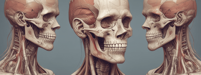
Hyoid Bone and Muscles
- The hyoid bone is the only bone that does not articulate with another bone.
- It is U-shaped, consisting of paired lesser horns laterally and paired greater horns posteriorly.
- It serves as a movable base for the tongue and is located at the level of C3.
Suprahyoid and Infrahyoid Muscles
- Suprahyoid muscles (digastric, stylohyoid, mylohyoid, geniohyoid) elevate the hyoid bone.
- Infrahyoid muscles (sternothyroid, thyrohyoid) depress the hyoid bone, except for the sternothyroid, which depresses the larynx.
Fascial Layers of the Neck
- Superficial cervical fascia:
- Encloses the platysma, a thin broad muscular sheet.
- Part of the muscles of facial expression, innervated by the facial nerve.
- Deep cervical fascia:
- Divides into anterior and posterior parts, attaching to the borders of the suprasternal notch.
- Has four layers: investing, pretracheal, prevertebral, and alar fascia.
Hyoid Muscles
- Investing/enveloping layer:
- Encircles the deeper parts of the neck.
- Splits to enclose the trapezius and sternocleidomastoid muscles.
- Encloses the submandibular and parotid glands.
- Suprahyoid muscles:
- Include digastric, stylohyoid, mylohyoid, and geniohyoid.
- Infrahyoid muscles:
- Include sternothyroid and thyrohyoid.
Sternocleidomastoid Muscle
- A strong, thick muscle crossing the neck.
- Anterior border covers the carotid arteries, IJV, deep cervical LN, and partly overlaps the thyroid gland.
- Posterior border relates to the cervical plexus of nerves, phrenic nerve, and upper part of the brachial plexus.
- Action: head rotation to the opposite side and flexion of the neck.
Trapezius Muscle
- A large triangular paired muscle located on the posterior aspect of the neck and thorax.
- Action:
- Upper part: elevates and upwardly rotates the scapula.
- Middle part: adducts the scapula.
- Lower part: depresses and helps in rotating the scapula.
Deep Fascia of the Neck
- Pretracheal fascia:
- Thin layer attached superiorly to the thyroid and cricoid cartilages and inferiorly to the pericardium.
- Surrounds the larynx and trachea, encloses the thyroid and parathyroid glands, and forms a sheath.
- Carotid sheath:
- Local condensation of the prevertebral, pretracheal, and investing layers of the deep cervical fascia.
- Contains the common carotid artery, internal carotid artery, and vagus nerve.
Retropharyngeal and Danger Space
- Potential space bounded by deep cervical fascia.
- Lies between the vertebral and visceral compartments and contains loose connective tissue.
- Anatomical relationships:
- Anterior: visceral part of the pretracheal fascia.
- Posterior: prevertebral fascia.
- Lateral: carotid sheaths.
Clinical Correlations
- Ludwig's angina:
- Infection of the floor of the mouth underneath the tongue.
- Derived from dental infections, especially the 2nd and 3rd molars.
- Infection spreads contiguously to other spaces.
- Torticollis / Wryneck:
- Contraction or shortening of the sternocleidomastoid muscle.
- Due to injury to the SCM or avulsion of the accessory nerve at birth.
- Twisting of the neck with the chin pointing upward and to the opposite side.
Studying That Suits You
Use AI to generate personalized quizzes and flashcards to suit your learning preferences.


