Podcast
Questions and Answers
What is the primary function of the papillary muscles in relation to the tricuspid valve?
What is the primary function of the papillary muscles in relation to the tricuspid valve?
- To prevent the valve from inverting during ventricular contraction (correct)
- To connect the valve to the aorta
- To facilitate the opening and closing of the valve
- To allow for greater blood flow through the valve
Why are the walls of the left ventricle thicker than those of the right ventricle?
Why are the walls of the left ventricle thicker than those of the right ventricle?
- To support the weight of the heart
- To accommodate less blood volume
- To enable more effective pumping to the lungs
- To allow for more forceful contractions pumping blood to the body (correct)
What prevents backflow of blood from the aorta to the left ventricle?
What prevents backflow of blood from the aorta to the left ventricle?
- The mitral valve
- The pulmonary semilunar valve
- The chordae tendineae
- The aortic semilunar valve (correct)
What role does the fibrous skeleton of the heart play?
What role does the fibrous skeleton of the heart play?
What happens when the mitral valve closes during left ventricular contraction?
What happens when the mitral valve closes during left ventricular contraction?
What is the primary role of the mitral valve during the cardiac cycle?
What is the primary role of the mitral valve during the cardiac cycle?
What hormone is produced by the stretched walls of the atria in response to increased blood volume?
What hormone is produced by the stretched walls of the atria in response to increased blood volume?
How does ANP affect sodium ion reabsorption in the kidneys?
How does ANP affect sodium ion reabsorption in the kidneys?
What prevents backflow of blood from the pulmonary artery into the right ventricle during ventricular relaxation?
What prevents backflow of blood from the pulmonary artery into the right ventricle during ventricular relaxation?
Which artery carries blood from the right ventricle to the lungs?
Which artery carries blood from the right ventricle to the lungs?
What is the function of chordae tendineae in the heart's ventricles?
What is the function of chordae tendineae in the heart's ventricles?
During the contraction of the right ventricle, which structure opens to allow blood flow to the lungs?
During the contraction of the right ventricle, which structure opens to allow blood flow to the lungs?
What is the primary effect of ANP on blood volume and pressure?
What is the primary effect of ANP on blood volume and pressure?
Which structure prevents the backflow of blood from the left ventricle to the left atrium during ventricular contraction?
Which structure prevents the backflow of blood from the left ventricle to the left atrium during ventricular contraction?
What is the function of the epicardium in the heart's anatomy?
What is the function of the epicardium in the heart's anatomy?
From where does the right atrium receive deoxygenated blood?
From where does the right atrium receive deoxygenated blood?
Which statement about the fibrous skeleton of the heart is true?
Which statement about the fibrous skeleton of the heart is true?
What is the role of papillary muscles and chordae tendineae in the heart?
What is the role of papillary muscles and chordae tendineae in the heart?
Which artery branches directly from the ascending aorta?
Which artery branches directly from the ascending aorta?
Where do coronary veins empty blood after it has traveled through coronary capillaries?
Where do coronary veins empty blood after it has traveled through coronary capillaries?
What prevents backflow of blood from the pulmonary artery to the right ventricle?
What prevents backflow of blood from the pulmonary artery to the right ventricle?
Flashcards are hidden until you start studying
Study Notes
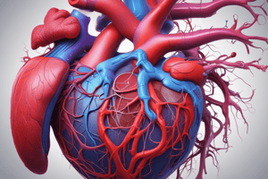
Anatomy of the Heart
- Epicardium: Serous membrane covering the myocardium.
- Myocardium: The muscular layer of the heart, responsible for forming the walls of all four chambers.
- Endocardium: Lining of the heart chambers and valves, smooth to prevent clotting.
- Right Atrium (RA): Receives deoxygenated blood from the body through superior and inferior caval veins.
- Tricuspid Valve: Right atrioventricular (AV) valve that prevents backflow of blood into the RA during RV contraction.
- Right Ventricle (RV): Pumps deoxygenated blood to the lungs via the pulmonary artery.
- Pulmonary Semilunar Valve: Prevents backflow from the pulmonary artery to the RV during relaxation of the RV.
- Left Atrium (LA): Receives oxygenated blood from the lungs through the four pulmonary veins.
- Mitral Valve: Left AV valve preventing backflow of blood into the LA when the LV contracts.
- Left Ventricle (LV): Pumps oxygenated blood to the body through the aorta, the largest artery.
- Aortic Semilunar Valve: Prevents backflow from the aorta into the LV during relaxation.
- Papillary Muscles and Chordae Tendineae: Work together in both ventricles to prevent AV valve inversion during contraction.
- Fibrous Skeleton of the Heart: Connective tissue that anchors heart valves and insulates electrical impulses between atria and ventricles.
Coronary Vessels
- Coronary Arteries: First branches of the ascending aorta; supply blood to the heart muscle.
- Blood Flow: Coronary arteries branch into smaller arteries, arterioles, and capillaries, merging to form coronary veins.
- Coronary Sinus: Collects blood from coronary veins and returns it to the right atrium.
Left Ventricle
- Wall Thickness: Thicker than the right ventricle for more powerful contractions to pump blood throughout the body.
- Aortic Valve: Located between the LV and aorta; opens during LV contraction and closes during relaxation to prevent backflow.
Left Atrium
- Function: Receives oxygenated blood and contributes to blood pressure regulation via hormone production.
- Atrial Natriuretic Peptide (ANP): Produced when atrial walls are stretched; promotes sodium excretion and lowers blood volume and pressure.
Right Ventricle
- Function: Pumps deoxygenated blood to the lungs through the pulmonary artery during contraction.
- Pulmonary Valve: Opens with RV contraction; prevents backflow into the RV during relaxation.
- Myocardium Columns: Papillary muscles and chordae tendineae project into the RV, supporting tricuspid valve function.
Studying That Suits You
Use AI to generate personalized quizzes and flashcards to suit your learning preferences.