Podcast
Questions and Answers
What is the primary function of the cornea in relation to light?
What is the primary function of the cornea in relation to light?
- To absorb light and convert it into energy
- To focus light exiting the eye
- To reflect light away from the eye
- To bend or refract light that enters the eye (correct)
What is the main composition of the cornea in primates?
What is the main composition of the cornea in primates?
- Fat cells
- Muscle cells
- Collagen (correct)
- Nerve cells
What is the purpose of the cornea beyond its optical function?
What is the purpose of the cornea beyond its optical function?
- To produce tears
- To regulate eye pressure
- To control the movement of the eye
- To provide a physical barrier for the eye (correct)
What is the location of the cornea in the eye?
What is the location of the cornea in the eye?
What is the meaning of the Latin word 'cornu' or 'cornea tela'?
What is the meaning of the Latin word 'cornu' or 'cornea tela'?
What is the primary goal of treatment for Fuch's dystrophy?
What is the primary goal of treatment for Fuch's dystrophy?
What is the role of keratocytes in the cornea?
What is the role of keratocytes in the cornea?
What is the characteristic shape of the cornea in Keratoconus?
What is the characteristic shape of the cornea in Keratoconus?
What is the treatment for Keratoconus?
What is the treatment for Keratoconus?
What is the underlying cause of Fuch's dystrophy?
What is the underlying cause of Fuch's dystrophy?
What is the function of the corneal epithelium?
What is the function of the corneal epithelium?
What is the primary component of the corneal stroma?
What is the primary component of the corneal stroma?
What is the function of Dua's layer?
What is the function of Dua's layer?
What is the characteristic of the Descemet's membrane?
What is the characteristic of the Descemet's membrane?
What is the function of the corneal endothelium?
What is the function of the corneal endothelium?
Study Notes
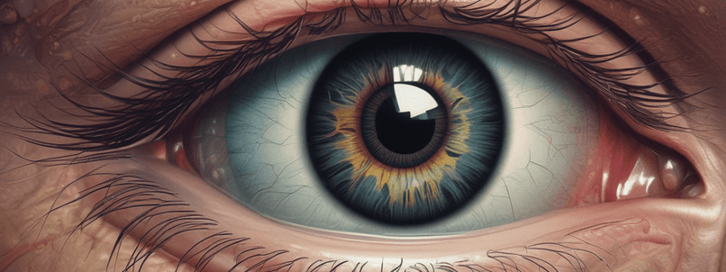
The Cornea of the Eye
- The cornea is the clear, firm outermost layer of the eye that encounters the external environment.
- It is derived from the Latin word "cornu" or "cornea tela", meaning horn or horny tissue.
Cornea Function
- The cornea has three important functions:
- to bend or refract light that enters the eye
- to focus light entering the eye
- to provide a physical barrier for the eye and protect it from germs, dust, etc.
Location and Structure of the Cornea
- The cornea is located at the anterior-most part of the eye at the surface that is exposed to the external environment.
- It covers the iris, pupil, and the anterior chamber of the eye.
- It is connected to the sclera and the junction between them is known as the corneal limbus.
- The structure of the cornea is mostly made of collagen and includes six distinct layers.
Layers of the Cornea
- The layers of the cornea from outermost to innermost are:
- Corneal epithelium
- Bowman's layer
- Corneal stroma
- Dua's layer
- Descemet's membrane
- Corneal endothelium
Corneal Epithelium
- The corneal epithelium is made of epithelial cells known as stratified squamous epithelium.
- It is non-keratinized tissue made of about six layers of cells.
- It regenerates quickly and easily at the basal layer allowing for quick healing of the outer surface of the eye.
- It stays wet due to lubrication from tears.
- Its function is to allow for refraction or bending of the light that enters the eye.
Bowman's Layer
- The Bowman's layer is a connective tissue layer which consists of randomly laid collagen fibrils.
- It also contains proteins like laminin, nidogen, and perlecan.
- It does not contain any cells and its function is to protect the corneal stroma underneath.
- This layer is not present in the eye of non-primates.
Stroma
- The corneal stroma is the thickest layer of the cornea making up approximately 90% of the entire corneal thickness.
- It contains connective tissue in the form of collagen fibrils that are laid down in a regular pattern.
- It contains keratocytes.
- These cells help with cellular regeneration.
- Its function is to provide bulk of corneal structure and to allow repair and regeneration of cells and tissue.
Dua's Layer
- The Dua's layer is a very thin layer of cornea.
- It is shown to be very strong withstanding high air pressure and having minimal effects to outside air.
- Its function is to provide protection to structures and layers underneath it.
- This layer was discovered in 2013 by Dr. Harminder Dua at University of Nottingham during transplant research.
Descemet's Membrane
- The Descemet's membrane is a layer of connective tissue and contains collagen Type 4 fibrils.
- Its function is to serve as basement membrane for the corneal endothelium underneath.
- The cells for the endothelium are derived from this layer.
- It is also known as the posterior limiting membrane.
Corneal Endothelium
- The corneal endothelium is the sixth and final layer of the cornea.
- It contains a single layer of simple squamous or cuboidal layer of cells.
- It is surrounded by fluid known as aqueous humor.
- Its function is to allow movement of fluid and salt between the stromal layer and the aqueous humor.
- This layer is different from the epithelium in that it does not regenerate.
Corneal Disorders
- The structure of the cornea is susceptible to physical damage due to its location on the surface of the eye.
- Some common problems associated with cornea include:
- Corneal abrasion: loss of surface of cornea, esp. the epithelial layer, due to a scratch or another trauma to the outer surface of the eye.
- Keratitis: corneal inflammation.
- Serious disorders that can cause permanent degeneration of the cornea include:
- Keratoconus: a degenerative disease where the cornea undergoes thinning and changes shape from a semicircle to a cone.
- Fuch's dystrophy: a genetic condition characterized by cloudy vision isolated to morning time only.
Studying That Suits You
Use AI to generate personalized quizzes and flashcards to suit your learning preferences.
Description
Discover the importance of the cornea, the outermost structure of the eye, and how it contributes to our sense of sight. Learn about the different parts of the eye and their functions. Test your knowledge of the human eye and its anatomy.


