Podcast
Questions and Answers
What is the key difference in movement between a uniaxial joint and a biaxial joint?
What is the key difference in movement between a uniaxial joint and a biaxial joint?
A uniaxial joint allows movement in one plane, while a biaxial joint allows movement in two planes.
What is the characteristic shape of the articular surfaces in a saddle joint?
What is the characteristic shape of the articular surfaces in a saddle joint?
Convex and concave regions that resemble the shape of a saddle.
What type of joint is the proximal radioulnar joint, and what movement does it permit?
What type of joint is the proximal radioulnar joint, and what movement does it permit?
A pivot joint, which permits the radius to rotate.
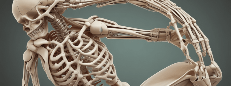
In which plane does a hinge joint, such as the elbow joint, allow movement?
In which plane does a hinge joint, such as the elbow joint, allow movement?
What is the name of the joint that permits the thumb to move toward the other fingers?
What is the name of the joint that permits the thumb to move toward the other fingers?
What type of joint is the intercarpal joint, and what type of movement does it allow?
What type of joint is the intercarpal joint, and what type of movement does it allow?
Which type of joint is considered the most freely mobile type of synovial joint?
Which type of joint is considered the most freely mobile type of synovial joint?
What is the name of the joint that permits the forearm to move only anteriorly toward the arm or posteriorly away from the arm?
What is the name of the joint that permits the forearm to move only anteriorly toward the arm or posteriorly away from the arm?
What is the name of the joint that pivots when you shake your head 'no'?
What is the name of the joint that pivots when you shake your head 'no'?
What is the main difference between a condylar joint and a hinge joint?
What is the main difference between a condylar joint and a hinge joint?
Explain the key structural difference between synovial joints and other types of joints, specifically highlighting the presence or absence of a specific anatomical feature.
Explain the key structural difference between synovial joints and other types of joints, specifically highlighting the presence or absence of a specific anatomical feature.
Describe the composition and function of synovial fluid within a synovial joint. Why is its presence crucial for joint functionality?
Describe the composition and function of synovial fluid within a synovial joint. Why is its presence crucial for joint functionality?
Elaborate on the role of the articular capsule in a synovial joint, focusing on its structure and contribution to joint stability.
Elaborate on the role of the articular capsule in a synovial joint, focusing on its structure and contribution to joint stability.
Explain the significance of articular cartilage in a synovial joint, focusing on its location and function. How does its structure contribute to its role?
Explain the significance of articular cartilage in a synovial joint, focusing on its location and function. How does its structure contribute to its role?
Describe the functional classification of synovial joints. How does this classification relate to their structural features?
Describe the functional classification of synovial joints. How does this classification relate to their structural features?
Discuss the importance of ligaments in a synovial joint, explaining their role in joint stability and movement. Provide an example of a ligament and its function in a specific joint.
Discuss the importance of ligaments in a synovial joint, explaining their role in joint stability and movement. Provide an example of a ligament and its function in a specific joint.
Explain the role of blood vessels and nerves in a synovial joint, focusing on their contribution to joint function and sensation.
Explain the role of blood vessels and nerves in a synovial joint, focusing on their contribution to joint function and sensation.
Compare and contrast the fibrous layer and the synovial membrane of the articular capsule, focusing on their structure and specific functions in a synovial joint.
Compare and contrast the fibrous layer and the synovial membrane of the articular capsule, focusing on their structure and specific functions in a synovial joint.
Describe the structural features that enable tendons to provide stability to joints.
Describe the structural features that enable tendons to provide stability to joints.
What is the function of a bursa in a synovial joint?
What is the function of a bursa in a synovial joint?
Explain how a tendon sheath differs from a bursa.
Explain how a tendon sheath differs from a bursa.
What is the role of fat pads in a synovial joint?
What is the role of fat pads in a synovial joint?
Describe the process of cavitation that occurs when you crack your knuckles.
Describe the process of cavitation that occurs when you crack your knuckles.
What are the three primary functions of synovial fluid within a joint?
What are the three primary functions of synovial fluid within a joint?
Define the terms uniaxial, biaxial, and multiaxial (or triaxial) as they relate to joint movement.
Define the terms uniaxial, biaxial, and multiaxial (or triaxial) as they relate to joint movement.
Identify and briefly describe the six types of synovial joints, from least to most mobile.
Identify and briefly describe the six types of synovial joints, from least to most mobile.
Compare and contrast the structure and movement permitted by a hinge joint and a ball-and-socket joint.
Compare and contrast the structure and movement permitted by a hinge joint and a ball-and-socket joint.
Explain how the structure of a saddle joint allows for a unique range of motion.
Explain how the structure of a saddle joint allows for a unique range of motion.
Explain the importance of the avascular nature of articular cartilage and its implications for healing.
Explain the importance of the avascular nature of articular cartilage and its implications for healing.
How does the structure of synovial fluid contribute to its lubricating and shock-absorbing functions?
How does the structure of synovial fluid contribute to its lubricating and shock-absorbing functions?
Describe the role of proprioceptors in joint function and their contribution to body movement.
Describe the role of proprioceptors in joint function and their contribution to body movement.
Explain the relationship between synovial fluid circulation and articular cartilage health.
Explain the relationship between synovial fluid circulation and articular cartilage health.
Compare and contrast the functions of ligaments and tendons, highlighting their respective roles in the musculoskeletal system.
Compare and contrast the functions of ligaments and tendons, highlighting their respective roles in the musculoskeletal system.
Explain the significance of Hilton's law in understanding the innervation of joints.
Explain the significance of Hilton's law in understanding the innervation of joints.
Discuss the role of nociceptors in joint function and their significance in pain perception.
Discuss the role of nociceptors in joint function and their significance in pain perception.
Explain how the structure of articular cartilage contributes to its function in reducing friction and absorbing compression.
Explain how the structure of articular cartilage contributes to its function in reducing friction and absorbing compression.
Explain the difference between intrinsic and extrinsic ligaments, and provide examples of each.
Explain the difference between intrinsic and extrinsic ligaments, and provide examples of each.
Describe the role of synovial fluid in nourishing articular cartilage and how this process is facilitated by joint movement.
Describe the role of synovial fluid in nourishing articular cartilage and how this process is facilitated by joint movement.
Flashcards are hidden until you start studying
Study Notes
Synovial Joints
- Synovial joints are freely mobile articulations (diarthroses) that permit a wide range of motion
- Examples of synovial joints include the glenohumeral (shoulder) joint, temporomandibular joint, elbow joint, and knee joint
General Anatomy of Synovial Joints
- Synovial joints have several basic features:
- Articular capsule (double-layered capsule)
- Joint cavity (space between bones)
- Synovial fluid (viscous, oily substance)
- Articular cartilage (hyaline cartilage on bone surfaces)
- Ligaments (connective tissue that connects bones)
- Nerves (sensory receptors that detect movement, stretch, and positioning)
- Blood vessels (transport oxygen and nutrients to tissue)
Articular Capsule
- Double-layered capsule:
- Outer layer: fibrous layer (dense connective tissue)
- Inner layer: synovial membrane (specialized connective tissue)
Articular Cartilage
- Thin layer of hyaline cartilage on bone surfaces
- Functions:
- Reduces friction during movement
- Acts as a cushion to absorb compression
- Prevents damage to articulating bone ends
- Lacks perichondrium (avascular, so no blood vessels to bring nutrients)
Synovial Fluid
- Viscous, oily substance within the joint cavity
- Functions:
- Lubricates articular cartilage
- Nourishes chondrocytes (cartilage cells)
- Acts as a shock absorber to distribute stresses
Ligaments
- Composed of dense regular connective tissue
- Functions:
- Stabilize, strengthen, and reinforce joints
- Can be intrinsic (thickenings of articular capsule) or extrinsic (outside the capsule)
Bursae and Fat Pads
- Bursae: saclike structures with synovial fluid that reduce friction
- Fat pads: packing material that provides protection and fills spaces between bones
Classification of Synovial Joints
- Classified by shape of articulating surfaces and movement allowed
- Six types of synovial joints:
- Plane joints (uniaxial, least mobile): e.g., intercarpal and intertarsal joints
- Hinge joints (uniaxial): e.g., elbow joint and finger joints
- Pivot joints (uniaxial): e.g., proximal radioulnar joint and atlantoaxial joint
- Condylar joints (biaxial): e.g., metacarpophalangeal joints (knuckles)
- Saddle joints (biaxial): e.g., carpometacarpal joint of the thumb
- Ball-and-socket joints (multiaxial, most mobile): e.g., coxal (hip) and glenohumeral (shoulder) joints
Studying That Suits You
Use AI to generate personalized quizzes and flashcards to suit your learning preferences.


