Podcast
Questions and Answers
Which muscle is divided into three parts - lumborum, thoracis, and cervicis?
Which muscle is divided into three parts - lumborum, thoracis, and cervicis?
- Multifidus
- Spinalis
- Longissimus
- Iliocostalis (correct)
What is the collective name for the three intermediate intrinsic back muscles?
What is the collective name for the three intermediate intrinsic back muscles?
- Erector spinae (correct)
- Deep back muscles
- Posterior muscles
- Intrinsic muscles
Which muscle attaches to the mastoid process and the occipital bone of the skull?
Which muscle attaches to the mastoid process and the occipital bone of the skull?
- Iliocostalis
- Splenius capitis (correct)
- Splenius cervicis
- Longissimus
What is the function of the deep muscles of the back?
What is the function of the deep muscles of the back?
How many layers can the deep back muscles be divided into?
How many layers can the deep back muscles be divided into?
Which muscle originates from the spinous processes of C7-T3/4 vertebrae?
Which muscle originates from the spinous processes of C7-T3/4 vertebrae?
What is the function of the splenius capitis muscle?
What is the function of the splenius capitis muscle?
What is the role of the deep fascia in relation to the deep muscles of the back?
What is the role of the deep fascia in relation to the deep muscles of the back?
What is the function of the spinalis muscle when it contracts bilaterally?
What is the function of the spinalis muscle when it contracts bilaterally?
Which of the following muscles is the largest of the three columns?
Which of the following muscles is the largest of the three columns?
What is the collective name of the deep intrinsic muscles?
What is the collective name of the deep intrinsic muscles?
Which of the following muscles is located medially within the erector spinae?
Which of the following muscles is located medially within the erector spinae?
What is the function of the spinalis muscle when it contracts unilaterally?
What is the function of the spinalis muscle when it contracts unilaterally?
Which of the following muscles is the most superficial of the deep intrinsic muscles?
Which of the following muscles is the most superficial of the deep intrinsic muscles?
What is the common innervation of the longissimus, spinalis, and iliocostalis muscles?
What is the common innervation of the longissimus, spinalis, and iliocostalis muscles?
Which of the following muscles is NOT part of the deep intrinsic muscles?
Which of the following muscles is NOT part of the deep intrinsic muscles?
Which muscle originates from the transverse processes of C4-T10?
Which muscle originates from the transverse processes of C4-T10?
What is the primary action of the multifidus muscle?
What is the primary action of the multifidus muscle?
Which muscle attaches to the spinous processes of the vertebrae?
Which muscle attaches to the spinous processes of the vertebrae?
What is the primary action of the rotatores muscle?
What is the primary action of the rotatores muscle?
Which muscle is located underneath the semispinalis muscle?
Which muscle is located underneath the semispinalis muscle?
What is the primary action of the interspinales muscle?
What is the primary action of the interspinales muscle?
Which muscle originates from the transverse processes of C7-T11?
Which muscle originates from the transverse processes of C7-T11?
What is the common innervation of the multifidus, rotatores, and spinalis muscles?
What is the common innervation of the multifidus, rotatores, and spinalis muscles?
What is the mortality rate of patients with aortic injuries within 1 week?
What is the mortality rate of patients with aortic injuries within 1 week?
What is the most common site of blunt aortic injuries?
What is the most common site of blunt aortic injuries?
What is the primary mechanism of blunt aortic injuries?
What is the primary mechanism of blunt aortic injuries?
What is the radiographic feature associated with thoracic aortic injury?
What is the radiographic feature associated with thoracic aortic injury?
What percentage of thoracic deaths are attributed to thoracic injury worldwide?
What percentage of thoracic deaths are attributed to thoracic injury worldwide?
What is the management approach for penetrating cardiac injuries?
What is the management approach for penetrating cardiac injuries?
What is a unique characteristic of the pediatric thorax that affects the mechanism of injury?
What is a unique characteristic of the pediatric thorax that affects the mechanism of injury?
What is the treatment for myocardial rupture?
What is the treatment for myocardial rupture?
Which type of injury is associated with shearing, tearing, and traction forces?
Which type of injury is associated with shearing, tearing, and traction forces?
What is the treatment for traumatic VSD?
What is the treatment for traumatic VSD?
What is the primary goal of initial evaluation in thoracic trauma patients?
What is the primary goal of initial evaluation in thoracic trauma patients?
What is the management approach for myocardial contusion?
What is the management approach for myocardial contusion?
What is the primary survey in the ATLS protocol?
What is the primary survey in the ATLS protocol?
What is the characteristic of a flail chest?
What is the characteristic of a flail chest?
What is the management approach for a simple pneumothorax?
What is the management approach for a simple pneumothorax?
What is the characteristic of a sternal fracture?
What is the characteristic of a sternal fracture?
What is the surgical approach for proximal tracheal injuries?
What is the surgical approach for proximal tracheal injuries?
What is the primary presenting symptom of a foreign body in the esophagus?
What is the primary presenting symptom of a foreign body in the esophagus?
What is the management of an acute foreign body in the airway?
What is the management of an acute foreign body in the airway?
What is a common complication of esophageal injuries?
What is a common complication of esophageal injuries?
What is the investigation of choice for diagnosing esophageal injuries?
What is the investigation of choice for diagnosing esophageal injuries?
What is the surgical approach for lower esophageal injuries?
What is the surgical approach for lower esophageal injuries?
What is the indication for surgical repair in tracheobronchial tree injuries?
What is the indication for surgical repair in tracheobronchial tree injuries?
What is the common presentation of diaphragmatic injuries?
What is the common presentation of diaphragmatic injuries?
What is the management of chronic foreign body in the esophagus?
What is the management of chronic foreign body in the esophagus?
What is the common site of diaphragmatic injuries?
What is the common site of diaphragmatic injuries?
Flashcards are hidden until you start studying
Study Notes
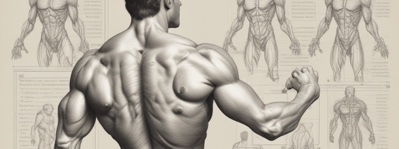
Back Muscles
- The back muscles can be categorized into three layers: superficial, intermediate, and deep.
Superficial Muscles
- The superficial muscles are also known as the spinotransversales.
- There are two muscles in this group: splenius capitis and splenius cervicis.
- They are associated with movements of the head and neck.
- Located on the posterolateral aspect of the neck, covering the deeper neck muscles.
Splenius Capitis
- Originates from the lower aspect of the ligamentum nuchae and the spinous processes of C7-T3/4 vertebrae.
- Fibers ascend, attaching to the mastoid process and the occipital bone of the skull.
- Innervated by posterior rami of spinal nerves C3 and C4.
- Actions: Rotate head to the same side.
Splenius Cervicis
- Originates from the spinous processes of T3-T6 vertebrae.
- Fibers ascend, attaching to the transverse processes of C1-3/4.
Intermediate Muscles
- There are three intermediate intrinsic back muscles: iliocostalis, longissimus, and spinalis.
- Together, these muscles form a column known as the erector spinae.
- The erector spinae is situated posterolaterally to the spinal column, between the vertebral spinous processes and the costal angle of the ribs.
Iliocostalis
- Located laterally within the erector spinae.
- Associated with the ribs.
- Can be divided into three parts: lumborum, thoracis, and cervicis.
Longissimus
- Situated between the iliocostalis and spinalis.
- Largest of the three columns.
- Can be divided into three parts: thoracic, cervicis, and capitis.
- Attachments: Arises from the common tendinous origin and attaches to the lower ribs, the transverse processes of C2-T12, and the mastoid process of the skull.
- Innervation: Posterior rami of the spinal nerves.
- Actions: Acts unilaterally to laterally flex the vertebral column, and acts bilaterally to extend the vertebral column and head.
Spinalis
- Located medially within the erector spinae.
- Smallest of the three muscle columns.
- Can be divided into the thoracic, cervicis, and capitis.
- Attachments: Arises from the common tendinous origin, and attaches to the spinous processes of C2, T1-T8, and the occipital bone of the skull.
- Innervation: Posterior rami of the spinal nerves.
- Actions: Acts unilaterally to laterally flex the vertebral column, and acts bilaterally to extend the vertebral column and head.
Deep Intrinsic Muscles
- Located underneath the erector spinae.
- Known collectively as the transversospinales.
- Three major muscles in this group: semispinalis, multifidus, and rotatores.
Semispinalis
- Most superficial of the deep intrinsic muscles.
- Can be divided by its superior attachments into thoracic, cervicis, and capitis.
- Attachments: Originates from the transverse processes of C4-T10.
- Fibers ascend 4-6 vertebral segments, attaching to the spinous processes of C2-T4 and the occipital bone of the skull.
- Innervation: Posterior rami of the spinal nerves.
- Actions: Extends and contralaterally rotates the head and vertebral column.
Multifidus
- Located underneath the semispinalis muscle.
- Best developed in the lumbar area.
- Attachments: Has a broad origin – arises from the sacrum, posterior iliac spine, common tendinous origin of the erector spinae, mamillary processes of lumbar vertebrae, transverse processes of T1-T3, and articular processes of C4-C7.
- Fibers ascend 2-4 vertebral segments, attaching to the spinous processes of the vertebrae.
- Innervation: Posterior rami of the spinal nerves.
- Actions: Stabilizes the vertebral column.
Rotatores
- Most prominent in the thoracic region.
- Attachments: Originates from the vertebral transverse processes.
- Fibers ascend, and attach to the lamina and spinous processes of the immediately superior vertebrae.
- Innervation: Posterior rami of the spinal nerves.
- Actions: Stabilizes the vertebral column and has a proprioceptive function.
Minor Deep Intrinsic Muscles
- Interspinales: Spans between adjacent spinous processes, and acts to stabilize the vertebral column.
- Intertranversari: Spans between adjacent transverse processes, and acts to stabilize the vertebral column.
- Levatores costarum: Originates from the transverse processes of C7-T11, and attaches to the rib immediately below, and acts to elevate the ribs.
Tracheobronchial Tree Injuries
- Can be conservative or surgical, depending on the severity of the injury
- Surgical approach is based on the location and extension of the injury
- Cervical collar incision for proximal trachea
- Right postero-lateral thoracotomy for lower trachea, carina, and proximal right main bronchus
- Left postero-lateral thoracotomy for distal left main bronchus
Foreign Body in the Airway
- Management depends on the severity of the injury
- Acute: Urgent bronchoscopy with or without bronchotomy
- Chronic: Bronchoscopy with precaution, with or without lung resection
Esophagus Injuries
- Rare, but can occur due to blunt or penetrating trauma
- Cervical esophageal injuries are the most common
- Symptoms include:
- Pneumothorax (left)
- Hemorrhage without rib fractures
- Lower sternum or epigastric pain (severe blunt trauma)
- Particulate matter in the ICD
- Penetrating injury that has crossed the mediastinum
- Odynophagia
- Dysphagia
- Surgical emphysema
- Mediastinitis
- Investigations:
- Combination of clinical suspicion, CXR, water-soluble contrast swallow, and oesophagoscopy
- Management:
- Timing: 24 hours, debride and drainage, surgical repair or resection with delayed reconstruction
- Approach: Right postero-lateral thoracotomy (RPLT) for upper esophagus, left postero-lateral thoracotomy (LPLT) for lower esophagus
Complications of Esophagus Injuries
- Mediastinal contamination
- Abscess formation
- Empyema thoracis
Foreign Body in the Esophagus
- Types: bone, meat, battery, coin
- Clinical presentation:
- Acute: dysphagia, choking, hematemesis
- Chronic: hemoptysis, coughing when feeding
- Management:
- Oesophagoscopy with or without mediastinal drainage and repair
Diaphragmatic Injuries
- Often occult, easily missed, especially on the left side
- Marker of severe thoracoabdominal trauma
- Causes:
- Blunt trauma
- Penetrating trauma (stab or iatrogenic)
- Clinical features:
- With or without signs of bowel obstruction, drainage of peritoneal content via chest drain
- NGT in the chest (CXR)
- Herniation of GIT
- Acute, delayed, common left
- Investigations:
- CXR: elevated hemidiaphragm, hemo-pneumothorax
- Swallow and follow through
- Contrast-enhanced CT scan
- Management:
- Surgical repair: thoracotomy, thoraco-abdominal incision, or laparotomy, laparoscopy
Cardiac Injuries
- Penetrating:
- Myocardial contusion, transient arrhythmias
- Valve injuries
- IVS rupture
- Blunt:
- Myocardial contusion, patchy areas of muscle necrosis, hemorrhagic infiltrate, rupture of small vessels
- Hemorrhage into the interstitium and around the muscle fibers
- Investigations:
- Admit to HCU/ICU for monitoring
- ECG, cardiac enzymes
- Treat dysarrhythmias and heart failure
- Formal ECHO/TEE
- Management:
- Elective or urgent surgery
- Myocardial rupture: simple cardiorrhaphy, pledgetted suture
- Mitral valve: repair or replacement
- Tricuspid valve: repair
- IVS: traumatic VSD, closure for larger ones, bypass or not
Surgery
- Elective or urgent
- Myocardial rupture: simple cardiorrhaphy, pledgetted suture
- Mitral valve: repair or replacement
- Tricuspid valve: repair
- IVS: traumatic VSD, closure for larger ones, bypass or not
Great Vessel Injuries
- Aorta is the most commonly injured in severe blunt or penetrating trauma
- 85-95% mortality
- Typically, patients survive the initial injury insult
- Mortality rates:
- 30% within 6 hours
- 50% within 24 hours
- 70% within 1 week
Blunt Aortic Injuries
- Mechanisms:
- Acceleration-deceleration
- Production of shearing forces
- Direct luminal compression - fixation points
- Site: isthmus, near ligamentum arteriosum
- Clinical features:
- Death on the scene - rapid exsanguination
- Expanding thoracic inlet hematoma, bruit, hypotension, pulse deficit
Aortic Disruption
- Radiographic features associated with thoracic aortic injury:
- Loss of aortic knuckle contour
- Widened mediastinum
- Obliteration of aorto-pulmonary window
- 1st/2nd rib fracture
- Depression of the right main bronchus
- Deviated NGT and tracheal displacement to the right
- Widened paratracheal stripe
- Left massive hemithorax
- Left pleural cap
Management
- Medical: endovascular stents
- Surgical: open surgical procedures
Thoracic Trauma
- Accounts for 20-25% of thoracic deaths worldwide
- Male vs female
- Age
- Blunt, penetrating, transfixing
Mechanism of Injury
-
- Penetrating:
- High velocity - gunshots
- Low velocity - stab wounds
-
- Blunt:
- Direct:
- Assault and blast
- Indirect:
- Falls, MVA (acceleration-deceleration injuries, crush injuries, and shearing forces)
-
- Transfixing
Special Factors
- Pediatric thorax: more cartilage, absorbs forces
- Geriatric thorax: calcification and osteoporosis, more fractures
Initial Evaluation
- Goal: prompt identification of life-threatening injuries
- Pathology:
- Airway obstruction
- Loss of oxygenation or ventilation
- Hypovolaemia
- Obstructive shock
- Ventilation-perfusion mismatch
- Physiological causes of death:
- Tissue hypoxia
- Hypercarbia
- Metabolic acidosis
Traumatic Rib Fractures
- Other bony fractures of the chest wall
- Sternal fractures:
- Up to 4%
- Transverse, in the upper or midportions
- Associated injuries: myocardium
- Cf: point of tenderness, swelling, and deformity
- Sternal fractures:
Studying That Suits You
Use AI to generate personalized quizzes and flashcards to suit your learning preferences.


