Podcast
Questions and Answers
What is the primary role of blood plasma in tissue perfusion?
What is the primary role of blood plasma in tissue perfusion?
- To transport hormones throughout the body.
- To generate heat for the body.
- To transport nutrients and oxygen while removing waste. (correct)
- To act as a buffer for pH control.
Which factor does NOT influence tissue perfusion?
Which factor does NOT influence tissue perfusion?
- Muscle mass of the patient. (correct)
- Peripheral vascular resistance.
- Cardiac pump function.
- Circulating blood volume.
How is tissue perfusion specifically measured?
How is tissue perfusion specifically measured?
- In ml/100g-min. (correct)
- In liters per hour.
- In grams per liter.
- In mmHg.
What is a primary condition that can lead to impaired tissue perfusion?
What is a primary condition that can lead to impaired tissue perfusion?
Which heart valve is responsible for preventing backflow from the left ventricle?
Which heart valve is responsible for preventing backflow from the left ventricle?
Which vessels carry oxygenated blood back to the heart?
Which vessels carry oxygenated blood back to the heart?
Which population is at higher risk for ineffective tissue perfusion?
Which population is at higher risk for ineffective tissue perfusion?
Which of the following can cause impaired blood flow due to a physical blockage?
Which of the following can cause impaired blood flow due to a physical blockage?
What leads to insufficient tissue perfusion, also known as ischemia?
What leads to insufficient tissue perfusion, also known as ischemia?
What sign may indicate that a patient is experiencing shock?
What sign may indicate that a patient is experiencing shock?
What is a consequence of chronic insufficient tissue perfusion?
What is a consequence of chronic insufficient tissue perfusion?
Which medication type can decrease distal perfusion causing risk to tissues?
Which medication type can decrease distal perfusion causing risk to tissues?
Which statement accurately describes the relationship between tissue perfusion and cellular function?
Which statement accurately describes the relationship between tissue perfusion and cellular function?
Which early warning sign is specifically associated with renal perfusion?
Which early warning sign is specifically associated with renal perfusion?
What treatment might be used to address volume loss in hemorrhagic shock?
What treatment might be used to address volume loss in hemorrhagic shock?
What is a crucial factor in diagnosing hemorrhagic shock?
What is a crucial factor in diagnosing hemorrhagic shock?
Which of the following conditions is NOT a result of inadequate tissue perfusion?
Which of the following conditions is NOT a result of inadequate tissue perfusion?
Which lifestyle factor is associated with an increased risk of ineffective tissue perfusion?
Which lifestyle factor is associated with an increased risk of ineffective tissue perfusion?
How is gastrointestinal perfusion indicated during a physical examination?
How is gastrointestinal perfusion indicated during a physical examination?
Which of the following is an evidence of reduced tissue perfusion?
Which of the following is an evidence of reduced tissue perfusion?
What effect does decreased tissue perfusion have on cellular energy production?
What effect does decreased tissue perfusion have on cellular energy production?
Which chambers of the heart receive deoxygenated blood?
Which chambers of the heart receive deoxygenated blood?
What physiological process is critically affected by decreased oxygenation due to inadequate tissue perfusion?
What physiological process is critically affected by decreased oxygenation due to inadequate tissue perfusion?
What can result from excessive perfusion (hyperemia)?
What can result from excessive perfusion (hyperemia)?
Study Notes
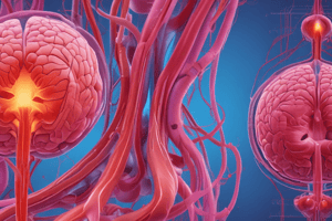
Tissue Perfusion
- Blood circulation through the vascular bed tissue
- Essential for organ functions: urine formation, muscle contraction, oxygen and carbon dioxide exchange
- Dependent on blood flow, influenced by circulating volume, cardiac pump function, and peripheral vascular resistance
Anatomy and Physiology of the Heart
- Veins: Carry blood towards the heart
- Arteries: Carry blood away from the heart
- Atria: Two upper chambers of the heart
- Ventricles: Two lower chambers of the heart
- 4 Main Valves: Prevent backflow of blood
- Tricuspid Valve: Exits right atrium
- Pulmonic Valve: Exits right ventricle
- Mitral (bicuspid) Valve: Exits left atrium
- Aortic Valve: Exits left ventricle
- SVC (Superior Vena Cava): Blood from upper body (head, neck, upper limbs, chest)
- IVC (Inferior Vena Cava): Blood from lower body (trunk, viscera, lower limbs)
- Blue: Deoxygenated blood (SVC, IVC, pulmonary artery)
- Red: Oxygenated blood
Blood Flow of the Heart
- Superior Vena Cava/Inferior Vena Cava → Right Atrium → Tricuspid Valve → Right Ventricle → Pulmonary Valve → Pulmonary Artery → Right & Left Pulmonary Arteries (branched from Pulmonary Arteries) → Lungs = Gas Exchange → Pulmonary Veins → Left Atrium → Mitral Valve → Left Ventricle → Aortic Semilunar Valve → Ascending Aorta → Aortic Arch (3 major branches) → Descending Aorta (chest to abdomen, splits to provide blood to pelvis and lower limbs)
Tissue Perfusion
- Actual gas exchange of oxygen and carbon dioxide occurs in capillaries (small blood vessels)
- When cells stop getting adequate oxygen supply, tissue perfusion is altered
Altered Tissue Perfusion
- Insufficient Perfusion (Ischemia): Demand exceeds supply, inducing oxygen debt and toxic cellular waste buildup, stressing tissue vitality. Untreated ischemia leads to cellular dysfunction, loss of organ function, and cell death (tissue infarction).
- Excessive Perfusion (Hyperemia): Supply exceeds demand, often associated with edema formation in the tissue
- Decreased tissue perfusion occurs when arteries lack proper nutrients or oxygen supply, leading to long-term (chronic) tissue damage, organ damage, or even death.
- Ineffective Tissue Perfusion: Insufficient blood flow, decreased oxygen, or nutrients on a cellular level
- 4 Early Warning Signs of Ineffective Tissue Perfusion:
- Renal perfusion: Regulates urine output
- Gastrointestinal perfusion: Change in bowel sounds, possibly nausea
- Peripheral perfusion: Blood flow to arms and legs
- Cerebral perfusion: Blood flow to the brain, leading to dizziness
- Consequences of Ineffective Tissue Perfusion:
- Temporary: Minimal to no consequences
- Acute: Destructive effect on the patient
- Chronic: Tissue organ damage
- Death
What Happens When Tissue Perfusion is Decreased?
- Decreased tissue perfusion is caused by changes in blood flow.
- Evidence of reduced tissue perfusion includes:
- Blood pressure changes
- Low pulse
- Swelling
- Labored breathing
- Abnormal heartbeat
- Mood swings
- Reduced urination
- Open blood vessels
- Central nervous system functioning
- Decreased oxygenation concentration in the blood
- Decreased tissue perfusion disrupts oxygen transport into cells, decreasing their ability to create ATP (adenosine triphosphate), the energy cells use for normal processes.
Causes of Inadequate Tissue Perfusion
- Any condition limiting blood flow can reduce perfusion to vital organs and distal extremities.
- Reduced blood flow can result in tissue death, leading to organ damage, loss of limb, or even patient death.
- Impaired tissue perfusion may be caused by:
- Hypovolemia: Caused by internal or external bleeding
- Decreased cardiac output: Cardiac shock, cardiac arrest, myocardial infarction (MI)
- Physical blockage: Internal (thrombus) or external (too tight cast)
- Medications: Vasopressors (pressers) can decrease distal perfusion to the point of tissue death.
Risk for Ineffective Tissue Perfusion
- Factors:
- Diabetes
- Anemia
- Smoking
- Sedentary lifestyle
- Obesity
- High blood pressure
- Inhibitors of oxygen perfusion into the blood:
- Smoking
- Vascular disease
- Other diseases
- Anemia
- Higher risk groups:
- Elderly
- African Americans
- Hispanics
Different Cases of Alterations in Tissue Perfusion
- Shock: A state where cellular oxygen demand outweighs supply, affecting both the cell and the organism.
- Post Traumatic Hemorrhagic Shock: A form of hypovolemic shock caused by severe traumatic blood loss leading to inadequate oxygen and nutrient delivery to tissues.
- Diagnosis: Symptoms often arise during the shock state, with no advance warnings. Physical exam reveals signs like low blood pressure and rapid heart rate. Patients may be less responsive to questions. X-rays, CBC, blood tests, ultrasound, CT scan, and MRI can be used for diagnosis.
- Medical Management:
- Volume expansion: Saline solution, lactated Ringer's solution, plasma proteins, or other plasma expanders can expand volume until whole blood can be matched.
- Pneumatic antishock garment: Used to improve blood flow and oxygen delivery to tissues.
Studying That Suits You
Use AI to generate personalized quizzes and flashcards to suit your learning preferences.
Related Documents
Description
Explore the essential concepts of tissue perfusion, focusing on blood circulation through the vascular system and its critical role in organ functions. This quiz will cover the anatomy of the heart, including the chambers, valves, and major blood vessels. Test your knowledge on how blood flow is influenced by various physiological factors.