Podcast
Questions and Answers
Where is the heart located within the chest cavity?
Where is the heart located within the chest cavity?
- Posterior to the vertebral column.
- Anterior to the sternum.
- Within the pleural cavity.
- Within the mediastinum. (correct)
What is the primary function of the heart within the cardiovascular system?
What is the primary function of the heart within the cardiovascular system?
- Transport of oxygen, hormones and nutrients. (correct)
- Regulation of electrolyte balance.
- Production of red blood cells.
- Filtration of cell wastes.
Which layer of the pericardium directly adheres to the surface of the heart?
Which layer of the pericardium directly adheres to the surface of the heart?
- Parietal layer of the serous pericardium.
- Visceral layer of the serous pericardium (epicardium). (correct)
- Mediastinal layer.
- Fibrous pericardium.
What is the primary effect of pericarditis on heart function?
What is the primary effect of pericarditis on heart function?
Which heart membrane is responsible for conducting electricity to coordinate contraction?
Which heart membrane is responsible for conducting electricity to coordinate contraction?
What type of tissue composes the endocardium, and what is its primary function?
What type of tissue composes the endocardium, and what is its primary function?
Which anatomical feature separates the atria from the ventricles?
Which anatomical feature separates the atria from the ventricles?
If a physician is examining the anterior surface of the heart, which chamber makes up most of this view?
If a physician is examining the anterior surface of the heart, which chamber makes up most of this view?
What is the role of the coronary sinus?
What is the role of the coronary sinus?
Which of the following best describes the function of the atria?
Which of the following best describes the function of the atria?
How do the walls of the ventricles compare to those of the atria, and why?
How do the walls of the ventricles compare to those of the atria, and why?
What is the primary function of the atrioventricular (AV) valves?
What is the primary function of the atrioventricular (AV) valves?
Which structures prevent the AV valve flaps from everting into the atria during ventricular contraction?
Which structures prevent the AV valve flaps from everting into the atria during ventricular contraction?
What is the primary function of the semilunar valves?
What is the primary function of the semilunar valves?
What causes the semilunar valves to open?
What causes the semilunar valves to open?
Of the pulmonary and systemic circuits, which originates from the right ventricle?
Of the pulmonary and systemic circuits, which originates from the right ventricle?
Which of the two circuits requires more pressure to function effectively?
Which of the two circuits requires more pressure to function effectively?
Why is the wall of the left ventricle significantly thicker than that of the right ventricle?
Why is the wall of the left ventricle significantly thicker than that of the right ventricle?
What is the primary difference between the pulmonary and systemic circuits in terms of blood oxygen levels?
What is the primary difference between the pulmonary and systemic circuits in terms of blood oxygen levels?
What vessels supply nutrition to the heart itself?
What vessels supply nutrition to the heart itself?
Where do the right and left coronary arteries originate?
Where do the right and left coronary arteries originate?
What are key characteristics of cardiac muscle cells?
What are key characteristics of cardiac muscle cells?
What is the function of intercalated discs in cardiac muscle?
What is the function of intercalated discs in cardiac muscle?
What specific structures are found within intercalated discs that allow ions to pass from cell to cell?
What specific structures are found within intercalated discs that allow ions to pass from cell to cell?
What is the role of desmosomes in cardiac muscle?
What is the role of desmosomes in cardiac muscle?
What does the term 'functional syncytium' refer to in the context of cardiac muscle?
What does the term 'functional syncytium' refer to in the context of cardiac muscle?
Which vessels return blood from body regions superior to the diaphragm to the right atrium?
Which vessels return blood from body regions superior to the diaphragm to the right atrium?
Where is the bicuspid valve located?
Where is the bicuspid valve located?
What is the purpose of the trabeculae carneae?
What is the purpose of the trabeculae carneae?
What is the annulus fibrosus?
What is the annulus fibrosus?
What part of the heart is most susceptible to damage from blocked coronary vessels?
What part of the heart is most susceptible to damage from blocked coronary vessels?
Where is the pulmonary semilunar valve located?
Where is the pulmonary semilunar valve located?
Which sequence correctly describes the flow of blood through the heart?
Which sequence correctly describes the flow of blood through the heart?
Flashcards
Heart Characteristics
Heart Characteristics
Cone shaped, size of a fist, less than a pound, sits within the mediastinum, in between two lungs, vertebral column on back, sternum on the front.
Cardiovascular System Function
Cardiovascular System Function
Major function is transport of oxygen, digested food, cell wastes, electrolytes and hormones. It is a muscular pump with one way valves.
Pericardium
Pericardium
A double wall fibro-serous sac around the heart composed of a superficial fibrous pericardium and a deep two-layer serous pericardium.
Pericarditis
Pericarditis
Signup and view all the flashcards
Heart membrane
Heart membrane
Signup and view all the flashcards
Epicardium
Epicardium
Signup and view all the flashcards
Myocardium
Myocardium
Signup and view all the flashcards
Endocardium
Endocardium
Signup and view all the flashcards
Heart Chambers
Heart Chambers
Signup and view all the flashcards
Interatrial Septum
Interatrial Septum
Signup and view all the flashcards
Interventricular Septum
Interventricular Septum
Signup and view all the flashcards
Coronary Sulcus
Coronary Sulcus
Signup and view all the flashcards
Anterior Interventricular Septum
Anterior Interventricular Septum
Signup and view all the flashcards
Atria
Atria
Signup and view all the flashcards
Three veins
Three veins
Signup and view all the flashcards
Superior Vena Cava
Superior Vena Cava
Signup and view all the flashcards
Inferior Vena Cava
Inferior Vena Cava
Signup and view all the flashcards
Coronary Sinus
Coronary Sinus
Signup and view all the flashcards
Ventricles
Ventricles
Signup and view all the flashcards
Ventricle Pumping Destinations
Ventricle Pumping Destinations
Signup and view all the flashcards
Heart Valves
Heart Valves
Signup and view all the flashcards
How Many Valves?
How Many Valves?
Signup and view all the flashcards
Atrioventricular (AV) Valves
Atrioventricular (AV) Valves
Signup and view all the flashcards
Chordae Tendineae
Chordae Tendineae
Signup and view all the flashcards
Papillary Muscles
Papillary Muscles
Signup and view all the flashcards
Semilunar (SL) Valves
Semilunar (SL) Valves
Signup and view all the flashcards
Aortic Semilunar Valve
Aortic Semilunar Valve
Signup and view all the flashcards
Pulmonary Semilunar Valve
Pulmonary Semilunar Valve
Signup and view all the flashcards
Semilunar Valves Function
Semilunar Valves Function
Signup and view all the flashcards
Blood Flow Circuits
Blood Flow Circuits
Signup and view all the flashcards
Pulmonary Circuit Origin
Pulmonary Circuit Origin
Signup and view all the flashcards
Systemic Circuit Origin
Systemic Circuit Origin
Signup and view all the flashcards
Pulmonary Circuit Function
Pulmonary Circuit Function
Signup and view all the flashcards
Systemic Circuit Function
Systemic Circuit Function
Signup and view all the flashcards
Coronary Arteries
Coronary Arteries
Signup and view all the flashcards
Study Notes
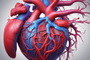
Anatomy of the Heart
- The heart is cone-shaped and about the size of a fist
- It weighs less than a pound
- The heart sits within the mediastinum, located between the two lungs
- The vertebral column is on the back side of the heart, and the sternum is located on the front
Cardiovascular System Functions
- The major purpose of the heart is to transport oxygen, digested food, cell wastes, electrolytes, and hormones
- The heart is a muscular pump made of cardiac muscle
- Contractions of the heart force blood through blood vessels
- It uses a one-way valve system
Coverings of the Heart
- The pericardium is a double-walled fibro-serous sac that surrounds the heart
- The fibrous pericardium is the superficial layer
- The deep two-layer serous pericardium has a parietal layer, which lines the internal surface of the fibrous pericardium
- The visceral layer, also known as the epicardium, lines the surface of the heart
- A fluid-filled pericardial cavity separates the layers
- Pericarditis is an inflammation of the pericardium due to adhesion between the serous pericardial layers, which interferes with heart movement
Heart Membrane Layers
- The heart membrane comprises a double-walled fibro-serous sac called the pericardium, positioned on top of the epicardium
- The epicardium is a thin layer of connective tissue and fat that provides an additional protective layer for the heart, underneath the pericardium
- The myocardium is the middle layer and the muscle tissue of the heart, composed of cardiac muscle cells that contract and conduct electricity to coordinate contraction
- The endocardium is the innermost layer of the heart and is composed of endothelial cells that provide a smooth, non-adherent surface for blood collection and pumping
Chambers of the Heart
- The heart has four chambers: two atria on the superior position and two ventricles on the inferior position
- The interatrial septum separates the atria
- The interventricular septum separates the ventricles
- The right ventricle makes up most of the anterior surface
- The left ventricle forms the apex of the heart and dominates the back surface
- Two grooves mark the boundaries of the four chambers
- The coronary sulcus is located at the junction between the atria and ventricle
- The anterior interventricular septum separates the right and left ventricles
Atria
- Atria are the receiving chambers for blood from circulation
- They are small and thin-walled, pushing blood minimally down to the ventricle
- Three veins enter blood to the right atrium
- The superior vena cava returns blood from body regions superior to the diaphragm
- The inferior vena cava returns blood from body regions inferior to the diaphragm
- The coronary sinus returns blood from the myocardium
Ventricles
- Ventricles make up most of the volume of the heart
- The right ventricle dominates the anterior surface
- The left ventricle dominates the posterior surface
- Ventricles are the actual pump of the heart
- Walls are very thick compared to the atria
- The right ventricle pumps blood into the pulmonary trunk
- The left ventricle pumps blood into the aorta, which is the largest artery in the body
Heart Valves
- A total of four valves ensure unidirectional blood flow through the heart
- Two atrioventricular (AV) valves between the atria and ventricular junctions prevent backflow into the atria when ventricles contract
- Flaps of the valves are made up of collagen cords called chordae tendineae or heart strings
- Chordae tendineae anchor AV valves to papillary muscles located on the inside surface of the heart
Semilunar Valves
- Two semilunar (SL) valves are between the ventricles and big arteries like the aorta and pulmonary artery
- The aortic semilunar valve lies between the left ventricle and the aorta
- The pulmonary semilunar valve lies between the right ventricle and pulmonary trunk
- Semilunar valves prevent backflow of blood into the ventricles and open and close in reaction to pressure in the ventricles
- Faulty valves can be replaced
Blood Flow Through the Heart
- There are two circuits: pulmonary and systemic
- Equal volumes of blood pump through both circuits
- Pulmonary circuits originate from the right ventricle and does not need much pressure because it travels to the lungs for a short trip
- The systemic circuit originates from the left ventricle requiring travelling all over the body needing more pressure
- The wall of the left ventricle is 3x thicker than the right
Pulmonary and Systemic Circuits
- Th right side of the heart receives O2 poor blood from throughout the body and pumps to the lungs to pick up O2 and drop CO2
- The blood vessels that carry blood to and from the lungs are called the “pulmonary circuit.”
- The left side of the heart receives O2-rich blood from the lungs and pumps it throughout the body to supply O2 and nutrients
- The blood vessels that carry blood to and from the body tissues are called the “systemic circuit.”
Coronary Circulation
- The heart receives nourishment from coronary arteries and its branches
- The right (R) and left (L) coronary arteries are the first branches of the ascending aorta
- The R coronary artery supplies R atrium and the back of both ventricles, branching into the marginal artery and others
- The L coronary artery supplies the L atrium and the L ventricle, branching into the circumflex artery and others
Cardiac Muscle Cells
- Cardiac muscle cells are short, fat, branched, and interconnected
- Like skeletal muscle, cardiac muscle is striated (less than skeletal) and contracts by the "sliding filament theory"
- Have thick and thin filament and Z, A, and I bands in sarcomere (like skeletal muscle)
- Each cardiac cell has one or two nuclei
- Mitochondria makes up 25-35% of cardiac cell volume
- Cardiac muscle cells (muscle fibers) are connected with next cell through “intercalated discs”
- Each disc has desmosomes for an adherence structure between two cells
- Each disc has gap junctions for plasma membrane channels for communication
- Desmosomes prevent adjacent cells from separating during contraction
- Gap junctions allow ions to pass from cell to cell, transmitting current across the entire heart called functional syncytium
Studying That Suits You
Use AI to generate personalized quizzes and flashcards to suit your learning preferences.