Podcast
Questions and Answers
In systemic AL amyloidosis, the heart rarely contributes to mortality.
In systemic AL amyloidosis, the heart rarely contributes to mortality.
False (B)
Localized amyloidosis in the lung parenchyma typically presents as single or multiple lung nodules known as amyloidomas.
Localized amyloidosis in the lung parenchyma typically presents as single or multiple lung nodules known as amyloidomas.
True (A)
Dysmotility and gastrointestinal bleeding are common manifestations of amyloidosis in the GI tract.
Dysmotility and gastrointestinal bleeding are common manifestations of amyloidosis in the GI tract.
True (A)
Typically, the liver and spleen show signs of focal amyloidomas in systemic amyloidosis.
Typically, the liver and spleen show signs of focal amyloidomas in systemic amyloidosis.
Amyloidosis in the mediastinum is often localized and presents primarily as plaques.
Amyloidosis in the mediastinum is often localized and presents primarily as plaques.
Involvement of the kidneys in systemic amyloidosis can lead to nephrotic syndrome.
Involvement of the kidneys in systemic amyloidosis can lead to nephrotic syndrome.
Localized amyloidosis in the pancreas is commonly associated with type 1 diabetes.
Localized amyloidosis in the pancreas is commonly associated with type 1 diabetes.
Involvement of the adrenal glands and testes typically indicates localized amyloidosis.
Involvement of the adrenal glands and testes typically indicates localized amyloidosis.
Systemic amyloidosis can cause calcification in the mesentery and retroperitoneum.
Systemic amyloidosis can cause calcification in the mesentery and retroperitoneum.
In Aβ2M amyloidosis, joint involvement typically results in unilateral joint effusions.
In Aβ2M amyloidosis, joint involvement typically results in unilateral joint effusions.
Localized Aβ amyloidosis does not typically lead to cerebral atrophy in the parietal lobe.
Localized Aβ amyloidosis does not typically lead to cerebral atrophy in the parietal lobe.
Muscle involvement in AL amyloidosis can result in pseudohypertrophy.
Muscle involvement in AL amyloidosis can result in pseudohypertrophy.
Cerebral amyloid angiopathy commonly causes multiple microhaemorrhages in a superficial distribution.
Cerebral amyloid angiopathy commonly causes multiple microhaemorrhages in a superficial distribution.
Involvement of the thyroid gland in systemic amyloidosis can lead to organ dysfunction.
Involvement of the thyroid gland in systemic amyloidosis can lead to organ dysfunction.
Focal masses in the oral cavity associated with systemic amyloidosis are characterized by microglossia.
Focal masses in the oral cavity associated with systemic amyloidosis are characterized by microglossia.
Isolated carpal tunnel syndrome can occur in mutant ATTR or both wild-type and Aβ2M amyloidosis.
Isolated carpal tunnel syndrome can occur in mutant ATTR or both wild-type and Aβ2M amyloidosis.
Flashcards
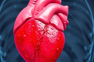
Amyloidosis in the Heart
Amyloidosis in the Heart
Amyloidosis in the heart is a common cause of death in systemic AL amyloidosis, manifesting as restrictive cardiomyopathy and potentially pericardial effusion. On MRI, it typically shows diffuse myocardial thickening with global subendocardial or transmural LGE, affecting all chambers.
Amyloidosis in the Airways
Amyloidosis in the Airways
Amyloidosis in the larynx, trachea, and bronchi is usually localized, presenting as diffuse airway thickening or multifocal plaques/masses with potential calcification.
Amyloidosis in the Lungs
Amyloidosis in the Lungs
In the lung parenchyma, localized amyloidosis often presents as solitary or multiple lung nodules (amyloidomas) with calcification. Systemic AL/AA disease can lead to interlobular septal thickening, reticulation, micronodules, consolidation, and punctate calcification with a basal and peripheral dominance, along with pleural effusions or thickening.
Amyloidosis in the Mediastinum
Amyloidosis in the Mediastinum
Signup and view all the flashcards
Amyloidosis in the GI Tract
Amyloidosis in the GI Tract
Signup and view all the flashcards
Amyloidosis in the Liver and Spleen
Amyloidosis in the Liver and Spleen
Signup and view all the flashcards
Amyloidosis in the Pancreas
Amyloidosis in the Pancreas
Signup and view all the flashcards
Amyloidosis in the Kidneys
Amyloidosis in the Kidneys
Signup and view all the flashcards
Mesentery and Retroperitoneum Amyloidosis
Mesentery and Retroperitoneum Amyloidosis
Signup and view all the flashcards
Amyloid Arthropathy
Amyloid Arthropathy
Signup and view all the flashcards
Bone and Soft Tissue Amyloidosis
Bone and Soft Tissue Amyloidosis
Signup and view all the flashcards
Peripheral Nerve Amyloidosis
Peripheral Nerve Amyloidosis
Signup and view all the flashcards
CNS Amyloidosis
CNS Amyloidosis
Signup and view all the flashcards
Cerebral Amyloid Angiopathy
Cerebral Amyloid Angiopathy
Signup and view all the flashcards
Cerebral Amyloidoma
Cerebral Amyloidoma
Signup and view all the flashcards
Head and Neck Amyloidosis
Head and Neck Amyloidosis
Signup and view all the flashcards
Study Notes
Amyloidosis Organ-Specific Manifestations
-
Heart: Systemic AL amyloidosis causes high mortality (ATTR better prognosis). Presents as restrictive cardiomyopathy, possibly with pericardial effusion. MRI shows diffuse myocardial thickening, affecting all heart chambers, with subendocardial/transmural late gadolinium enhancement (LGE).
-
Larynx, Trachea, Bronchi: Usually localized amyloidosis. Symptoms include airway thickening, multifocal plaques/masses (sometimes calcified).
-
Lung Parenchyma: Localized form shows nodules (amyloidomas), often calcified. Systemic AL/AA amyloidosis involves interlobular septal thickening, reticulation, micronodules, consolidation, and punctate calcification, mostly in the base and periphery, with pleura involvement possible. AA amyloidosis may coexist with lymphangioleiomyomatosis (LIP).
-
Mediastinum: Typically systemic amyloidosis, characterized by lymphadenopathy (possibly calcified) or a focal mass.
-
GI Tract: Very common in systemic amyloidosis. Presents with dysmotility, bleeding, diarrhea, obstruction, or perforation. Most common involvement is in the duodenum then stomach. CT may show luminal dilation, diffuse/nodular wall thickening, and calcification. Focal amyloidomas are rare.
-
Liver and Spleen: Common in systemic amyloidosis, causing diffuse organomegaly and possible punctate calcifications. Liver may be hypoattenuating on unenhanced CT; spleen shows reduced arterial enhancement. Gallbladder wall thickening is possible. Rare focal amyloidomas.
-
Pancreas: Usually localized amyloidosis, leading to Type 2 diabetes. Diffuse infiltration and enlargement of the pancreas are common, with loss of T1 hyperintensity on MRI, and possible punctate calcification mimicking chronic pancreatitis.
-
Renal Parenchyma: Frequent in systemic amyloidosis, causing nephrotic syndrome and renal failure. Kidneys may be enlarged and echogenic in early disease, with focal masses. Atrophy and amorphous calcification are seen in chronic disease. Renal sinus fat infiltration may occur.
-
Urothelium: Localized, most commonly affecting bladder>ureter>renal pelvis>urethra. Focal thickening, stricture, or mass (possibly calcified). Isolated involvement of the seminal vesicles is also possible, presenting with diffuse wall thickening.
-
Adrenals and Testes: Systemic amyloidosis. Characterized by diffuse infiltration and enlargement, leading to potential organ dysfunction.
-
Mesentery and Retroperitoneum: Systemic amyloidosis. Infiltrative soft-tissue replacement of normal fat, often with progressive calcification. Low-attenuation lymphadenopathy may also be present; ascites is rare.
-
Joints (Amyloid Arthropathy): Typically in Aβ2M amyloidosis. Bilateral erosive polyarthropathy affecting wrists, shoulders, hips, and knees. MRI shows T1/T2 hypointense synovial thickening (no blooming, differentiating from pigmented villonodular synovitis—PVNS) along with effusions, and without joint space narrowing. Surrounding muscle swelling (shoulder pad sign) may be present. Cervical spine involvement is common.
-
Bones and Soft Tissues: Mostly systemic AL amyloidosis. Focal soft-tissue masses are common; osseous lesions are lytic. Muscle involvement can mimic pseudohypertrophy or inflammatory myopathy. Breast involvement can resemble carcinoma.
-
Peripheral Nerves: Often in mutant ATTR or AL amyloidosis, marked by diffuse nerve enlargement, with potential enhancement on MRI. Isolated carpal tunnel syndrome is possible in wild-type ATTR or Aβ2M amyloidosis.
-
Central Nervous System (CNS):
-
Alzheimer's Disease: Most common localized Aβ amyloidosis form. Associated with cerebral atrophy, especially in the parietal and mesial temporal lobes.
-
Cerebral Amyloid Angiopathy: Common, causing numerous microhemorrhages in subcortical areas. Larger hemorrhages (intracerebral/subarachnoid) or focal inflammation (vasogenic edema) may occur.
-
Cerebral Amyloidoma: Rare, characterized by enhancing nodular masses with surrounding vasogenic edema.
-
Pituitary: Amyloid deposits can be seen in pituitary adenomas.
-
Head and Neck:
-
Orbits: Localized disease affecting lacrimal glands and other orbital structures.
-
Nasal Cavity/Paranasal Sinuses: Localized disease involving a focal mass, potentially obstructing drainage.
-
Oral Cavity: Systemic disease. Macroglossia is a hallmark.
-
Thyroid and Salivary Glands: Systemic disease; may cause organ dysfunction.
Studying That Suits You
Use AI to generate personalized quizzes and flashcards to suit your learning preferences.
Description
Explore the role of amyloidosis in various organs, including the heart, lungs, and GI tract. Understand the specific symptoms and diagnostic techniques associated with systemic and localized forms. This quiz provides insights into the clinical implications and outcomes of amyloidosis manifestations.