Podcast
Questions and Answers
What is the average age of patients diagnosed with ameloblastic fibroma?
What is the average age of patients diagnosed with ameloblastic fibroma?
- 25 years
- 5 years
- 35 years
- 14 years (correct)
Where is ameloblastic fibroma most commonly found?
Where is ameloblastic fibroma most commonly found?
- Mandibular Molar area (correct)
- Maxillary Incisor area
- Mandibular Incisor area
- Maxillary Molar area
What histopathological feature characterizes ameloblastic fibroma?
What histopathological feature characterizes ameloblastic fibroma?
- Thin strands and cords of odontogenic epithelium (correct)
- Presence of mature odontoblasts
- Thick collagen fibers
- Extensive necrosis
Which of the following is a radiographic feature of ameloblastic fibroma?
Which of the following is a radiographic feature of ameloblastic fibroma?
What type of cells are primarily found in the connective tissue background of ameloblastic fibroma?
What type of cells are primarily found in the connective tissue background of ameloblastic fibroma?
Flashcards
Ameloblastic Fibroma
Ameloblastic Fibroma
A benign tumor of the jaw that consists of both epithelial and connective tissue, resembling the dental follicle.
Age of Ameloblastic Fibroma
Age of Ameloblastic Fibroma
The typical age group affected by this tumor is around 14 years old.
Location of Ameloblastic Fibroma
Location of Ameloblastic Fibroma
The lower jaw near the molar teeth is the most common location for this tumor.
Radiographic Appearance of Ameloblastic Fibroma
Radiographic Appearance of Ameloblastic Fibroma
Signup and view all the flashcards
Microscopic Features of Ameloblastic Fibroma
Microscopic Features of Ameloblastic Fibroma
Signup and view all the flashcards
Study Notes
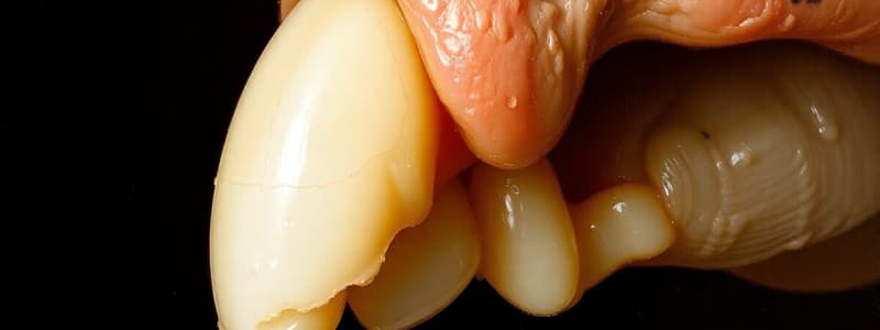
Ameloblastic Fibroma
- Benign mixed odontogenic neoplasm
- Consists of neoplastic odontogenic epithelium in actively proliferating connective tissue resembling tooth follicle or dental papilla.
- Average age of onset is 14 years.
- Commonly located in the mandibular molar area.
- Painless and slow-growing lesion.
- Causes slight buccal and lingual cortical expansion.
- Radiographic appearance: unilocular or multilocular radiolucent areas, resembling ameloblastoma.
- Histological features: Thin strands and cords of odontogenic epithelium.
- Epithelial component includes peripheral cuboidal or columnar cells enclosing stellate cells.
- Resembles the dental lamina.
- Connective tissue is composed of embryonic, proliferating fibroblastic cells with minimal collagen.
- Often exhibits zones of hyalinization, especially juxta-epithelial hyalinization, surrounding the epithelial component.
Studying That Suits You
Use AI to generate personalized quizzes and flashcards to suit your learning preferences.
Description
This quiz delves into the characteristics of Ameloblastic Fibroma, a benign mixed odontogenic neoplasm. It explores its histological features, common locations, and radiographic appearances, providing insight into its clinical significance. Understand the pathology of this condition and how it resembles other odontogenic tumors.


