Podcast
Questions and Answers
What is the estimated rate of actinic keratosis in men in the United States?
What is the estimated rate of actinic keratosis in men in the United States?
- 5%
- 10.2%
- 15%
- 26.5% (correct)
Which of the following is a common assessment finding in actinic keratosis?
Which of the following is a common assessment finding in actinic keratosis?
- Blue-tinged, smooth plaques
- Deep, draining ulcers
- Fluctuant, fluid-filled bullae
- Round or oval-shaped raised, scaly papules (correct)
Which of the following is a FIRST-LINE treatment for actinic keratosis when a patient presents with 10 or fewer AK lesions?
Which of the following is a FIRST-LINE treatment for actinic keratosis when a patient presents with 10 or fewer AK lesions?
- Cryotherapy with liquid nitrogen (correct)
- Photodynamic therapy
- Curettage
- Topical retinoids
Which strength of topical fluorouracil cream is typically effective for treating actinic keratosis?
Which strength of topical fluorouracil cream is typically effective for treating actinic keratosis?
What is the primary focus when assessing risk factors for actinic keratosis?
What is the primary focus when assessing risk factors for actinic keratosis?
A patient with actinic keratosis is prescribed topical fluorouracil (5-FU) cream for facial lesions. What potential side effect should the patient be educated about?
A patient with actinic keratosis is prescribed topical fluorouracil (5-FU) cream for facial lesions. What potential side effect should the patient be educated about?
Which of the following is included in the differential diagnosis of actinic keratosis?
Which of the following is included in the differential diagnosis of actinic keratosis?
What is the most common presentation of atopic dermatitis (eczema)?
What is the most common presentation of atopic dermatitis (eczema)?
Which of the following is a known risk factor for atopic dermatitis?
Which of the following is a known risk factor for atopic dermatitis?
What percentage of children with atopic dermatitis experience remission by puberty?
What percentage of children with atopic dermatitis experience remission by puberty?
In which areas of the body does atopic dermatitis commonly affect infants?
In which areas of the body does atopic dermatitis commonly affect infants?
What is the primary goal in the management of atopic dermatitis?
What is the primary goal in the management of atopic dermatitis?
Which of the following nonpharmacologic measures is recommended to prevent dry skin in patients with atopic dermatitis?
Which of the following nonpharmacologic measures is recommended to prevent dry skin in patients with atopic dermatitis?
How should moisturizers be applied after bathing to maximize their effectiveness in patients with atopic dermatitis?
How should moisturizers be applied after bathing to maximize their effectiveness in patients with atopic dermatitis?
Under what circumstances should topical treatment for atopic dermatitis be stopped?
Under what circumstances should topical treatment for atopic dermatitis be stopped?
Which of the following best describes contact dermatitis?
Which of the following best describes contact dermatitis?
What is a key initial step in managing contact dermatitis?
What is a key initial step in managing contact dermatitis?
Which of the following is more likely to be a cause of irritant contact dermatitis rather than allergic contact dermatitis?
Which of the following is more likely to be a cause of irritant contact dermatitis rather than allergic contact dermatitis?
A patient presents with a rash characterized by burning, pruritus, and pain, predominantly on their hands. What type of contact dermatitis is most likely?
A patient presents with a rash characterized by burning, pruritus, and pain, predominantly on their hands. What type of contact dermatitis is most likely?
While both atopic and contact dermatitis can involve eczema, which statement is most accurate regarding their causes?
While both atopic and contact dermatitis can involve eczema, which statement is most accurate regarding their causes?
What is the most common causative organism in cases of folliculitis, furunculosis, and carbunculosis?
What is the most common causative organism in cases of folliculitis, furunculosis, and carbunculosis?
Which of the following factors is specifically associated with an increased risk of furunculosis?
Which of the following factors is specifically associated with an increased risk of furunculosis?
A patient presents with a cluster of furunculosis that develops slowly with fever and sloughing. Which condition is most likely?
A patient presents with a cluster of furunculosis that develops slowly with fever and sloughing. Which condition is most likely?
Which diagnostic study is most likely to be required in cases of folliculitis to assess for Pityrosporum folliculitis?
Which diagnostic study is most likely to be required in cases of folliculitis to assess for Pityrosporum folliculitis?
What is the recommended nonpharmacologic management for folliculitis?
What is the recommended nonpharmacologic management for folliculitis?
If MRSA is suspected or identified in a patient with a bacterial skin infection, what topical treatment should be considered in addition to an oral agent?
If MRSA is suspected or identified in a patient with a bacterial skin infection, what topical treatment should be considered in addition to an oral agent?
In cases of furunculosis on the face, scalp, or neck, what is the most appropriate course of action?
In cases of furunculosis on the face, scalp, or neck, what is the most appropriate course of action?
If Pityrosporum folliculitis is present, what type of medication should be avoided because it may worsen the condition?
If Pityrosporum folliculitis is present, what type of medication should be avoided because it may worsen the condition?
Which of the following is a typical presenting symptom in cellulitis?
Which of the following is a typical presenting symptom in cellulitis?
Which of the following organisms is the most common pathogen for erysipelas, a superficial form of cellulitis?
Which of the following organisms is the most common pathogen for erysipelas, a superficial form of cellulitis?
Which statement is most accurate about the areas of the body typically affected by cellulitis?
Which statement is most accurate about the areas of the body typically affected by cellulitis?
What is a common finding in the assessment of a patient with cellulitis?
What is a common finding in the assessment of a patient with cellulitis?
A patient presents with cellulitis of the lower extremity. What nonpharmacologic intervention is most appropriate?
A patient presents with cellulitis of the lower extremity. What nonpharmacologic intervention is most appropriate?
A patient presents with suspected cellulitis, cellulitis. Which diagnostic test should be ordered FIRST?
A patient presents with suspected cellulitis, cellulitis. Which diagnostic test should be ordered FIRST?
A patient develops acute herpes zoster involving the ophthalmic branch of the trigeminal nerve. What is the most important next step in management?
A patient develops acute herpes zoster involving the ophthalmic branch of the trigeminal nerve. What is the most important next step in management?
What is the usual presentation of dermatomal rash of Herpes Zoster?
What is the usual presentation of dermatomal rash of Herpes Zoster?
Which intervention is most crucial for reducing the likelihood of postherpetic neuralgia (PHN) in a patient diagnosed with herpes zoster?
Which intervention is most crucial for reducing the likelihood of postherpetic neuralgia (PHN) in a patient diagnosed with herpes zoster?
Which of the following is a common patient complaint associated with herpes zoster?
Which of the following is a common patient complaint associated with herpes zoster?
According to the reference article, which vaccine is preferred for the immunization of healthy adults 50 years and older against herpes zoster?
According to the reference article, which vaccine is preferred for the immunization of healthy adults 50 years and older against herpes zoster?
What is the most appropriate management strategy to decrease the risk of disseminated varicella-zoster virus (VZV)?
What is the most appropriate management strategy to decrease the risk of disseminated varicella-zoster virus (VZV)?
What is the most common type of psoriasis?
What is the most common type of psoriasis?
Which of the following is a common site for psoriasis?
Which of the following is a common site for psoriasis?
What percentage of patients demonstrates pits in their nails?
What percentage of patients demonstrates pits in their nails?
If the elbows and knees are affected, which condition is indicated?
If the elbows and knees are affected, which condition is indicated?
Regarding topical steriods as a treatment, which consideration is MOST important?
Regarding topical steriods as a treatment, which consideration is MOST important?
Flashcards
Actinic Keratosis (AK)
Actinic Keratosis (AK)
Actinic keratosis (AK) are keratotic scaly lesions, the most common precancerous epithelial skin lesions that can progress to squamous cell carcinoma (SCC)
Etiology of Actinic Keratosis
Etiology of Actinic Keratosis
Cumulative UV radiation, immunocompromised status, and immunosuppressive therapy.
Assessment Findings of AK
Assessment Findings of AK
Round or oval-shaped raised, scaly papules that are flesh-colored, red, pink, brown or black. Size varies from 2 mm to 5 mm; larger lesions are possible
Differential Diagnosis of AK
Differential Diagnosis of AK
Signup and view all the flashcards
Prevention of AK
Prevention of AK
Signup and view all the flashcards
Treatment of AK
Treatment of AK
Signup and view all the flashcards
Atopic Dermatitis (Eczema)
Atopic Dermatitis (Eczema)
Signup and view all the flashcards
Areas Affected by Eczema
Areas Affected by Eczema
Signup and view all the flashcards
Etiology of Atopic Dermatitis
Etiology of Atopic Dermatitis
Signup and view all the flashcards
Prevention of Atopic Dermatitis
Prevention of Atopic Dermatitis
Signup and view all the flashcards
Nonpharmacologic Management for Eczema
Nonpharmacologic Management for Eczema
Signup and view all the flashcards
Bathing Recommendations for Eczema
Bathing Recommendations for Eczema
Signup and view all the flashcards
Pharmacologic Management for Eczema
Pharmacologic Management for Eczema
Signup and view all the flashcards
Follow-Up for Eczema
Follow-Up for Eczema
Signup and view all the flashcards
Contact Dermatitis
Contact Dermatitis
Signup and view all the flashcards
Treatment for Contact Dermatitis
Treatment for Contact Dermatitis
Signup and view all the flashcards
Tinea/ Fungal Infections
Tinea/ Fungal Infections
Signup and view all the flashcards
Area fungal infections affect
Area fungal infections affect
Signup and view all the flashcards
Populations where fungal infections are more common
Populations where fungal infections are more common
Signup and view all the flashcards
Treatments for Tinea
Treatments for Tinea
Signup and view all the flashcards
Candidiasis (Yeast)
Candidiasis (Yeast)
Signup and view all the flashcards
Clinical Presentation of Candidiasis
Clinical Presentation of Candidiasis
Signup and view all the flashcards
Etiology: Bacterial Infections
Etiology: Bacterial Infections
Signup and view all the flashcards
What is folliculitis
What is folliculitis
Signup and view all the flashcards
cluster of furuncles
cluster of furuncles
Signup and view all the flashcards
Furunculosis Treatments
Furunculosis Treatments
Signup and view all the flashcards
Skin moisture
Skin moisture
Signup and view all the flashcards
Mupirocin (Bactroban)
Mupirocin (Bactroban)
Signup and view all the flashcards
Cellulitis
Cellulitis
Signup and view all the flashcards
Etiology of cellulitis
Etiology of cellulitis
Signup and view all the flashcards
Parts of the body that are commonly affected
Parts of the body that are commonly affected
Signup and view all the flashcards
Expected outcome= cellulitis
Expected outcome= cellulitis
Signup and view all the flashcards
Herpes Zoster (Shingles)
Herpes Zoster (Shingles)
Signup and view all the flashcards
Pharmacotherapy of shingles can provide..
Pharmacotherapy of shingles can provide..
Signup and view all the flashcards
NONPHARMACOLOGIC MANAGEMENT- Shingles
NONPHARMACOLOGIC MANAGEMENT- Shingles
Signup and view all the flashcards
Psoriasis
Psoriasis
Signup and view all the flashcards
Factor associated with Psoriasis?
Factor associated with Psoriasis?
Signup and view all the flashcards
Psoriasis outcome
Psoriasis outcome
Signup and view all the flashcards
Scabies- NONPHARMACOLOGIC MANAGEMENT
Scabies- NONPHARMACOLOGIC MANAGEMENT
Signup and view all the flashcards
Pharmacologic treatment
Pharmacologic treatment
Signup and view all the flashcards
Study Notes
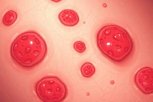
- Actinic keratosis (AK) presents on sun-exposed skin as keratotic scaly lesions and erythematous papules or plaques.
- AK lesions are precancerous epithelial skin lesions that can progress to squamous cell carcinoma (SCC).
- AK is considered pre-malignant cells and precursors to squamous cell carcinoma, though most do not become cancerous.
Etiology
- Cumulative exposure to ultraviolet radiation (sunlight, tanning beds) and arsenic can cause actinitic keratosis
- Immunocompromised status (HIV/AIDS, solid organ or bone marrow transplant) can cause actinitic keratosis
- Immunosuppressive therapy can cause actinitic keratosis
Incidence
- Actinic keratosis is common in middle-aged and older adults.
- Men are affected more often than women.
- It typically presents in younger adults (30 to 40 years old).
- The estimated rate of AK in the United States is 10.2% in women and 26.5% in men.
Assessment Findings
- Actinic keratosis presents as round or oval-shaped raised, scaly papules.
- Lesions are flesh-colored, red, pink, brown, or black.
- It may present as papules or plaques with a rough texture.
- The size varies from 2 mm to 5 mm; larger lesions are possible.
Differential Diagnosis
- Seborrheic keratosis
- Verruca
- Solar lentigo
- Basal cell carcinoma
- Squamous cell carcinoma
- Melanoma
Diagnostic Studies
- Diagnosis is usually made on clinical presentation due to the characteristic appearance.
- Skin biopsy is reserved for individuals who fail to respond to treatment.
- Skin biopsy is only used when it must be determined whether an AK has progressed to squamous cell carcinoma.
Prevention
- Sunscreen should be worn daily (even on cloudy days), along with hats/gloves and protective clothing.
- SPF of 30 should be used for dark skin and 40 to 50 for fair skin.
- Mineral sunscreens are physical blockers that contain zinc or titanium dioxide.
- Chemical sunscreens contain chemicals that absorb UV radiation.
- Lip balms containing SPF are recommended.
Nonpharmacologic Management
- Treatment options for AK include destructive therapies for isolated lesions (cryotherapy, curettage) and topical medications for multiple lesions.
- Cryotherapy with liquid nitrogen is a first-line treatment for patients with 10 or fewer AK lesions.
- Tissue scraping (curettage) removes tissue via a surgical instrument (curette).
- Tissue scraping (curettage) is immediately followed by electrodesiccation with an electric cautery instrument, requires topical anesthesia
- Curettage is an effective treatment for patients with a limited number of AK lesions, particularly hyperkeratotic lesions.
Pharmacologic Treatment / Management
- Topical fluorouracil (5-FU) cream: 5% strength is effective.
- Application of topical fluorouracil to facial lesions causes significant erythema, blistering and crusting of erosions, which may result in temporary cosmetic effects
- For face: 0.5% apply topical fluorouracil BID for 2-4 weeks.
- For lips: 1-2% 5-FU once daily to BID for 3 weeks.
- Topical imiquimod 5% cream is used 2-3 times weekly for up to 16 weeks depending on the skin field being treated.
- Topical imiquimod is helpful for small lesions but is more expensive than 5-FU and may require longer duration of treatment on thicker skin.
- Use topical treatments sparingly and treat small areas, such as lower arms or face, at the same time.
- If a patient experiences flu-like symptoms, topical treatment should be stopped.
- Photodynamic therapy (including lips) combines topical treatment and light therapy, delivered in the office setting.
- Trichloroacetic acid peels.
- Tretinoin 0.02-0.1% or salicylic acid 6% may enhance effects of 5-FU.
Consultation Referral
- A dermatologist should be consulted if AK lesions appear frequently, a biopsy of lesions or more intensive treatment may be needed
- Any lesion unresponsive to topical therapy warrants referral.
Follow up
- Follow up frequency depends on the appearance of AK lesions and whether associated malignancy is present.
Expected Course
- Excellent
- Patients may experience erythema, edema, blistering, or eschar formation at sites treated with liquid nitrogen therapy; may last for 3-5 days after treatment.
Complications
- Squamous cell carcinoma in situ or squamous cell carcinoma.
Epidemiology
- Increased age: Actinic keratosis increasingly affect the older population due to the high cumulative lifetime exposure to the sun and inadequate sun protection measures.
- Pale or light skin individuals are more at risk.
- Those in Australia are at a higher risk due to its proximity to the equator.
- Excessive sun exposure increases risk.
History and Physical
- Ask about any symptoms associated with the lesions, such as pruritus (itching), pain, or bleeding with minor trauma.
- If there are symptoms associated with the lesions, it may indicate an increased risk of invasive squamous cell carcinoma progression.
- Determine if the patient has a history of prior skin cancer treatments or surgeries.
- Conduct a thorough assessment of all the risk factors associated with actinic keratosis.
- The assessment of all the risk factors associated with actinic keratosis includes duration and history of sun exposure, past occurrences of sunburn, regular use of sunscreen, sun protection habits, and the patient's occupation, especially if it involves prolonged outdoor activities.
- The focus is mainly on the sun-exposed areas of the body, including the head, face, scalp, neck, dorsal forearms, and hands.
- The presence of any ulceration and bleeding should be noted.
Atopic Dermatitis (Eczema)
- A chronic pruritic skin eruption that presents as an erythematous, patchy, papular, plaque-like rash with inflammation; acute exacerbations appear in characteristic sites; severity varies widely.
- The terms “eczema” and “atopic dermatitis” are often used interchangeably.
- Eczema is most common in children, but it affects many adults.
- May cause itchy, dry skin or a rash, or cause the skin to blister, ooze, crust, or flake off.
Eczematous Dermatitis
- Common in young children, though it can occur at any age.
- Exacerbations and remissions of dry, itchy red skin.
- Areas affected: hands, feet, ankles, wrists, neck, upper chest, eyelids, inside the bend of the elbows and knees, and in infants, the face and scalp.
- If untreated, lichenification occurs.
- Associated with other atopic diseases (IgE): asthma, urticaria, allergic rhinitis
- Personal or family history.
- Dry skin, sweating, heat, dry environments, and occlusion.
- Soaps, laundry detergents, wool.
- Exacerbated by stress, infection, and allergies.
Etiology
- Multifactorial: genetic, physiologic, immunologic, environmental
- Eosinophilia and elevated serum IgE levels
- Family history of allergies, asthma, allergic rhinitis
Incidence
- Affects almost 10.7% of children in U.S.
- 60% have first episode between infancy and 12 years.
- Typically begins after age 2 months; when it develops before then, symptoms tend to be persistent.
- 90% experience remission by puberty.
- Affects both sexes equally.
- More common in Black and Asian patients.
- Starting after age 30 is uncommon; diagnosis in such presentations should be made after consultation with a dermatologist or skin biopsy.
Risk Factor
- Family history of atopic diseases
- Skin infections
- Stress
- Temperature extremes
- Contact with irritating substances (harsh soaps, scented skin products, wearing new clothing before washing).
History and Physical
- General: pruritus, dry & scaly, erythema, Lichenification, dry skin, erythema on flexural folds, linear excoriations, antecubital fossa, posterior patella, scalp.
- Infants: Lesions on flexural surfaces of arms, legs, and on trunk, face (especially cheeks)
- Infant lesions are erythematous and papular, and vesicles may ooze and form crusts
- Children: Lesions common on wrists, ankles and flexural surfaces
- Children Presence of scales and plaques occur, lichenification occurs as a result of scratching
- Adults: Flexural surfaces are common sites (dorsa of the hands and feet)
- Adult conditions often reappear in adulthood after absence since childhood, and lichenification and scaling are typical
Differential Diagnosis
- Contact dermatitis (allergic and irritant)
- Seborrheic dermatitis
- Scabies
- Psoriasis
Diagnostic Test
- Usually none needed
- Skin biopsy to rule out other skin disorders
- 80% of patients may have eosinophilia during episodes of disease activity
- Serum allergy testing available
- Bacterial culture may be indicated in severe cases
Prevention
- Liberal use of emollients to prevent dry skin
- Avoid known precipitating factors (stress, wool clothing, detergents with fragrance, etc.)
- Wash clothing with fragrance-free detergents, fabric softeners and dryer sheets
- Keep environments as free of dust as possible
- Use of air purifiers and humidifiers
- Eliminate carpets; clean bedding weekly; and use mattress protectors to reduce dust mites
- Wash bedding in water that is 120° to 130°F
- Humidity in home should be no more than 50%
Note
- Serum allergy testing reveals that dust mites in the environment pose a high threat for skin allergies.
- Dermatophagoides farinae: American dust mite
- Dermatophagoides pteronyssinus: foreign dust mites
Nonpharmacologic Management
- Patient education
- Decrease inflammation and controlling itching, control exacerbating factors
- Cool humidified environment
- Tepid water for showers (avoid baths), pat skin dry
- Lubrication of skin (aquphor, cetaphil)
- Control emotional factors
- Antihistamines- Blocks activity at histamine receptor sites
- Topical corticosteroids
- The key to management of atopic dermatitis is maintenance of skin hydration.
- Bathing is recommended but not more than once daily; moisturizer should be applied 1 to 3 minutes after patting skin dry, while skin is slightly moist.
- Super fatted soaps are best; soap-free cleansers should have a neutral or low pH; use soap only on scalp, armpits, groin, feet
- Prevent skin trauma (sunburns, scrub brushes, etc.)
- When possible, soak in warm water for 20 minutes before applying emollient
- Apply cool wet compresses (Burow's solution) to oozing or weeping lesions
- Wet wrap therapy for moderate to severe atopic conditions
- Avoid ointments with oozing atopic dermatitis, to allow lesions to dry and promote healing
- Sunshine or UVB treatments
- Bleach baths: three times weekly (1/4 cup household bleach in full tub of water; soak 2-3 minutes); atopic patients are predisposed to secondary skin infections
- Fabrics such as wool or acrylics may irritate skin
- Patient education about disease process, self-care, precipitating factors; importance of hydrating skin with moisturizers 1-2 times daily or as often as needed to keep skin moist
Pharmacologic Management
- Pediatric patients may be more susceptible to topical corticosteroid-induced hypothalamic-pituitary-adrenal (HPA) axis suppression than older patients due to larger ratio of skin surface area to body weight.
- Limit use to lowest effective potency and duration.
- Topical corticosteroids (creams preferred) are the mainstay of therapy (use lowest potency that controls symptoms).
- Antihistamines (oral and topical) for itching
- Vital to taper off corticosteroids to avoid side effects and rebound flares of dermatitis
- Emollients 2-3 times per day or as needed to correct dry skin (Eucerin, Lubriderm, Cetaphil, Vaseline); steroid-sparing emollients recommended
- Consider oral corticosteroids for severe cases; use only in short bursts
- Intralesional steroid injections
- Topical phosphodiesterase-4 inhibitor in place of ineffective steroid dose: crisaborole (Eucrisa) 2% ointment.
- Apply thin layer BID in patients >2 years
- Topical calcineurin inhibitors: pimecrolimus topical cream 1% (Elidel) or tacrolimus ointment 0.03% (Protopic) and 0.1%; maintenance dosing twice weekly for 12 months; use moisturizers on other days.
- May reduce or delay exacerbations; not for patients <2 years.
- Dry skin treated with corticosteroid therapy will exhibit minimal response; however, use for 7 days or less may ease erythema and pruritus.
Consultation Referral
- Dermatologist
- Allergist
Follow Up
- Initial treatment for 2 weeks with follow-up at 6 to 8 weeks
- Follow-up is important to assure that patient is improving, and steroid medication is not being overused for prolonged periods.
- If flare is severe, prescribe a stronger steroid for 2 weeks, then decrease potency or change to a topical phosphodiesterase-4 inhibitor in place of ineffective steroid dose: crisaborole (Eucrisa) 2% ointment, or topical calcineurin inhibitors such as Elidel or Protopic
Expected Course
- Exacerbations and remission; expect flareups
Complications
- Secondary infection: excoriations in children are more likely; may need treatment; eczema slow to improve if infection is not treated
- Steroids can cause atrophy
Contact Dermatitis
- A red, itchy rash caused by direct contact with a substance or an allergic reaction to it
- The rash isn't contagious or life-threatening but can be very uncomfortable.
- Many substances can cause such reactions, including soaps, cosmetics, fragrances, jewelry and plants.
- The cause must be identified and avoided so the rash can clear up in two to four weeks with the following care
Treatment
- Soothing your skin with cool, wet compresses
- Special Soaps (Ivy soap)
- Antihistamines
- Topical corticosteroids
| Feature | Irritant | Allergic |
|---|---|---|
| Causes | Solvents, bleach, alcohol, chemicals in personal products | Nickel (jewelry), medications (topical cram/ointments, poison oak, personal products |
| Location | Usually hands | Exposed areas, usually hands |
| Symptoms | Burning, pruritus, pain | Predominantly pruritus |
| Surface appearance | Dry fissured skin | Vesicles and bullae |
| Lesion Borders | Less distinct | Distinct angles, line and borders |
Fungal Infections
- Fungal infections are a common dermatologic problem with exposure to fungal pathogens among common environments
- Most prevalent diseases of fungal infections include: Nails, Tinea (Ringworm), yeast (candida), mouth, throat, esophagus and vagina
- There is a higher incidence of fungal infections with immunocompromised patients (Autoimmune, HIV, immunosuppressant drugs), patients on antibiotics, and diabetics particularly if uncontrolled
Nail Fungus - Onychomycosis
- Caused by dermatophyte (tinea), yeast or mold and begins as a white or yellow spot under the tip of fingernail or toenail
- May cause nails to discolor, thicken and crumble at the edge, possibly affecting several nails
- If the condition is mild, it may not require treatment
- If nail fungus is painful and has caused thickened nails, treatment may be best
- Even with adequate treatment, nail fungus often comes back
Onychomycosis
- Is differentated from the following: malignancy, psoriasis, trauma
- Requires treatment
Treatment
- Topical treatment with ciclopirox (Penlac) which is a nail lacquer for usually at least 12 weeks or more (max 48 weeks)
- Oral terbinafine (Lamisil): 12 weeks for toenails, 6 weeks for fingernails
- LFT's are required at baseline and every 6 weeks while on treatment
- For yeast fluconazole (Diflucan) is preferred
Tinea (Ringworm)
-
Dermatophyte superficial skin infection
-
There is greater exposure in gym going population and there is greater risk with immunocompromised and diabetics
-
Includes Tinea capitis (scalp), Tinea corporis (trunk, extremities, feet, groin, face or hands), Tinea manuum (palms and spaces between fingers) Tinea cruris (inguinal area), Tinea pedis (feet and spaces between toes)
-
Diagnosies is easiest way to diagnos using KOH microscopy (used for dx fungal ringworm)
-
Differentials include: atopic dermatitis, contact dermatitis, folliculitis, psoriasis
-
Requires treatment that focuses on the following
Treatment
- Tinea on the skin is usually treated with non-prescription antifungal creams, lotions, or powders applied to the skin for 2 to 4 weeks with the following: Clotrimazole (Lotrimin AF, Mycelex), Miconazole (Desenex, Micatin, Monistat), Terbinafine (Lamisil), Ketoconazole (Xolegel, Nizoral)
- Tinea on the scalp can be treated with medicated shampoos with the following: Selenuium sulfide (Selsun) Ketoconzole shampoo (Nizoral)
- Widepsread tinea particulalry on the scalp or nails that doesn't respond to other treatments usually needs to be treated with prescription antifungal medication taken orally for 1 to 3 months: (Creams, lotions, or powders don't work for ringworm on the scalp) Griseofulvin (Grifulvin V, Gris-PEG), Terbinafine (Lamisil), Itraconazole (Onmel, Sporanox), Fluconazole (Diflucan)
- Oral antifungals require liver monitoring
Candidiasis (Yeast)
- Infection with the yeast Candida which has more than 150 varieties
- Usually occurs in moist areas of the skin-skin
- Is usually opportunistic with - obesity, certain medications (antibiotics), corticosteroids, malnutrition, diabetes, immunocompromised states like HIV, and can occur in warm, humid environments
Clinical presentation: depends on the location - may cause rashes, scaling, itching, plaques, swelling and vaginal discharge Diagnosies: by exam, sometimes by scraping (KOH slide) and culture
- Introduces the following Bacterial Infections (FOLLICULITIS, FURUNCULOSIS, CARBUNCULOSIS)
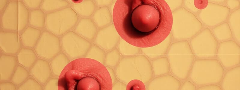
Folliculitis
Superficial infection/irritation of the hair follicles; lesions occur as small raised erythematous, occasionally pruritic, pustules <5 mm in size
Furunculosis (boils)
Deep infection of the hair follicle that is an acute, round, tender nodule that becomes a pustule or perifollicular abscess; common sites are the neck, face, buttocks, waistline and breasts
Carbunculosis (a cluster of furuncles)
Deep suppurative lesion with extension into subcutaneous area; coalescence of several inflamed follicles; produces purulent drainage
-
Notes than the following can be relevant to Etiology, Incidence, Risk Factors, and assessment findings:
-
Folliculitis, furunculosis, carbunculosis: Staphylococcus aureus is the most common causative organism; methicillin-resistant Staphylococcus aureus (MRSA) can be a common outpatient pathogen; "Hot tub" folliculitis: Pseudomonas aeruginosa , Pityrosporum folliculitis
Studying That Suits You
Use AI to generate personalized quizzes and flashcards to suit your learning preferences.