Podcast
Questions and Answers
What is the primary function of the cranial part of the accessory nerve?
What is the primary function of the cranial part of the accessory nerve?
- Innervating the trapezius muscle
- Contributing to the pharyngeal plexus (correct)
- Controlling eye movement
- Regulating blood flow
The spinal part of the accessory nerve exits the skull through the jugular foramen.
The spinal part of the accessory nerve exits the skull through the jugular foramen.
True (A)
Which cervical spinal cord segments does the spinal part of the accessory nerve arise from?
Which cervical spinal cord segments does the spinal part of the accessory nerve arise from?
C1 to C5
The accessory nerve innervates the ___ muscle.
The accessory nerve innervates the ___ muscle.
Match the following branches of the accessory nerve with their respective functions:
Match the following branches of the accessory nerve with their respective functions:
Which muscle is NOT affected by the pharyngeal plexus?
Which muscle is NOT affected by the pharyngeal plexus?
The glossopharyngeal nerve is involved in the formation of the pharyngeal plexus.
The glossopharyngeal nerve is involved in the formation of the pharyngeal plexus.
What anatomical structures lie adjacent to the vagus and accessory nerves in the carotid sheath?
What anatomical structures lie adjacent to the vagus and accessory nerves in the carotid sheath?
The accessory nerve unites with the cranial part after entering the skull via the ___ ___.
The accessory nerve unites with the cranial part after entering the skull via the ___ ___.
Which of the following muscles is NOT innervated by the internal branch of the accessory nerve?
Which of the following muscles is NOT innervated by the internal branch of the accessory nerve?
What muscles are weakened due to dysfunction in the accessory nerve?
What muscles are weakened due to dysfunction in the accessory nerve?
The precentral gyrus contains the cell bodies of lower motor neurons.
The precentral gyrus contains the cell bodies of lower motor neurons.
Which cervical spinal nerves innervate the sternocleidomastoid muscle?
Which cervical spinal nerves innervate the sternocleidomastoid muscle?
Lesions to the accessory nerve may result in weakness of the __________ muscle.
Lesions to the accessory nerve may result in weakness of the __________ muscle.
Match the following conditions with their causes:
Match the following conditions with their causes:
Where do cortical bulbar fibers originate?
Where do cortical bulbar fibers originate?
Most fibers supplying the trapezius muscle remain ipsilateral.
Most fibers supplying the trapezius muscle remain ipsilateral.
What is the effect of damage to the accessory nerve proximal to the posterior triangle of the neck?
What is the effect of damage to the accessory nerve proximal to the posterior triangle of the neck?
If cervical lymph nodes are __________, they may compress the accessory nerve and lead to dysfunction.
If cervical lymph nodes are __________, they may compress the accessory nerve and lead to dysfunction.
Which muscle is primarily responsible for scapular elevation?
Which muscle is primarily responsible for scapular elevation?
Which part of the accessory nerve is responsible for innervating the sternocleidomastoid muscle?
Which part of the accessory nerve is responsible for innervating the sternocleidomastoid muscle?
The cranial part of the accessory nerve supplies the trapezius muscle directly.
The cranial part of the accessory nerve supplies the trapezius muscle directly.
What structures do the internal carotid artery and internal jugular vein lie adjacent to in the carotid sheath?
What structures do the internal carotid artery and internal jugular vein lie adjacent to in the carotid sheath?
The accessory nerve provides motor supply to the __________ muscle, which is involved in scapular movement.
The accessory nerve provides motor supply to the __________ muscle, which is involved in scapular movement.
Match the part of the accessory nerve to its origin or main function:
Match the part of the accessory nerve to its origin or main function:
Which of the following nerves contribute to the pharyngeal plexus?
Which of the following nerves contribute to the pharyngeal plexus?
The function of the accessory nerve is solely sensory.
The function of the accessory nerve is solely sensory.
Which cervical spinal cord segments contribute to the innervation of the trapezius muscle?
Which cervical spinal cord segments contribute to the innervation of the trapezius muscle?
The accessory nerve exits the skull through the __________.
The accessory nerve exits the skull through the __________.
What muscle is innervated by the external branch of the accessory nerve?
What muscle is innervated by the external branch of the accessory nerve?
What can dysfunction in the accessory nerve lead to?
What can dysfunction in the accessory nerve lead to?
The precentral gyrus contains the cell bodies of lower motor neurons.
The precentral gyrus contains the cell bodies of lower motor neurons.
What are the cervical spinal segments that innervate the trapezius muscle?
What are the cervical spinal segments that innervate the trapezius muscle?
The accessory nerve is closely associated with __________, which can cause compression leading to dysfunction.
The accessory nerve is closely associated with __________, which can cause compression leading to dysfunction.
Match the following lesions with their potential causes:
Match the following lesions with their potential causes:
Which of the following statements is true about the cortical bulbar fibers?
Which of the following statements is true about the cortical bulbar fibers?
Most fibers supplying the trapezius muscle are ipsilateral.
Most fibers supplying the trapezius muscle are ipsilateral.
What are the possible causes of lesions affecting the accessory nerve?
What are the possible causes of lesions affecting the accessory nerve?
Damage to trapezius fibers can lead to __________ issues and scapular winging.
Damage to trapezius fibers can lead to __________ issues and scapular winging.
Which of the following is a clinical manifestation of accessory nerve palsy?
Which of the following is a clinical manifestation of accessory nerve palsy?
What are the functions of the sternocleidomastoid muscle in the context of accessory nerve damage?
What are the functions of the sternocleidomastoid muscle in the context of accessory nerve damage?
The precentral gyrus is part of the primary motor cortex that contains the cell bodies of upper motor neurons.
The precentral gyrus is part of the primary motor cortex that contains the cell bodies of upper motor neurons.
What cervical spinal nerves are primarily involved in innervating the trapezius muscle?
What cervical spinal nerves are primarily involved in innervating the trapezius muscle?
Dysfunction in the accessory nerve can particularly lead to difficulties in __________ and arm abduction.
Dysfunction in the accessory nerve can particularly lead to difficulties in __________ and arm abduction.
Match the following potential causes of lesions affecting the accessory nerve with their descriptions.
Match the following potential causes of lesions affecting the accessory nerve with their descriptions.
What consequence may arise from lesions affecting the accessory nerve?
What consequence may arise from lesions affecting the accessory nerve?
Most fibers supplying the trapezius muscle cross to the contralateral side.
Most fibers supplying the trapezius muscle cross to the contralateral side.
Where do cortical bulbar fibers descend to reach for motor control?
Where do cortical bulbar fibers descend to reach for motor control?
Cervical nerve supply functions especially from the spinal roots C2, C3, and C4 to innervate the __________ muscle.
Cervical nerve supply functions especially from the spinal roots C2, C3, and C4 to innervate the __________ muscle.
Which of the following statements about cortical fibers is accurate?
Which of the following statements about cortical fibers is accurate?
What is the primary role of the cranial part of the accessory nerve?
What is the primary role of the cranial part of the accessory nerve?
The spinal part of the accessory nerve transmits sensory information.
The spinal part of the accessory nerve transmits sensory information.
Which anatomical structure does the accessory nerve exit through in the skull?
Which anatomical structure does the accessory nerve exit through in the skull?
The accessory nerve originates from the motor nucleus in the __________.
The accessory nerve originates from the motor nucleus in the __________.
Match the following muscles with their innervating branches of the accessory nerve:
Match the following muscles with their innervating branches of the accessory nerve:
Which of the following is NOT a muscle supplied by the pharyngeal plexus?
Which of the following is NOT a muscle supplied by the pharyngeal plexus?
The internal branch of the accessory nerve supplies the trapezius muscle.
The internal branch of the accessory nerve supplies the trapezius muscle.
What spinal cord segments contribute to the spinal part of the accessory nerve?
What spinal cord segments contribute to the spinal part of the accessory nerve?
The __________ plexus comprises fibers from the accessory, vagus, and glossopharyngeal nerves.
The __________ plexus comprises fibers from the accessory, vagus, and glossopharyngeal nerves.
Which adjacent structures are located near the accessory nerve within the carotid sheath?
Which adjacent structures are located near the accessory nerve within the carotid sheath?
What is the primary origin of the accessory nerve?
What is the primary origin of the accessory nerve?
The accessory nerve contains only a cranial part.
The accessory nerve contains only a cranial part.
Name one muscle innervated by the external branch of the accessory nerve.
Name one muscle innervated by the external branch of the accessory nerve.
The accessory nerve traverses the __________ foramen to exit the medulla.
The accessory nerve traverses the __________ foramen to exit the medulla.
Match the following branches of the accessory nerve with their functions:
Match the following branches of the accessory nerve with their functions:
Which of the following muscles is included in the pharyngeal plexus?
Which of the following muscles is included in the pharyngeal plexus?
The accessory nerve exits the skull through the foramen magnum.
The accessory nerve exits the skull through the foramen magnum.
Which cranial nerve contributes to the formation of the pharyngeal plexus alongside the accessory nerve?
Which cranial nerve contributes to the formation of the pharyngeal plexus alongside the accessory nerve?
The cervical spinal cord segments involved in innervating the trapezius muscle include C2, C3, and __________.
The cervical spinal cord segments involved in innervating the trapezius muscle include C2, C3, and __________.
What anatomical structures lie adjacent to the vagus nerve in the carotid sheath?
What anatomical structures lie adjacent to the vagus nerve in the carotid sheath?
Which cervical spinal nerves are responsible for innervating the trapezius muscle?
Which cervical spinal nerves are responsible for innervating the trapezius muscle?
Weakness in the sternocleidomastoid and trapezius muscles is a potential consequence of accessory nerve dysfunction.
Weakness in the sternocleidomastoid and trapezius muscles is a potential consequence of accessory nerve dysfunction.
What crucial structure do cortical bulbar fibers reach to aid motor control?
What crucial structure do cortical bulbar fibers reach to aid motor control?
Access to the accessory nerve can be compromised by inflammation or enlargement of cervical __________.
Access to the accessory nerve can be compromised by inflammation or enlargement of cervical __________.
Match the following muscles to their innervating cervical spinal nerves:
Match the following muscles to their innervating cervical spinal nerves:
Which of the following conditions can lead to lesions affecting the accessory nerve?
Which of the following conditions can lead to lesions affecting the accessory nerve?
Most fibers supplying the trapezius muscle remain ipsilateral.
Most fibers supplying the trapezius muscle remain ipsilateral.
What is a clinical manifestation of accessory nerve palsy?
What is a clinical manifestation of accessory nerve palsy?
Cortical fibers activate motor neurons in the cervical region, specifically C2, C3, and __________.
Cortical fibers activate motor neurons in the cervical region, specifically C2, C3, and __________.
What is the role of the precentral gyrus in relation to motor control?
What is the role of the precentral gyrus in relation to motor control?
What is a common clinical manifestation of accessory nerve dysfunction?
What is a common clinical manifestation of accessory nerve dysfunction?
Cervical spinal nerves C2 and C3 are responsible for innervating the trapezius muscle.
Cervical spinal nerves C2 and C3 are responsible for innervating the trapezius muscle.
Name the two muscles that are primarily affected by dysfunction of the accessory nerve.
Name the two muscles that are primarily affected by dysfunction of the accessory nerve.
Most fibers supplying the trapezius muscle cross to the __________ side.
Most fibers supplying the trapezius muscle cross to the __________ side.
Match the following conditions with their potential causes:
Match the following conditions with their potential causes:
Which part of the accessory nerve is vital for neck movement and shoulder stabilization?
Which part of the accessory nerve is vital for neck movement and shoulder stabilization?
Damage to the trapezius fibers can lead to scapular elevation issues.
Damage to the trapezius fibers can lead to scapular elevation issues.
What is the anatomical structure closely associated with the accessory nerve that can cause dysfunction when inflamed?
What is the anatomical structure closely associated with the accessory nerve that can cause dysfunction when inflamed?
Cortical bulbar fibers activate motor neurons in the cervical region, specifically __________, __________, and __________.
Cortical bulbar fibers activate motor neurons in the cervical region, specifically __________, __________, and __________.
What potential cause can lead to lesions affecting the accessory nerve?
What potential cause can lead to lesions affecting the accessory nerve?
Which cranial nerve originates from the motor nucleus located in the medulla?
Which cranial nerve originates from the motor nucleus located in the medulla?
The spinal part of the accessory nerve enters the skull via the jugular foramen.
The spinal part of the accessory nerve enters the skull via the jugular foramen.
Which muscles are primarily innervated by the internal branch of the accessory nerve?
Which muscles are primarily innervated by the internal branch of the accessory nerve?
The _______________ muscle is innervated by the external branch of the accessory nerve.
The _______________ muscle is innervated by the external branch of the accessory nerve.
Match the following parts of the accessory nerve to their functions:
Match the following parts of the accessory nerve to their functions:
Which structures are adjacent to the vagus and accessory nerves in the carotid sheath?
Which structures are adjacent to the vagus and accessory nerves in the carotid sheath?
The pharyngeal plexus is comprised solely of fibers from the accessory nerve.
The pharyngeal plexus is comprised solely of fibers from the accessory nerve.
What cervical spinal cord segments contribute to the spinal part of the accessory nerve?
What cervical spinal cord segments contribute to the spinal part of the accessory nerve?
The accessory nerve exits the skull through the __________.
The accessory nerve exits the skull through the __________.
Match the following muscles to their supply from the accessory nerve:
Match the following muscles to their supply from the accessory nerve:
Flashcards are hidden until you start studying
Study Notes
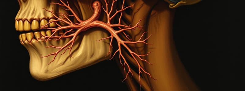
Accessory Nerve (Cranial Nerve XI)
- Originates from the motor nucleus located in the medulla, particularly from the nucleus ambiguus.
- Nucleus ambiguus also gives rise to the glossopharyngeal (cranial nerve IX) and vagus nerves (cranial nerve X).
- Consists of two parts: the cranial part and the spinal part.
Cranial Part of Accessory Nerve
- Exits the medulla and traverses the jugular foramen.
- Travels alongside the vagus nerve through the carotid sheath.
- Primarily forms branches that contribute to the pharyngeal plexus, which innervates muscles of the pharynx (superior, middle, and inferior constrictor muscles) and soft palate muscles.
Pharyngeal Plexus
- Comprised of fibers from the accessory nerve, vagus nerve, and glossopharyngeal nerve.
- Provides motor supply to pharyngeal constrictors and muscles of the soft palate including:
- Tensor veli palatini
- Palatoglossus
- Palatopharyngeus
- Levator veli palatini
- Muscles of the uvula
Spinal Part of Accessory Nerve
- Arises from the cervical spinal cord (C1 to C5) and moves upwards through the lateral funiculus.
- Enters the cranial cavity via the foramen magnum.
- After entering the skull, unites with the cranial part and exits through the jugular foramen.
Branches of the Accessory Nerve
-
Internal Branch:
- Runs with the vagus nerve within the carotid sheath.
- Supplies motor fibers to muscles associated with the pharyngeal plexus.
-
External Branch:
- Emerges from the accessory nerve and innervates the sternocleidomastoid muscle.
- Passes beneath the styloid process, occipital artery, and digastric muscle.
- Supplies the trapezius muscle after traversing the posterior cervical triangle.
- Receives contributions from the ventral rami of C2 and C3 for the sternocleidomastoid and from C3 and C4 for the trapezius.
Anatomical Relationships
- The internal carotid artery and internal jugular vein lie adjacent to the vagus and accessory nerves in the carotid sheath.
- The spinal part is positioned between the anterior root and dorsal root of the cervical spinal nerves.
Clinical Relevance
- Dysfunction in the accessory nerve can lead to deficits in shoulder elevation and head rotation due to weakness in the sternocleidomastoid and trapezius muscles.### Cortical Bulbar Fibers and Their Functions
- Cortical bulbar fibers originate from the cortex and connect to the accessory nerve, aiding motor control.
- The precentral gyrus contains the cell bodies of upper motor neurons, making it part of the primary somatosensory and motor cortex.
- Cortical bulbar fibers descend to reach the nucleus ambiguus, which is crucial for motor function.
Cervical Nerve Supply
- Cortical fibers activate motor neurons in the cervical region, specifically C2, C3, and C4.
- C2 and C3 innervate the sternocleidomastoid muscle; C3 and C4 innervate the trapezius muscle.
- Most fibers supplying the trapezius cross to the contralateral side, while those for the sternocleidomastoid remain ipsilateral.
Lesions and Associated Conditions
- Lesions can occur at various points: cortical bulbar fibers, the nucleus ambiguus, spinal cord, or peripheral processes.
- Potential causes of lesions include infarctions, tumors, abscesses, ALS, polio, and trauma to the neck region.
- Accessory nerve damage can also stem from metastatic lesions or compressions from enlarged cervical lymph nodes.
Clinical Manifestations of Accessory Nerve Palsy
- Damage to the accessory nerve proximal to the posterior triangle of the neck results in sternocleidomastoid (SCM) weakness, affecting head movement.
- Damage to trapezius fibers leads to scapular elevation issues, scapular winging, and arm abduction difficulties.
- Lesions affecting nerve fibers in the posterior triangle may spare SCM function while impairing trapezius function.
Important Anatomical Structures
- The accessory nerve is closely associated with cervical lymph nodes, which can cause compression and lead to dysfunction if inflamed or enlarged.
- The medial branches of the spinal accessory nerve are integral for neck movement and shoulder stabilization.
Conclusion
- Understanding the accessory nerve's pathways and its muscular innervation is vital for diagnosing associated neuromuscular disorders and assessing clinical implications of nerve damage.
Accessory Nerve (Cranial Nerve XI)
- Originates from the motor nucleus in the medulla, particularly from the nucleus ambiguus, which also gives rise to cranial nerves IX and X.
- Composed of a cranial part and a spinal part.
Cranial Part of Accessory Nerve
- Exits the medulla through the jugular foramen and travels alongside the vagus nerve in the carotid sheath.
- Contributes branches to the pharyngeal plexus, innervating pharyngeal muscles and soft palate muscles.
Pharyngeal Plexus
- Formed by fibers from the accessory nerve, vagus nerve, and glossopharyngeal nerve.
- Supplies motor innervation to pharyngeal constrictors and soft palate muscles, including:
- Tensor veli palatini
- Palatoglossus
- Palatopharyngeus
- Levator veli palatini
- Muscles of the uvula
Spinal Part of Accessory Nerve
- Arises from cervical spinal cord segments C1 to C5 and ascends through the lateral funiculus.
- Enters the cranial cavity via the foramen magnum, uniting with the cranial part before exiting through the jugular foramen.
Branches of the Accessory Nerve
- Internal Branch:
- Follows the vagus nerve in the carotid sheath; provides motor fibers to the pharyngeal plexus.
- External Branch:
- Innervates the sternocleidomastoid muscle, traverses several anatomical structures, and subsequently supplies the trapezius muscle.
- Receives contributions from C2 and C3 (sternocleidomastoid) and C3 and C4 (trapezius).
Anatomical Relationships
- Positioned between the internal carotid artery and internal jugular vein within the carotid sheath.
- The spinal part lies between the anterior and dorsal roots of cervical spinal nerves.
Clinical Relevance
- Accessory nerve dysfunction can impair shoulder elevation and head rotation due to weakness in sternocleidomastoid and trapezius muscles.
Cortical Bulbar Fibers and Their Functions
- Originate from the cortex, facilitate motor control for the accessory nerve.
- Upper motor neuron cell bodies are located in the precentral gyrus, part of the primary motor cortex.
Cervical Nerve Supply
- Cortical fibers activate motor neurons in cervical regions C2, C3, and C4, specifically innervating the sternocleidomastoid and trapezius muscles.
- Trapezius motor fibers primarily cross to the contralateral side, while sternocleidomastoid fibers remain ipsilateral.
Lesions and Associated Conditions
- Lesions may arise in several areas: cortical bulbar fibers, nucleus ambiguus, spinal cord, or peripheral processes.
- Causes of lesions include infarctions, tumors, abscesses, ALS, polio, and neck trauma.
- Accessory nerve damage may also be due to metastatic lesions or compression from enlarged cervical lymph nodes.
Clinical Manifestations of Accessory Nerve Palsy
- Proximal damage results in sternocleidomastoid weakness, hindering head movement.
- Damage to trapezius fibers leads to scapular elevation issues, winging, and difficulties in arm abduction.
- Lesions may spare sternocleidomastoid function while impairing trapezius function in the posterior triangle.
Important Anatomical Structures
- The accessory nerve is closely linked to cervical lymph nodes, which can cause compression and lead to dysfunction if inflamed or enlarged.
- Medial branches of the spinal accessory nerve are crucial for neck movement and shoulder stabilization.
Conclusion
- Knowledge of the accessory nerve's pathway and muscular innervation is essential for diagnosing neuromuscular disorders and evaluating nerve damage implications.
Studying That Suits You
Use AI to generate personalized quizzes and flashcards to suit your learning preferences.


