Podcast
Questions and Answers
What is the main process involved in Acantholysis?
What is the main process involved in Acantholysis?
- Rupture of desmosomes (correct)
- Destruction of keratinocytes
- Formation of new desmosomes
- Separation of keratinocytes
Where are desmosomes primarily located?
Where are desmosomes primarily located?
- In the epidermis (correct)
- Between blood cells
- In nerve tissue
- In muscle tissue
Which antibody is associated with Pemphigus Foliaceous?
Which antibody is associated with Pemphigus Foliaceous?
- Desmoglein-1-3 antibodies
- Desmoglein-3 antibody
- Desmoglein-A antibody
- Desmoglein-1 antibody (correct)
Which clinical sign involves applying tangential pressure to the skin?
Which clinical sign involves applying tangential pressure to the skin?
What happens during the Bulla Spread Sign test?
What happens during the Bulla Spread Sign test?
What is a key characteristic of Pemphigus Vulgaris compared to Pemphigus Foliaceous?
What is a key characteristic of Pemphigus Vulgaris compared to Pemphigus Foliaceous?
What sign applies to smaller, intact bullae?
What sign applies to smaller, intact bullae?
Where is Desmoglein-3 primarily found in the skin?
Where is Desmoglein-3 primarily found in the skin?
Which lab method can be used to investigate Acantholysis?
Which lab method can be used to investigate Acantholysis?
In which condition do you typically find a subcorneal split in the skin?
In which condition do you typically find a subcorneal split in the skin?
Study Notes
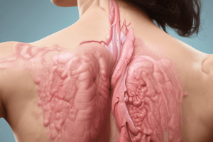
Acantholysis Overview
- Acantholysis refers to the disruption of desmosomes, leading to cell separation.
- Desmosomes serve as vital cell-to-cell junctions in the epidermis, connecting keratinocytes.
Pemphigus Classification
- Pemphigus Foliaceous: Associated with Desmoglein-1 antibody; manifests primarily in the upper epidermis.
- Pemphigus Vulgaris: Linked to Desmoglein-3 and Desmoglein-1 antibodies; affects both the upper and lower epidermal layers.
Desmoglein Expression in Skin and Mucosa
- Desmoglein-1: Predominantly found in the upper epidermis; related to pemphigus foliaceous.
- Desmoglein-3: Located in the lower epidermis; associated with pemphigus vulgaris.
- In oral mucosa, Desmoglein-1 has low or absent concentration, while Desmoglein-3 is present at high levels.
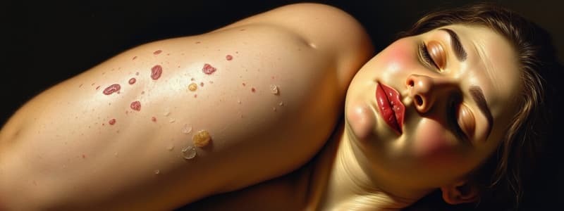
Lesional Characteristics
- Pemphigus Foliaceous leads to subcorneal splits due to Desmoglein-1 involvement.
- Pemphigus Vulgaris results in suprabasal splits, primarily influenced by Desmoglein-3-1.
Clinical Signs to Identify Acantholysis
- Nikolsky Sign: Applying tangential pressure on the skin causes upper epidermal layers to separate.
- Bulla Spread Sign: Lateral pressure on a bulla results in the extension of bulla margins, displaying irregular borders typical of pemphigus vulgaris.
- Asboe-Hansen Sign: Variation of Bulla Spread Sign; pressure applied to the center of intact tense bullae leads to spreading.
Laboratory Techniques for Diagnosis
- Tzanck Smear: Microscopic examination of cell samples to identify acantholysis.
- Histopathology: Tissue examination to study the microscopic changes related to pemphigus diseases.
Studying That Suits You
Use AI to generate personalized quizzes and flashcards to suit your learning preferences.
Description
Explore the mechanisms of acantholysis and how it relates to pemphigus, including the role of desmosomes and keratinocytes. This quiz will test your understanding of these important concepts in dermatology and cell biology. Diagram insights will enhance your grasp of the subject.