🎧 New: AI-Generated Podcasts Turn your study notes into engaging audio conversations. Learn more

Abdominal Wall Masses and Hernias Quiz
33 Questions
0 Views

Abdominal Wall Masses and Hernias Quiz

Created by
@DefeatedSagacity

Podcast Beta

Play an AI-generated podcast conversation about this lesson

Questions and Answers

Match the following descriptions with the correct type of hernia:

Defect in abdominal musculature after surgery = Incisional hernia Protrusion of the navel = Umbilical hernia Occurs in the midline of the epigastrium with fat content = Midline hernia Nonreducible and may require immediate surgery = Incarcerated hernia

Match the following situations with the corresponding conditions:

Develops during pregnancy = Umbilical hernia Occurs in chronic respiratory disease = Diaphragmatic hernia Small fascial defects in linea alba = Midline hernia Contains a bit of fat and feels as a small, tender nodule = Incarcerated hernia

Match the following statements with the correct condition:

Increased abdominal pressure causes protrusion = Hernias Separation of rectus abdominis muscles = Diastasis recti Obstructed blood supply to protruded contents = Incarcerated hernia Smooth, even movement with respiration = Normal abdominal findings

Match the following characteristics with the appropriate condition:

<p>Fatigue, weakness, and bloating = Paralytic ileus Defect in diaphragm muscle leading to abdominal contents entering chest cavity = Diaphragmatic hernia Develops after a surgical incision = Incisional hernia Protrusion at the umbilicus due to weakness in abdominal wall = Umbilical hernia</p> Signup and view all the answers

Match the following findings with the correct condition:

<p>Visible superficial abdominal wall masses = Hernias Little clinical significance, common in pregnancy/postpartum period = Diastasis recti Intrathoracic pressure repeatedly increased = Diaphragmatic hernia Rectus abdominis muscles separation on head raising from table = Diastasis recti</p> Signup and view all the answers

Match the following descriptions with the correct condition: 1. Bowel sounds hypoactive or absent, no masses, patient on diuretics for hypertension. 2. Commonly caused by diuretics, narcotics, and hypothyroidism. 3. Bulges or masses may appear upon deep breath or head raise maneuver. 4. Muscles contract producing prominence in thin or athletic adults.

<p>Paralytic ileus = Commonly caused by diuretics, narcotics, and hypothyroidism Diaphragmatic hernia = Bulges or masses may appear upon deep breath or head raise maneuver Umbilical hernia = Muscles contract producing prominence in thin or athletic adults</p> Signup and view all the answers

Match the following conditions with their potential causes: 1. Distended abdomen due to tumor, pancreatic cyst, or gastric dilation. 2. Asymmetric distention indicating hernia, tumor, cysts, bowel obstruction, etc.

<p>Diaphragmatic hernia = Distended abdomen due to tumor, pancreatic cyst, or gastric dilation Incisional hernia = Asymmetric distention indicating hernia, tumor, cysts, bowel obstruction</p> Signup and view all the answers

Match the following conditions with their characteristic features: 1. Abdomen significantly distended with hypoactive bowel sounds and diminished deep tendon reflexes. 2. Contour remains smooth and symmetric upon deep breath or head raise maneuver.

<p>Paralytic ileus = Abdomen significantly distended with hypoactive bowel sounds and diminished deep tendon reflexes Umbilical hernia = Contour remains smooth and symmetric upon deep breath or head raise maneuver</p> Signup and view all the answers

Match the following findings with their respective implications in neonates:

<p>Distended veins across the abdomen = Suggestive of vascular obstruction Spider nevi = May indicate liver disease Single umbilical artery = Alerts to the possibility of congenital anomalies Granulomatous tissue nodule in umbilical folds = May indicate a granuloma</p> Signup and view all the answers

Match the following descriptions with the correct conditions in newborns:

<p>Dry and odorless umbilical stump area = Normal after separation Persistent drainage after cord separation = Possible patent urachal cyst or remnant Protrusion through umbilicus or rectus abdominis when infant strains = May suggest umbilical hernia Inverted umbilicus with visible and palpable bulge = Characteristic of small umbilical hernia</p> Signup and view all the answers

Match the following symptoms with their potential diagnoses in infants:

<p>Intestinal structure visible through umbilical area membrane = Suggestive of omphalocele Serosanguineous discharge after cord separation = Indicative of granuloma if no signs of infection present Redness, induration, and skin warmth in umbilical stump inspection = Signs of possible infection Protrusion of omentum and intestine through umbilical opening = Characteristic of incisional hernia</p> Signup and view all the answers

Match the following physical exam findings with their implications in newborns:

<p>Umbilical cord with only two arteries present = Associated with congenital anomalies Visible protrusion through rectus abdominis muscles on straining = Suggestive of diaphragmatic hernia Dry and odorless umbilical stump area inspection = Normal post-separation at 2 weeks of age Serous discharge seen after cord separation = Consideration for a patent urachal cyst or remnant</p> Signup and view all the answers

Match the following findings in infants with their potential diagnoses:

<p>Visible granulomatous tissue nodule in umbilical folds = Indicative of a granuloma Distended veins across the abdomen examination finding = Suggestive of vascular obstruction Protrusion of omentum and intestine through umbilical opening = Characteristic of incisional hernia Umbilical cord with a single artery present = Signals possibility of congenital anomalies</p> Signup and view all the answers

Match the following terms with their correct descriptions:

<p>Paralytic ileus = Condition where there is a temporary cessation of intestinal peristalsis Diaphragmatic hernia = Hernia that occurs when abdominal organs push through an opening in the diaphragm Umbilical hernia = Hernia that occurs when part of the intestine protrudes through the umbilical opening in the abdominal muscles Incisional hernia = Hernia that develops at the site of a surgical incision in the abdominal wall</p> Signup and view all the answers

Match the following signs/symptoms with their associated condition:

<p>Periumbilical discoloration (Cullen sign) = Intra-abdominal bleeding Sister Mary Joseph’s nodule = Intra-abdominal malignancy Symmetric distention from umbilicus to symphysis = Ovarian tumor, pregnancy, uterine fibroids Striae of recent origin turning silvery white over time = Result from pregnancy or weight gain</p> Signup and view all the answers

Match the following findings with their potential causes:

<p>Generalized symmetric distention = Obesity, enlarged organs, fluid or gas Enlarged and sometimes painful umbilical nodule = Cancer metastasis Striae of Cushing disease remaining purplish = Cushing disease Lesions as secondary skin changes = Gastrointestinal diseases</p> Signup and view all the answers

Match the following descriptions with their corresponding term:

<p>Temporary cessation of intestinal peristalsis = Paralytic ileus Hernia at the site of a surgical incision = Incisional hernia Organ protrusion through diaphragm opening = Diaphragmatic hernia Intestine protruding through umbilical opening = Umbilical hernia</p> Signup and view all the answers

Match the following conditions with their typical characteristics:

<p>Symmetric abdomen from seated position to standing position = Contralateral areas of abdomen should be symmetric in appearance and contour Signs of intra-abdominal bleeding = Periumbilical discoloration (Cullen sign) Striae turning silvery white over time = Result from pregnancy or weight gain Pink or blue striae of recent origin = Abdominal tumors or ascites</p> Signup and view all the answers

Match the following conditions with their descriptions:

<p>Paralytic ileus = A condition characterized by the temporary cessation of bowel peristalsis Diaphragmatic hernia = A defect in the diaphragm that allows abdominal organs to move into the chest cavity Hernias = Protrusion of an organ or tissue through an abnormal opening in the body wall Umbilical hernia = A type of hernia that occurs near the belly button in infants</p> Signup and view all the answers

Match the following terms with their correct definitions:

<p>Incisional hernia = A type of hernia that develops at the site of a surgical scar Diastasis rectus abdominis = A separation between the rectus abdominis muscles in the midline Rectus abdominis muscles = Muscles located in the abdomen that may become separated in certain conditions Peristaltic waves = Wave-like contractions of the intestines to move contents along the digestive tract</p> Signup and view all the answers

Match the following symptoms with their corresponding conditions:

<p>Frequent vomiting in infants = May indicate a problem like herniation through rectus abdominis muscles Visible peristaltic waves in thin infants = Could suggest an intestinal obstruction such as pyloric stenosis Potbellied appearance in young children = Common when standing, sitting, and supine, due to a slight protrusion of the abdomen Tangential lighting observation of abdomen = Used to look for peristaltic waves and abnormalities in infants</p> Signup and view all the answers

Match the following age-related information with their corresponding conditions:

<p>Most umbilical hernias close spontaneously by 1 to 2 years of age = Developmental timeline for closure of this type of hernia Maximum size reached by umbilical hernias generally by 1 month of age = Indication of growth progression for this hernia type Diastasis rectus abdominis commonly found between xiphoid and umbilicus = Typical location for this separation in children Rectus abdominis muscle non-approximation is common in diastasis rectus abdominis = Explanation for the separation between these muscles</p> Signup and view all the answers

Match the following visual characteristics with their corresponding conditions:

<p>Herniation through rectus abdominis muscles visible under tangential lighting = Visual clue to a rare but problematic condition Potbellied appearance in young children = Physical presentation due to protrusion of the abdomen Umbilical hernia in an infant (Fig. 18.31) = Visible example of a specific type of hernia at a certain age Midline separation between xiphoid and umbilicus (Diastasis rectus abdominis) = Anatomical feature commonly observed in children</p> Signup and view all the answers

Match the following types of hernias with their descriptions:

<p>Diaphragmatic hernia = Herniation of abdominal organs through a defect in the diaphragm Umbilical hernia = Protrusion of abdominal contents through a weakness in the umbilical ring Incisional hernia = Bulging of tissue through a surgical incision in the abdominal wall Paralytic ileus = Non-mechanical obstruction of the intestines due to lack of peristalsis</p> Signup and view all the answers

Match the following anatomical structures with their descriptions:

<p>Peritoneum = Serous membrane lining the abdominal cavity and protecting abdominal organs Greater and lesser omentum = Double folds of peritoneum around the stomach providing protective covering Mesentery = Fan-shaped fold of peritoneum covering most of the small intestine and anchoring it to the posterior abdominal wall Linea alba = Tendinous band in the midline of the abdomen between rectus abdominis muscles</p> Signup and view all the answers

Match the following abdominal muscles with their locations:

<p>Rectus abdominis muscles = Anterior muscles protecting the abdominal cavity Internal oblique muscles = Muscles located laterally to protect the abdomen External oblique muscles = Muscles located laterally to protect the abdomen Inguinal ligament (Poupart ligament) = Extends from ilium to pubis on each side</p> Signup and view all the answers

Match the following alimentary tract components with their functions:

<p>Esophagus = Transports food from mouth to stomach Stomach = Site for food digestion with gastric juices Small intestine = Primary site for nutrient absorption Large intestine = Absorbs water and forms solid waste for excretion</p> Signup and view all the answers

Match the following movements with their control mechanisms:

<p>Peristalsis = Movement of food along alimentary tract under autonomic nervous system control Ingestion and digestion = Functions performed by alimentary tract for nutrient absorption Absorption of nutrients, electrolytes, and water = Essential function carried out by small intestine Excretion of waste products = End function of alimentary tract under autonomic nervous system control</p> Signup and view all the answers

Match the abdominal structure with its correct description:

<p>External oblique aponeurosis = Forms the outermost layer of the anterior abdominal wall Transverse abdominis = Deep muscle layer that helps compress the abdominal contents Rectus sheath = Covering of the rectus abdominis muscle Internal oblique = Muscle layer that lies between the external oblique and transverse abdominis</p> Signup and view all the answers

Match the abdominal organ with its correct location:

<p>Gallbladder = Underneath the liver Spleen = Located in the left upper quadrant of the abdomen Pancreas = Adjacent to the duodenum Appendix = Attached to the cecum in the lower right abdomen</p> Signup and view all the answers

Match the abdominal structure with its function:

<p>Cystic duct = Connects the gallbladder to the common bile duct Common bile duct = Carries bile from the liver to the small intestine Falciform ligament = Attaches liver to anterior abdominal wall and diaphragm Hepatic portal vein = Transports nutrient-rich blood from digestive organs to liver</p> Signup and view all the answers

Match the abdominal feature with its characteristic:

<p>Vermiform appendix = Blind pouch extension from cecum Hepatic flexure = Bend in colon near liver Ileocecal valve = Prevents backflow of fecal material from large intestine to small intestine Pyloric sphincter = Regulates food passage from stomach to small intestine</p> Signup and view all the answers

Match the abdominal structure with its associated artery or vein:

<p>Superior mesenteric artery and vein = Supplies blood to and drains blood from small intestine and part of colon Inferior vena cava = Returns deoxygenated blood from lower body to heart Celiac trunk = Branches into common hepatic, splenic, and left gastric arteries Renal artery and vein = Supply and drain blood from kidneys</p> Signup and view all the answers

Study Notes

Abdominal Wall Masses

  • Superficial abdominal wall masses may become visible due to increased abdominal pressure.
  • An incisional hernia is caused by a defect in the abdominal musculature that develops after a surgical incision, resulting in a protrusion in the area of the surgical scar.
  • Protrusion of the navel indicates an umbilical hernia, which can occur in adults during pregnancy, long-standing ascites, or chronic respiratory disease.

Hernias

  • Hernias may occur in the midline of the epigastrium due to small fascial defects in the linea alba.
  • Most hernias are reducible, meaning the contents of the hernia can be pushed back into place.
  • If not reducible, the hernia is nonreducible or incarcerated, which may require immediate surgery.

Diastasis Recti

  • Diastasis recti is the separation of the rectus abdominis muscles, which can become apparent when the patient raises their head from the table.
  • It is more common in pregnancy and the postpartum period and is of little clinical significance.

Abdominal Distention

  • Abdominal distention can be caused by tumor, pancreatic cyst, or gastric dilation in the upper half of the abdomen.
  • Asymmetric distention or protrusion may indicate hernia, tumor, cysts, bowel obstruction, muscle or soft tissue hematoma, or enlargement of abdominal organs.

Abdominal Examination

  • Inspect the abdomen for movement, symmetry, and any lesions or nodules.
  • Smooth, even movement should occur with respiration.
  • Distended veins across the abdomen are an unexpected finding, suggestive of vascular obstruction or abdominal distention or obstruction.
  • Spider nevi may indicate liver disease.

Umbilical Cord

  • Inspect the umbilical cord of the newborn, counting the number of vessels (2 arteries and 1 vein).
  • A single umbilical artery should alert you to the possibility of congenital anomalies.
  • Any intestinal structure present in the umbilical cord or protruding into the umbilical area and visible through a thick transparent membrane suggests an omphalocele.

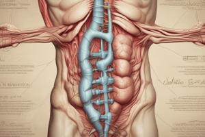
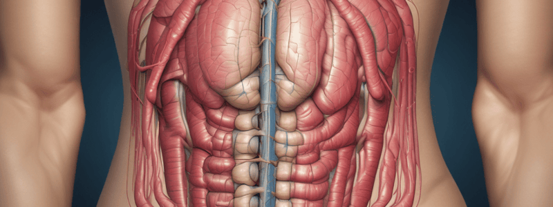
Anatomy and Physiology

  • The abdominal cavity contains several of the body's vital organs.
  • The peritoneum—a serous membrane—lines the cavity and forms a protective cover for many of the abdominal organs.
  • The rectus abdominis muscles anteriorly and the internal and external oblique muscles laterally form and protect the abdominal cavity.
  • The alimentary tract—a tube approximately 27 feet long—runs from the mouth to the anus and includes the esophagus, stomach, small intestine, and large intestine.

Studying That Suits You

Use AI to generate personalized quizzes and flashcards to suit your learning preferences.

Quiz Team

Related Documents

Chapter 18 403 - 431.pdf

Description

Test your knowledge on superficial abdominal wall masses and hernias, including incisional hernias, umbilical hernias, and adult type hernias. Learn about the causes and characteristics of different types of abdominal wall protrusions.

More Quizzes Like This

Abdominal Wall Anatomy Quiz
40 questions

Abdominal Wall Anatomy Quiz

FelicitousCognition avatar
FelicitousCognition
Abdominal Wall Masses and Hernias Quiz
40 questions
Abdominal Wall Masses and Hernias Quiz
40 questions
Anatomy of the Abdominal Wall Part I
9 questions
Use Quizgecko on...
Browser
Browser