Podcast
Questions and Answers
What information is crucial to include when ordering an X-ray to ensure proper interpretation?
What information is crucial to include when ordering an X-ray to ensure proper interpretation?
- The patient's preferred time of day for the procedure
- Patient's insurance policy number for billing purposes
- Detailed family medical history to assess genetic predispositions
- Relevant clinical history and reason for the X-ray (correct)
What is a primary advantage of using fluoroscopy over standard X-rays in a clinical setting?
What is a primary advantage of using fluoroscopy over standard X-rays in a clinical setting?
- Fluoroscopy eliminates the need for contrast agents.
- Fluoroscopy allows for real-time visualization during procedures like lumbar punctures. (correct)
- Fluoroscopy can penetrate dense tissues, such as bones, more effectively.
- Fluoroscopy provides a higher resolution image with less radiation exposure.
Which of the following is a relative contraindication for performing fluoroscopy, particularly concerning image quality and procedure safety?
Which of the following is a relative contraindication for performing fluoroscopy, particularly concerning image quality and procedure safety?
- Hypertension
- Pregnancy (correct)
- Mild anxiety
- Previous allergic reaction to iodinated contrast
Which of the following best describes the primary utility of an angiogram?
Which of the following best describes the primary utility of an angiogram?
Which patient condition would be an absolute contraindication for undergoing an angiogram?
Which patient condition would be an absolute contraindication for undergoing an angiogram?
A patient with a known allergy to contrast dye and chronic kidney disease is being considered for an angiogram. Which of the following is the MOST appropriate course of action?
A patient with a known allergy to contrast dye and chronic kidney disease is being considered for an angiogram. Which of the following is the MOST appropriate course of action?
How does ultrasound imaging primarily generate images of internal body structures?
How does ultrasound imaging primarily generate images of internal body structures?
What is a significant limitation of ultrasound imaging that may necessitate the use of an alternative imaging modality?
What is a significant limitation of ultrasound imaging that may necessitate the use of an alternative imaging modality?
Which of the following is a key advantage of using ultrasound over other imaging techniques like CT scans or X-rays?
Which of the following is a key advantage of using ultrasound over other imaging techniques like CT scans or X-rays?
In computed tomography (CT), what technological advancement contributes to improved image resolution?
In computed tomography (CT), what technological advancement contributes to improved image resolution?
A patient is scheduled for a CT scan to evaluate a suspected abdominal mass. What initial question should be addressed before proceeding with IV contrast?
A patient is scheduled for a CT scan to evaluate a suspected abdominal mass. What initial question should be addressed before proceeding with IV contrast?
After a CT scan with contrast, a patient experiences a rise in creatinine levels within 24-48 hours. Which of the following interventions is MOST appropriate to prevent further kidney damage?
After a CT scan with contrast, a patient experiences a rise in creatinine levels within 24-48 hours. Which of the following interventions is MOST appropriate to prevent further kidney damage?
Why is magnetic resonance imaging (MRI) particularly effective at distinguishing subtle differences in soft tissues?
Why is magnetic resonance imaging (MRI) particularly effective at distinguishing subtle differences in soft tissues?
What is the MOST significant safety concern that must be addressed before a patient undergoes an MRI examination?
What is the MOST significant safety concern that must be addressed before a patient undergoes an MRI examination?
What is the most accurate description of optimization in the context of radiation exposure during medical imaging?
What is the most accurate description of optimization in the context of radiation exposure during medical imaging?
Flashcards
What is Fluoroscopy?
What is Fluoroscopy?
Imaging technique using low-dose radiation to produce real-time, moving images on a screen.
What radiation is used for fluoroscopy?
What radiation is used for fluoroscopy?
A continuous beam of low-dose radiation focused on a specific body area. Commonly for lumbar punctures or epidurals.
What is an Angiogram?
What is an Angiogram?
A combination of radiocontrast and fluoroscopy for visualizing blood flow.
What are the angiogram's relative contraindications?
What are the angiogram's relative contraindications?
Signup and view all the flashcards
What is Ultrasound?
What is Ultrasound?
Signup and view all the flashcards
What is the Doppler shift?
What is the Doppler shift?
Signup and view all the flashcards
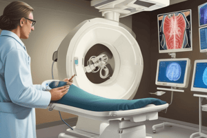
What is Computed Tomography (CT)?
What is Computed Tomography (CT)?
Signup and view all the flashcards
What are CT scan plane orientations?
What are CT scan plane orientations?
Signup and view all the flashcards
What contrasts does CT use?
What contrasts does CT use?
Signup and view all the flashcards
What are CT's absolute contraindications?
What are CT's absolute contraindications?
Signup and view all the flashcards
What is Magnetic Resonance Imaging (MRI)?
What is Magnetic Resonance Imaging (MRI)?
Signup and view all the flashcards
What are the absolute contraindications to MRI?
What are the absolute contraindications to MRI?
Signup and view all the flashcards
What is Nuclear Medicine?
What is Nuclear Medicine?
Signup and view all the flashcards
What is Positron Emission Tomography (PET)?
What is Positron Emission Tomography (PET)?
Signup and view all the flashcards
What is Interventional Radiology?
What is Interventional Radiology?
Signup and view all the flashcards
Study Notes
X-Rays
- A relatively inexpensive imaging method used for insurance purposes.
- X-rays emit a minimal amount of ionizing radiation.
- Used to visualize bones, metal, soft tissue, air, and fat.
- When ordering, avoid ruling out diagnoses and provide a brief patient history for context.
- Require at least two views.
- Used to view bone fractures, assess joints and the spine, and evaluate cardiopulmonary issues.
- A signed order with the reason and relevant information is needed.
- There are no absolute contraindications.
- Relative contraindications include pregnancy and obesity.
- The risk is minimal, and the benefit is high
Fluoroscopy
- Uses low-dose radiation to display a continuous image on a screen.
- Allows for a real-time view, especially useful during needle procedures like lumbar punctures and epidurals.
- Uses a rotating C-arm.
- Prerequisites include a signed order with relevant clinical details, and someone who can interpret the real-time image.
- There are no absolute contraindications.
- Relative contraindications include pregnancy and obesity, as it can be difficult to visualize the needle.
- Adverse effects are the same as with X-rays.
Angiogram
- Combines a small radioactive agent with fluoroscopy.
- Used to visualize blood flow and identify blockages.
- Can be used with MRI or CT scans.
- Vascular access occurs through the femoral artery, femoral vein, or jugular vein.
- Used for coronary, cerebral, pulmonary, renal, and peripheral vascular imaging.
- Prerequisites include knowing the patient's hydration status and underlying health conditions, especially if they are on blood thinners.
- Absolute contraindications include the patient's weight being too high
- Relative contraindications include pregnancy, contrast allergies, chronic kidney disease, and blood thinners.
- Adverse effects include bleeding at the insertion site, contrast-induced kidney injury, allergic reactions, and blood vessel damage.
Ultrasound
- Uses sound waves to create real-time images.
- Uses "Doppler shift,” where changes in frequency occur when sound waves are reflected by moving blood.
- Limited by its inability to travel through bones, deep tissues, and air.
- Used for OB viewing and gallbladder imaging.
- Can also image the thyroid, heart (echocardiogram), scrotum, and eyes.
- Has the advantages of having no radiation, portability, and use in multiple planes (transverse and sagittal).
- Provides real-time visualization for biopsies and needle aspirations.
- Disadvantages include being operator and patient dependent.
- Does not work well on patients with a lot of fat or those who cannot hold their breath.
- Requires a signed order with relevant clinical information.
- There are no absolute contraindications.
- Relative contraindications include obesity and skin infections.
- There are no adverse effects.
Commutated Tomography (CT)
- Uses a rotating X-ray tube to capture multiple slices for more detailed imaging.
- Employs numerous detectors.
- It can be performed with or without contrast.
- Most images are in the axial plane, which is a horizontal plane from the foot of the bed.
- Coronal images cut anterior and posterior, while sagittal images divide left and right.
- CT reformations create 3D images from 2D slices.
- More detectors provide better resolution.
- Ideally used for imaging body cavities such as the lungs, heart, liver, spleen, brain, and coronary arteries.
- Uses iodine-based IV contrast or barium sulfate (oral) if needed.
- Great for visualizing blood vessels and organs.
- Be mindful that patients can be allergic.
- Ask questions to determine if IV contrast is needed.
- Not needed for routine head CT, renal stones, or follow-ups.
- Used for masses, malignancy, clots, and GI infections.
- Contrast allergies can be pretreated with Benadryl/steroids.
- Renal dysfunction, with an increased neuropathy risk with lower GFR
- Screening GFR, lower doses, hydration and avoiding NSAIDs can prevent AKI.
- Assess if a CT is necessary and if an ultrasound can be used as an alternative.
- Absolute contraindications are pregnancy, contrast allergies, and renal impairment.
- Relative contraindications include obesity.
- Adverse effects include radiation, AKI, allergies, and thyroid-iodine issues.
MRI
- Protons (H) in the body emit radio waves in the presence of a magnetic.
- They are able to distinguish tiny differences.
- T1-T2 standard settings are used.
- Patients may have to hold their breath
- Gadolinium is used for MRI with contrast.
- Used for masses and cancer
- Very slow and costly.
- Doesn’t use radiation.
- Least available.
- Used for outpatient follow-up, additional evaluation of joints/MSK, and is great for the brain.
- Requires a signed MRI safety form.
- Absolute contraindications include recent surgery, aneurysm clips, cardiac pacing, cochlear implants, and foreign metals.
- Relative contraindications include joint implants, neurostimulators, and claustrophobia.
- Adverse effects are rare.
Radiation
- Ionizing radiation involves detaching electrons from subatomic particles or electromagnetic waves.
- Found in the environment.
- Even low doses can cause cancer.
- Measured by: Radioactivity (amount released by a material), Exposure (amount in the air), Absorbed dose, Effective dose (potential harms), and Units (mSv).
- The exposure of a CT scan is equal to 80 x-rays.
- Optimization is vital.
- Radiation should be "as low as reasonably available"
Nuclear Medicine
- Evaluates both structure and function using a radioisotope.
- Used for metastatic bone disease.
- Areas taking up radionuclide = "hot spot", indicating increased metabolic activity (usually bad)
- Areas not absorbing radionuclide = "cold spot", indicating standard level of metabolic activity (usually good)
PET Scan
- Positron Emission Tomography (PET) is used for diagnosing, staging, and re-staging cancer.
- Displays molecular activity.
- Glucose marker: Most cancers are highly metabolically active and require increased rates of glucose metabolism.
- Involves significant radiation.
Interventional Radiology
- Interventional radiologists use imaging guidance to perform minimally invasive, non-surgical procedures for diagnosis or treatment.
- These procedures include thoracentesis, paracentesis, lumbar puncture, arthrogram, and biopsies.
Viral Assays
- Used to look at the antibody's antigen, DNA or RNA.
Nucleoid Acid Amplification Test
- Amplifies specific DNA or RNA strands for identification.
PCR
- A subset of NAT is called a polymerase chain reaction.
- PCR amplifies specific genes and can be used to monitor a variety of diseases, including bacterial infections.
Immunologic Essay (ELISA)
- Uses antigen antibody testing to look at a specific antigen, sometimes as a point-of-care test.
Antibody Test
- Looks for specific antibodies for pathogens/disease in serum to know what you're looking for (Acute vs Titer).
Blood Culture
- Main aspects include obtaining to detect the bacteria in blood
- Need multiple samples for blood culture, to make sure it is not contaminated (one aerobic and one anaerobic sample)
- Collect the specimen properly, do not collect from IV.
- Check for antibiotic testing requirements and multiple cultures requirements.
- Results come back slow as the culture must grow.
- Ideally the patient is not on antibiotics, as that can create false negatives
- Get 20 ml of blood volume.
- Indications include SEPSIS, pneumonia, or ventilator acquired pneumonia.
- Get more than one to ensure it's not contamination
- Staph on skin normally causes contamination.
- When reading cultures: Consider the number of positive cultures against total cultures obtained, the organism recovered, the length of time for cultures to become positive, the site of culture collection, and the likelihood of bacteremia based on clinical assessment.
- Full results from blood culture usually takes 48–72 hours, but 5 days may be needed
- Procalcitonin: Acute phase reactants
- Marker of inflammation in lung, intestines due to bacteria.
- Used to differentiate SEPSIS from SIRS.
- Correlate to server values on patient.
Lactate Test
- A test for oxygen exchange problems.
- Indications if oxygen supply to tissue has been is diminished, which is generally a measure of tissue hypoxia.
- Common causes are shock, cardiac issues, mesenteric issues, carbon monoxide poisoning liver failure.
D-Dimer
- Oxygen exchange problem.
- Product of fibrin degranulation
- Used for; pulmonary embolism or venous thromboembolism
- Best used in conjugation with clinical probability assessments.
- If you have high pre test probability, skip it and go to diagnostic imaging
Arterial Blood Gas
- Test oxygen exchange and acid base
- Provides information on cardiopulmonary and metabolic status.
- Oxygen for Lungs.
- Acid base for kidney.
- For VERY SICK PATIENTS.
- Critical illness or ventilation.
- Gotta do Allens test.
- Performed on radial artery.
- Measure of ventilation and respiratory component of acid base levels.
- Look at bicarbonate HCO3, it's measured with metabolic component of acid base equilibrium, and can identify increased PH or decreased PH.
- PH, CO2, Bicarb, are most important
- Asses ventilation status and acid base status
- PaCO2, levels are inversely related to PH (respiratory)
- Hco3, are directly related to PH
X-rays
- Good to evaluate pneumonia, lung tumors, COPD, collapsed lung, and air in the lungs.
- Chest CT is used for infection, oxygen exchange issues, and masses or other structural problems.
- If a chest X-ray is not enough.
- CTPA (CT pulmonary angiography) Used to produce images of blood vessels in the lungs
- Used for pulmonary embolism.
- Highly sensitive and specific for P.E.
- Shows a filling defect
- Ventilation perfusion scan (VQ scan) scans the oxygen exchange areas.
- Can show embolism
- Not as good as a CT
- Chest X-ray
- Posterior-anterior and lateral are the best views
- Correct patient, date, time, examination, indication and prior studies
- Important to verify the image quality
- Should be during inspiration (10 posterior ribs)
- Should have the reverent anatomy
- Rotation can be observed by the equal distance between the medical clavicular head and the spinal process
- Film placement will determine if it is in PA (back on patient)
- In AP, the front of the patient in on the film, with a larger width and mediastinum.
- ABCDE …L
- Airway: trachea, carina, mediastinum
- Bones: fractures, spine
- Cardiac: size and borders
- Cardiac silhouette should be half of the ribs
- Diaphragm: shape border/angles
- Angles sharp, right should be one rib space higher than left
- Lungs should be darker as they move down
- Spine sign increased density of lower spine
- Silhouette sign: elimination of the silhouette of loss of lung soft/tissue interface caused by a mass or fluid in the normally air-filled lungs
- Air bronchogram: tubular outline of an airway made visible by filling of the surrounding alveoli by fluid
- Nodules: spherical opacity within the lung parenchyma
Studying That Suits You
Use AI to generate personalized quizzes and flashcards to suit your learning preferences.