Podcast
Questions and Answers
Which of the following anatomical structures does NOT serve as an attachment point for the flexor retinaculum?
Which of the following anatomical structures does NOT serve as an attachment point for the flexor retinaculum?
- Capitate (correct)
- Scaphoid tubercle
- Pisiform
- Hook of hamate
A patient presents with pain and paresthesia in their wrist. Given the location between the pisiform and hook of hamate, which structure is MOST likely involved?
A patient presents with pain and paresthesia in their wrist. Given the location between the pisiform and hook of hamate, which structure is MOST likely involved?
- Radial artery
- Ulnar nerve (correct)
- Extensor pollicis longus tendon
- Median nerve
Why is the midcarpal joint important for wrist range of motion?
Why is the midcarpal joint important for wrist range of motion?
- It lacks interosseous ligaments, allowing for increased mobility between the proximal and distal carpal rows. (correct)
- It contains strong interosseous ligaments which stabilize the wrist during forceful movements.
- It houses the triangular fibrocartilage complex (TFCC), which cushions the wrist joint.
- It is a fixed joint that provides a stable base for radiocarpal movements.
Which motion and loading pattern is MOST likely to aggravate a TFCC injury?
Which motion and loading pattern is MOST likely to aggravate a TFCC injury?
In wrist biomechanics, the radiocarpal joint includes the articulation between the TFCC and which carpal bones?
In wrist biomechanics, the radiocarpal joint includes the articulation between the TFCC and which carpal bones?
During wrist flexion and extension, which carpal bone serves as the axis of rotation?
During wrist flexion and extension, which carpal bone serves as the axis of rotation?
A patient has limited wrist extension. Which mobilization technique would be MOST appropriate to improve this motion?
A patient has limited wrist extension. Which mobilization technique would be MOST appropriate to improve this motion?
During ulnar deviation, how do the proximal and distal rows of carpals glide?
During ulnar deviation, how do the proximal and distal rows of carpals glide?
During finger flexion, which of the following actions occurs at the wrist to ensure optimal function of the long finger flexors?
During finger flexion, which of the following actions occurs at the wrist to ensure optimal function of the long finger flexors?
Which of the following statements accurately describes the relationship between wrist position and grip strength?
Which of the following statements accurately describes the relationship between wrist position and grip strength?
Which of the following best describes the position of the wrist that results in the weakest interphalangeal flexion force?
Which of the following best describes the position of the wrist that results in the weakest interphalangeal flexion force?
Which of the following structures serves as the focal point for the transverse metacarpal arch of the hand?
Which of the following structures serves as the focal point for the transverse metacarpal arch of the hand?
In the context of hand biomechanics, what is the primary role of the wrist?
In the context of hand biomechanics, what is the primary role of the wrist?
What is the 'cascade sign' and how does it relate to finger flexion?
What is the 'cascade sign' and how does it relate to finger flexion?
During prehension, which type of grip typically involves the object making contact with the palm of the hand?
During prehension, which type of grip typically involves the object making contact with the palm of the hand?
Which nerve is primarily involved in the release of a gripped object?
Which nerve is primarily involved in the release of a gripped object?
Which of the following best describes the joint positioning observed in a Swan Neck deformity?
Which of the following best describes the joint positioning observed in a Swan Neck deformity?
A patient presents with their fourth and fifth digits resting in a position opposite to the lumbricals action. Which nerve is MOST likely affected?
A patient presents with their fourth and fifth digits resting in a position opposite to the lumbricals action. Which nerve is MOST likely affected?
What is the PRIMARY cause of a Boutonniere deformity?
What is the PRIMARY cause of a Boutonniere deformity?
Which condition is characterized by a fixed flexion deformity at the MCP and PIP joints, primarily affecting digits 4 and 5?
Which condition is characterized by a fixed flexion deformity at the MCP and PIP joints, primarily affecting digits 4 and 5?
A patient has developed arthritic changes on the dorsal surfaces of their PIP joints. What are these changes called?
A patient has developed arthritic changes on the dorsal surfaces of their PIP joints. What are these changes called?
Distal phalanx flexion, resulting from the rupture or avulsion of the extensor tendon at its distal insertion, BEST describes which condition?
Distal phalanx flexion, resulting from the rupture or avulsion of the extensor tendon at its distal insertion, BEST describes which condition?
In the context of thumb deformities, what combination of joint positions defines a Z deformity of the thumb?
In the context of thumb deformities, what combination of joint positions defines a Z deformity of the thumb?
A patient with thenar muscle wasting presents with their thumb resting in line with the hand. Which nerve is MOST likely affected and what is this presentation called?
A patient with thenar muscle wasting presents with their thumb resting in line with the hand. Which nerve is MOST likely affected and what is this presentation called?
A patient presents with an inability to oppose the thumb and flex the 4th and 5th digits. Which nerve(s) is/are MOST likely affected?
A patient presents with an inability to oppose the thumb and flex the 4th and 5th digits. Which nerve(s) is/are MOST likely affected?
A patient exhibits a resting finger extension deformity and is unable to cup their hand. Dysfunction of which nerve is the MOST likely cause?
A patient exhibits a resting finger extension deformity and is unable to cup their hand. Dysfunction of which nerve is the MOST likely cause?
What is the MOST prominent symptom that differentiates carpal tunnel syndrome (CTS) from pronator teres syndrome related to median nerve compression?
What is the MOST prominent symptom that differentiates carpal tunnel syndrome (CTS) from pronator teres syndrome related to median nerve compression?
During an evaluation, a patient is noted to have their MCP and IP joints of digits 4 and 5 stuck in a flexed position due to fascial shortening. Palpation reveals tender, thick nodules in the palmar fascia in line with digits 4 & 5. This is MOST indicative of which condition?
During an evaluation, a patient is noted to have their MCP and IP joints of digits 4 and 5 stuck in a flexed position due to fascial shortening. Palpation reveals tender, thick nodules in the palmar fascia in line with digits 4 & 5. This is MOST indicative of which condition?
Which structure is MOST directly affected in Skier's Thumb?
Which structure is MOST directly affected in Skier's Thumb?
A patient presents with their wrist flexed and fingers postured in flexion. They are unable to extend their wrist and fingers. Which condition is MOST likely causing these symptoms?
A patient presents with their wrist flexed and fingers postured in flexion. They are unable to extend their wrist and fingers. Which condition is MOST likely causing these symptoms?
In treating Dupuytren's contracture, why is a long-term and consistent treatment approach emphasized?
In treating Dupuytren's contracture, why is a long-term and consistent treatment approach emphasized?
How does CMC joint laxity contribute to thumb positioning?
How does CMC joint laxity contribute to thumb positioning?
A patient presents with a finger that is stuck in a flexed position and unable to extend without assistance, accompanied by a painful snapping sensation. Which of the following interventions is MOST appropriate in the initial management of this condition?
A patient presents with a finger that is stuck in a flexed position and unable to extend without assistance, accompanied by a painful snapping sensation. Which of the following interventions is MOST appropriate in the initial management of this condition?
When differentiating between Rheumatoid Arthritis (RA) and Osteoarthritis (OA) in the fingers, which of the following characteristics is MOST indicative of RA?
When differentiating between Rheumatoid Arthritis (RA) and Osteoarthritis (OA) in the fingers, which of the following characteristics is MOST indicative of RA?
A patient with finger arthritis exhibits significant ulnar drift at the MCP joints. Which of the following exercise approaches should be AVOIDED to prevent further exacerbation of this condition?
A patient with finger arthritis exhibits significant ulnar drift at the MCP joints. Which of the following exercise approaches should be AVOIDED to prevent further exacerbation of this condition?
A patient diagnosed with trigger finger is undergoing rehabilitation. Which of the following interventions is MOST appropriate to reduce flexor tone and promote extensor function during the initial phase of treatment?
A patient diagnosed with trigger finger is undergoing rehabilitation. Which of the following interventions is MOST appropriate to reduce flexor tone and promote extensor function during the initial phase of treatment?
Which statement BEST describes why intrinsic hand muscle strengthening is preferred over extrinsic strengthening in managing finger arthritis?
Which statement BEST describes why intrinsic hand muscle strengthening is preferred over extrinsic strengthening in managing finger arthritis?
A patient presents with pain and locking in their finger. Conservative treatment, including splinting and massage, has not resolved the issue. What is the MOST appropriate next step in managing this patient's condition?
A patient presents with pain and locking in their finger. Conservative treatment, including splinting and massage, has not resolved the issue. What is the MOST appropriate next step in managing this patient's condition?
A patient is diagnosed with osteoarthritis affecting the PIP and DIP joints of their fingers. Which of the following clinical presentations would be MOST indicative of this condition?
A patient is diagnosed with osteoarthritis affecting the PIP and DIP joints of their fingers. Which of the following clinical presentations would be MOST indicative of this condition?
A therapist is treating a patient with Rheumatoid Arthritis (RA) in the hands. Which of the following interventions should be implemented with caution?
A therapist is treating a patient with Rheumatoid Arthritis (RA) in the hands. Which of the following interventions should be implemented with caution?
A patient presents with pain in the anatomical snuffbox. Which activity would MOST likely exacerbate their pain, indicating De Quervain's tenosynovitis?
A patient presents with pain in the anatomical snuffbox. Which activity would MOST likely exacerbate their pain, indicating De Quervain's tenosynovitis?
A physical therapist is evaluating a patient with suspected TFCC injury. Which combination of movements would MOST likely reproduce the patient's symptoms?
A physical therapist is evaluating a patient with suspected TFCC injury. Which combination of movements would MOST likely reproduce the patient's symptoms?
A patient reports deep, ulnar-sided wrist pain that worsens when carrying groceries. Edema is minimal. Which of the following conditions should the therapist suspect FIRST?
A patient reports deep, ulnar-sided wrist pain that worsens when carrying groceries. Edema is minimal. Which of the following conditions should the therapist suspect FIRST?
Following immobilization for a TFCC injury, a patient is ready to begin active wrist ROM exercises. Which of the following exercises should be initiated with assistance from the therapist?
Following immobilization for a TFCC injury, a patient is ready to begin active wrist ROM exercises. Which of the following exercises should be initiated with assistance from the therapist?
During an evaluation, the therapist performs the Finkelstein test. Which presentation would MOST warrant the use of this test?
During an evaluation, the therapist performs the Finkelstein test. Which presentation would MOST warrant the use of this test?
A patient is diagnosed with De Quervain’s tenosynovitis. After acute inflammation subsides, which of the following strengthening exercises is MOST appropriate to begin with?
A patient is diagnosed with De Quervain’s tenosynovitis. After acute inflammation subsides, which of the following strengthening exercises is MOST appropriate to begin with?
When designing a home exercise program for a patient recovering from a wrist injury, what principle should the therapist prioritize regarding muscle imbalances?
When designing a home exercise program for a patient recovering from a wrist injury, what principle should the therapist prioritize regarding muscle imbalances?
A therapist wants to isolate and strengthen the intrinsic muscles of the hand. Which exercise BEST achieves this goal?
A therapist wants to isolate and strengthen the intrinsic muscles of the hand. Which exercise BEST achieves this goal?
Flashcards
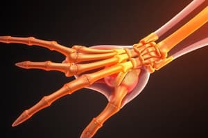
Carpal Tunnel Landmarks
Carpal Tunnel Landmarks
Bounded by the scaphoid tubercle, trapezium tubercle, hook of hamate, and pisiform, it's where the flexor retinaculum attaches.
Anatomical Snuffbox Landmarks
Anatomical Snuffbox Landmarks
Lateral: Abductor pollicis longus and extensor pollicis brevis. Medial: Extensor pollicis longus. Floor: Scaphoid (radial artery inside).
Tunnel of Guyon
Tunnel of Guyon
Located between the pisiform and hook of hamate, covered by the pisohamate ligament; contains the ulnar nerve.
Midcarpal Joint
Midcarpal Joint
Signup and view all the flashcards
TFCC
TFCC
Signup and view all the flashcards
Radiocarpal Joint (Biomechanics)
Radiocarpal Joint (Biomechanics)
Signup and view all the flashcards
Flexion-Extension Wrist Joints
Flexion-Extension Wrist Joints
Signup and view all the flashcards
Radial/Ulnar Deviation Glides
Radial/Ulnar Deviation Glides
Signup and view all the flashcards
Functional arches of the hand
Functional arches of the hand
Signup and view all the flashcards
Key Longitudinal Arch
Key Longitudinal Arch
Signup and view all the flashcards
Transverse Carpal Arch
Transverse Carpal Arch
Signup and view all the flashcards
Transverse Metacarpal Arch
Transverse Metacarpal Arch
Signup and view all the flashcards
Cascade Sign in Finger Flexion
Cascade Sign in Finger Flexion
Signup and view all the flashcards
Wrist Stabilization During Finger Flexion
Wrist Stabilization During Finger Flexion
Signup and view all the flashcards
Optimal Wrist Position for Flexion Force
Optimal Wrist Position for Flexion Force
Signup and view all the flashcards
Dorsal Digital Expansion (DDE)
Dorsal Digital Expansion (DDE)
Signup and view all the flashcards
Claw Fingers
Claw Fingers
Signup and view all the flashcards
Claw Fingers (Resting)
Claw Fingers (Resting)
Signup and view all the flashcards
Wrist Drop
Wrist Drop
Signup and view all the flashcards
Carpal Tunnel Syndrome
Carpal Tunnel Syndrome
Signup and view all the flashcards
Dupuytren’s Contracture
Dupuytren’s Contracture
Signup and view all the flashcards
Skier’s Thumb
Skier’s Thumb
Signup and view all the flashcards
Ulnar Nerve Claw
Ulnar Nerve Claw
Signup and view all the flashcards
Wrist Drop
Wrist Drop
Signup and view all the flashcards
Trigger Finger
Trigger Finger
Signup and view all the flashcards
Trigger Finger Splinting
Trigger Finger Splinting
Signup and view all the flashcards
RA in Fingers
RA in Fingers
Signup and view all the flashcards
OA in Fingers
OA in Fingers
Signup and view all the flashcards
Swan Neck Deformity
Swan Neck Deformity
Signup and view all the flashcards
Hypermobility in RA
Hypermobility in RA
Signup and view all the flashcards
Boutonniere Deformity
Boutonniere Deformity
Signup and view all the flashcards
Hypomobility in OA
Hypomobility in OA
Signup and view all the flashcards
Ulnar Drift
Ulnar Drift
Signup and view all the flashcards
Dupuytren's Contracture
Dupuytren's Contracture
Signup and view all the flashcards
Lumbrical Strengthening
Lumbrical Strengthening
Signup and view all the flashcards
Heberden's Nodes
Heberden's Nodes
Signup and view all the flashcards
Bouchard's Nodes
Bouchard's Nodes
Signup and view all the flashcards
Ulnar Drift
Ulnar Drift
Signup and view all the flashcards
Mallet Finger
Mallet Finger
Signup and view all the flashcards
Ape Hand
Ape Hand
Signup and view all the flashcards
De Quervain's Tenosynovitis
De Quervain's Tenosynovitis
Signup and view all the flashcards
De Quervain's Cause
De Quervain's Cause
Signup and view all the flashcards
TFCC Injury
TFCC Injury
Signup and view all the flashcards
TFCC Injury Symptoms
TFCC Injury Symptoms
Signup and view all the flashcards
TFCC Treatment
TFCC Treatment
Signup and view all the flashcards
Orthopedic Test Indication
Orthopedic Test Indication
Signup and view all the flashcards
Home Care Principles
Home Care Principles
Signup and view all the flashcards
Hand Muscle Isolation
Hand Muscle Isolation
Signup and view all the flashcards
Study Notes
- These notes cover the anatomy, biomechanics, pathologies, orthopedic tests, and home care for the wrist and hand
Carpal Tunnel Landmarks
- The carpal tunnel landmarks include the scaphoid tubercle, trapezium tubercle, hook of hamate, and pisiform.
- The flexor retinaculum attaches to these four points.
Anatomical Snuffbox Landmarks
- The lateral tendons are the abductor pollicis longus and extensor pollicis brevis.
- The medial tendon is the extensor pollicis longus.
- The scaphoid forms the floor.
- The radial artery exists inside the snuffbox space
Tunnel of Guyon
- Is formed between the pisiform and hook of hamate, covered by the pisohamate ligament
- Contains the ulnar nerve.
Joints and Ligaments of the Wrist
- The distal radioulnar joint is a synovial, pivot joint with articulation between the ulnar notch of the radius and the head of the ulna.
- The radiocarpal joint is a synovial, ellipsoidal joint with articulation between the distal end of the radius and the proximal surfaces of the scaphoid and lunate.
- The ulnomeniscotriquetral joint is a synovial ellipsoidal joint is between the meniscus (triangular piece of fibrocartilage distal to ulnar head) and the proximal surface of the triquetrum.
- Capsular strength/coaptation is weak in all three joints: distal radioulnar, radiocarpal, and ulnomeniscotriquetral.
- Key ligaments include the flexor and extensor retinacula and radial and ulnar collateral ligaments
- The flexor and extensor retinacula have six tunnels.
- The radial collateral ligament limits excessive abduction and ulnar deviation.
- The ulnar collateral ligament limits excessive adduction and radial deviation.
Functional Anatomy
- The distal radioulnar joint allows 1 degree of freedom (supination-pronation).
- The radiocarpal and ulnocarpal (UMT) joints allow 2 degrees of freedom (flexion-extension, radial deviation-ulnar deviation).
- Head of the ulna is convex, ulnar notch of the radius is concave.
- Carpals are convex adn the Radius and meniscus are concave
- Resting and closed packed positions require 10 degrees supination and 5 degrees supination resepectively.
- Limitation is equal in all directions for capuslar pattern of restriction
- ROM and end feel with supination at 0-90° firm, pronation at 0-70/90° firm/hard
Triangular Fibrocartilage Complex (TFCC)
- The triangular fibrocartilage complex includes the disc of the UMT.
- The TFCC is an important structure for wrist stability, has poor vascularization with slow healing.
- It's most aggravated by extension with pronation under load.
Wrist Biomechanics
- The wrist complex involves the distal radioulnar, radiocarpal, and midcarpal joints.
- Radiocarpal joint: articulation between the TFCC and carpals (lunate, triquetrum), extends the ulna, UMT joint included when discussing biomechanics
- Axis of flexion-extension: capitate
- Flexion primarily occurs at the midcarpal joint.
- Extension primarily occurs at the radiocarpal joint.
- Radiocarpal closed pack (full extension) results from asymmetry of scaphoid movement and relies on movement of the lunate on the scaphoid
- Convex-on-concave: movement is opposite in radial-ulnar deviation
- Full ulnar or radial deviation requires mobility between carpal rows.
- In ulnar deviation, both rows of carpals glide radially
Hand Biomechanics
- There's 1 longitudinal arch per digit.
- Digit 3 and the capitate are the most important
- There are 2 transverse arches.
- The transverse carpal arch runs through the distal row of carpals and is also known as the proximal transverse arch
- the distal transverse arch runs through the head of the metacarpals.
- Heads of the metacarpals consist of the distal transverse arch.
- Focal point locations: MC 3, capitate and lunate.
- With Cascade Sign and finger flexion, only the index finger flexes in the sagittal plane, but all other fingers flex in an oblique plane towards the scaphoid
Length-Tension Relationships
- Extrinsic muscles of the hand: wrist provides a stable base, wrist position controls the length of extrinsic hand muscle. Movements of the wrist are usually in reverse of the movements of the fingers and reinforce the action of extrinsic muscle of the fingers
Extrinsic Muscle Actions
- Wrist extensors stabilize the wrist and prevent long finger flexors from simultaneously flexing the wrist during finger flexion
- Wrist flexors activate to stabilize the wrist so long finger extensors can function effectively during finger extension
- As grip strength increases, extensors slacken allowing flexors to achieve a strong contraction.
- Greatest interphalangeal flexion force occurs with ulnar deviation and neutral flexion-extension.
- The weakest interphalangeal flexion force occurs when the wrist is in full flexion due to its lack of ability to generate forve
Dorsal Digital Expansion (DDE)
- DDE originates on the posterior, medial, and lateral surfaces of proximal phalanges 1-5.
- Extrinsic tendons are ED and EPL with intrinsic tendons are lumbricals and interossei,
- There's trifurcation on the dorsal aspect.
Prehension (grip) and Nerve Involvement
- Power grips contact the palm and are isometric.
- Precision grips do not contact the palm and are isotonic.
- Grabbing usually involves the median and ulnar nerves.
- Release involves the radial nerve.
Swan Neck Deformity
- MCP: Flexion
- PIP: Extension
- DIP: Flexion
- Causes: Muscle contracture or tearing of the volar plate at the PIP. Commonly seen with RA or post-trauma
Boutonniere Deformity
- MCP: Extension
- PIP: Flexion
- DIP: Extension
- Causes: Rupture of the central slip at the PIP of DDE. Commonly seen with RA or post-trauma
Dupuytren's Contracture
- Contracture of palmar fascia, which includes skin.
- The middle and ring fingers (digits 4, 5) are affected.
- There is development of fixed flexion deformity at MCP and PIP joints
Heberden's and Bouchard's Nodes
- Heberden's Nodes: arthritic changes on dorsal surfaces of DIPs.
- Bouchard's Nodes: arthritic changes on dorsal surfaces of PIPs
Drifts & Mallet Finger
- Ulnar Drift: with RA, changes in MCP result in pull on long tendons.
- Radial Drift: with OA
- Mallet Finger is when distal phalanx is flexed due to avulsion or rupture of the extensor tendon at the site of insertion
Thumb Deformities
- Zigzag deformity and Z deformity can affect the thumb.
- The CMC is flexed, MCP hyperextended, and IP partially flexed in a Zigzag deformity. Associated with RA.
- The MCP is flexed, and the IP is hyperextended in a Z deformity, which is is familial
Ulnar Nerve Deformities
- Bishop's Hand (Benediction Hand): Occurs at rest. Loss of lumbricals leads to digits 4 & 5 resting opposite the lumbricals' action.
- Claw Hand: Occurs at rest. Similar to Bishop's hand but may have some abduction of digits 4 & 5.
- Froment's Sign: Active. Loss of the adductor pollicis (innervated by ulnar nerve) leads to compensatory recruitment of the flexor pollicis longus (median nerve)
Median Nerve Deformities
- Ape Hand: Occurs at rest. Thenar wasting causes the thumb to rest in line with other digits with inability to oppose or flex thumb.
- Oath Hand: Active. Unsuccessful attempts to make a fist, ulnar digits 4 & 5 flex while median digits 1, 2, 3 do not.
Claw Fingers
- Is categorized under both Median and Ulnar combined nerve damage
- Fingers are often hyper-extended at the MCP joints and flexed at the interphalangeal joints, the
- Patients cannot cup hand
Radial Nerve Deformities
- Wrist Drop: Occurs at rest. Loss of extensors leads to flexed wrist and fingers with inability to extend wrist or fingers.
Carpal Tunnel Syndrome
- Median nerve compression: Decreased space in tunnel or increased contents; external pressure on wrist
- The hallmark sign is nocturnal pain, especially in digits 1-3, as opposed to the skin over the thenar eminence
- Differentiate from pronator teres as alternate compression site.
Dupuytren's Contracture
- Affects the palmar fascia with 3 layers (longitudinal, transverse, vertical). Palmaris longus inserts into palmar fascia.
- It is slowly progressive with no effect on tendon, muscle, or joint.
- Signs: Tender, thick, nodular palmar fascia and MCP & IP joints stuck in flexed position with digits 4 & 5 affected.
- Treat with heat, MFR, and stretching.
Skier's Thumb/Gamekeeper's Thumb
- This is a sprain of the ulnar collateral ligament (UCL) of the first MCP joint.
- The MCP joint sits between mobile CMC and rigid IP joint.
- UCL dysfunction may result from acute trauma or repetitive stress.
- The thumb's ulnar-sided MCP will be painful at digit 1, with gripping and pinching being difficult and painful
- Additionally, there will be a positive UCL stress test.
Flexor Pollicis Longus Tenosynovitis
- The tendinous sheath of the FPL extends from radial & superficial extending to the carpal tunnel before angling around the scaphoid to thumb.
- The site is a potential location of irritation to the FPL sheath
- History: Palmar thumb pain increases especially with movement that is caused by repeated thumb use
Symptoms and Differential Diagnosis
Symptoms show pain with AROM or concentric RROM but pain-free MMT Isometric contraction does not cause movement and isn't painful. Distinguish from median nerve & C6 nerve issues
1st Carpometacarpal (CMC) Osteoarthritis (OA)
- It is characterized as, a long term degenerative process; causes first CMC relative instability that predispose UCL injury of thumb
- It is characterized by thumb pain in the area of the anatomical snuffbox that gets worse during movement.
- Pain is generally worse in morning or after prolonged disuse, heat helps
- Provoking activities causes compression (radial deviation, pinching, forceful gripping),
- Pain from these types of activities can create disability and a loss of function,
- Radial nerve irritation can be a differential
- Thenar muscle MFR, stretching into abduction, & massage is helpful
- Strengthening dorsal interosseous muscle and other hand muscles is helpful
Fractures and Dislocations
- Colles fracture will show the "Dinner Fork" sign- the radius fragment will be displaced dorsally just proximal to wrist from FOOSH force.
- Galeazzi, radius fracture the distal radioulnar joint could be dislocated with complications of ulnar nerve injury
- the commonly fractured carpal occurs in the Scaphoid fracture, will be difficult diagnose by xray and commonly misdiagnosed as sprain
Trigger Finger (Digital Tenovaginitis)
- Is categorized as stenosis/ tenosynovitis
- It is induced by, Thickening of flexor tendon sheath (FDS) by Nodules along affected tendon, usually at distal MCP to palm crease
Symptoms and treatment
- When the finger is flexed, the nodule moves proximally and a palpable can be heard when the finger is extended again.
- The finger becomes locked or triggered in flexed position, sometimes a passive pull is needed to extend finger (passive)
- Idiopathic cause, increased finger flexion force, palpable lump
Finger Arthritis
- RA mainly affects the MCPs
- PIPs and DIPs (Heberden's nodes) mostly suffer from OA
- RA typically presents as hypermobile with inflammation, OA is more of hypomobile issue
- Gout should be ruled out by medical exam and joint distraction (massage) are beneficial
Wartenberg Syndrome
- Compression of the superficial radial nerve (between the ECLR and BR in the forearm) causes tingling sensation
De Quervain's Syndrome
- Includes: Abductor pollicis longus & extensor pollicis brevis. Tenosynovitis (inflammation of tendon sheath) is due to repetitive ulnar-radial deviation and forceful gripping.
Treatment
- MFR performed in anatomical snuffbox, strengthen the circumcenteric muscles (surrounding the injured muscles)
TFCC Injury
- This injury occurs with a FOOSH incident involving hyperextension or rotation injury. However, it can come slowly with repetitive stress.
Symptoms
- The injury, will be felt, deep ulnar sided in wrist, weight bearing, and can often be worse with end range pronation and extension, wrist instability
- If there is no injuries to other tissues, edema is rare in injury alone
- With all injuries immobilzing the injury is priority, and you can add bracing/compression to support wrist joint in addition to (medical management), adding light ROM and mobility to the wrist can help
- Strengthening forearm muscles and massaging those muscles supports function in the wrist by mobilizing those muscles
Home Care Principles
- Focus on strengthening weak muscles and stretching short muscles
- Creativiely strengthen different finger and limb movements
- Isolate hand muscles (no wrist movement) from extrinsic muscles if possible
Studying That Suits You
Use AI to generate personalized quizzes and flashcards to suit your learning preferences.