Podcast
Questions and Answers
Which heart sound is associated with the closure of the atrioventricular (AV) valves at the onset of ventricular systole?
Which heart sound is associated with the closure of the atrioventricular (AV) valves at the onset of ventricular systole?
- S3
- S2
- S4
- S1 (correct)
Which heart sound is typically NOT auscultated in normal small animals?
Which heart sound is typically NOT auscultated in normal small animals?
- S3 (correct)
- S2
- Both S1 and S2
- S1
An S4 heart sound is associated with which condition?
An S4 heart sound is associated with which condition?
- Rapid ventricular filling
- Congestive heart failure
- Semilunar valve closure
- Atrial systole filling non-compliant ventricles (correct)
What does a Grade I murmur signify?
What does a Grade I murmur signify?
A clinician auscultates a very loud murmur on a canine patient. Upon palpation, they can feel a thrill on the thorax. What grade of murmur is the clinician most likely hearing?
A clinician auscultates a very loud murmur on a canine patient. Upon palpation, they can feel a thrill on the thorax. What grade of murmur is the clinician most likely hearing?
What does it mean if a murmur is described as 'continuous'?
What does it mean if a murmur is described as 'continuous'?
Which of the following best describes a 'plateau' intensity profile in the context of describing heart murmurs?
Which of the following best describes a 'plateau' intensity profile in the context of describing heart murmurs?
Which of the following conditions can cause turbulent blood flow, leading to a heart murmur?
Which of the following conditions can cause turbulent blood flow, leading to a heart murmur?
Which of the following abnormalities can lead to a heart murmur?
Which of the following abnormalities can lead to a heart murmur?
Which of the following statements regarding the severity of cardiac disease and heart murmurs is most accurate?
Which of the following statements regarding the severity of cardiac disease and heart murmurs is most accurate?
What two factors directly determine cardiac output?
What two factors directly determine cardiac output?
What mechanisms can directly alter stroke volume?
What mechanisms can directly alter stroke volume?
In mitral insufficiency, what is the initial response of the body?
In mitral insufficiency, what is the initial response of the body?
In mitral insufficiency, how do the kidneys compensate as the disease progresses?
In mitral insufficiency, how do the kidneys compensate as the disease progresses?
What is a frequent end result of mitral insufficiency if left untreated?
What is a frequent end result of mitral insufficiency if left untreated?
In the context of the provided schematic drawing of the heart, what does 'Pulmonary edema' refer to?
In the context of the provided schematic drawing of the heart, what does 'Pulmonary edema' refer to?
Which term refers to the accumulation of fluid in the peritoneal space?
Which term refers to the accumulation of fluid in the peritoneal space?
If there is fluid accumulation in the plural space, what condition would that be called?
If there is fluid accumulation in the plural space, what condition would that be called?
Which dog breed is predisposed to tricuspid valve disease (congenital)?
Which dog breed is predisposed to tricuspid valve disease (congenital)?
Which dog breed is most commonly affected by dilated cardiomyopathy?
Which dog breed is most commonly affected by dilated cardiomyopathy?
In dilated cardiomyopathy, what is the initial compensatory mechanism?
In dilated cardiomyopathy, what is the initial compensatory mechanism?
In dilated cardiomyopathy, which valve is eventually affected?
In dilated cardiomyopathy, which valve is eventually affected?
What dietary deficiency is directly related to dilated cardiomyopathy in cats?
What dietary deficiency is directly related to dilated cardiomyopathy in cats?
Which of the following best describes a key characteristic of feline hypertrophic cardiomyopathy?
Which of the following best describes a key characteristic of feline hypertrophic cardiomyopathy?
What is the typical shape of a normal cardiac silhouette on a ventrodorsal (VD) radiograph?
What is the typical shape of a normal cardiac silhouette on a ventrodorsal (VD) radiograph?
Canine vertebral heart size (VHS) is calculated by measuring which parameters on a lateral thoracic radiograph?
Canine vertebral heart size (VHS) is calculated by measuring which parameters on a lateral thoracic radiograph?
In canine radiography, what vertebral landmark is typically used as the cranial starting point to measure heart size on a lateral view for VHS calculation?
In canine radiography, what vertebral landmark is typically used as the cranial starting point to measure heart size on a lateral view for VHS calculation?
Which statement is most accurate regarding the use of radiographs to assess cardiac size in dogs versus cats?
Which statement is most accurate regarding the use of radiographs to assess cardiac size in dogs versus cats?
What is the physiological basis for the heart's ability to compensate initially in dilated cardiomyopathy (DCM)?
What is the physiological basis for the heart's ability to compensate initially in dilated cardiomyopathy (DCM)?
In the progression of mitral valve insufficiency, what is the mechanism by which increased blood volume contributes to pulmonary edema?
In the progression of mitral valve insufficiency, what is the mechanism by which increased blood volume contributes to pulmonary edema?
What is the underlying cause of mitral valve malalignment as a sequela of dilated cardiomyopathy (DCM)?
What is the underlying cause of mitral valve malalignment as a sequela of dilated cardiomyopathy (DCM)?
In veterinary cardiology, what is the paradox regarding the auscultation and grading of heart murmurs?
In veterinary cardiology, what is the paradox regarding the auscultation and grading of heart murmurs?
What is the pathophysiologic rationale for why animals with tricuspid valve insufficiency may develop ascites and pleural effusion?
What is the pathophysiologic rationale for why animals with tricuspid valve insufficiency may develop ascites and pleural effusion?
What is the primary physiological consequence of hypertrophic cardiomyopathy (HCM) that leads to diastolic dysfunction?
What is the primary physiological consequence of hypertrophic cardiomyopathy (HCM) that leads to diastolic dysfunction?
What is the relationship between increased preload and regurgitation in mitral insufficiency, and how does this contribute to a 'snowball effect'?
What is the relationship between increased preload and regurgitation in mitral insufficiency, and how does this contribute to a 'snowball effect'?
How does a taurine deficiency cause dilated cardiomyopathy in cats at a cellular level? What is the mechanism of action?
How does a taurine deficiency cause dilated cardiomyopathy in cats at a cellular level? What is the mechanism of action?
Flashcards
Cardiac Auscultation
Cardiac Auscultation
Listening to the sounds of the heart using a stethoscope.
S1 Heart Sound
S1 Heart Sound
The closure of the atrioventricular valves at the start of ventricular systole.
S2 Heart Sound
S2 Heart Sound
The closure of the semilunar valves (pulmonary and aortic) at the end of ventricular systole.
S3 Heart Sound
S3 Heart Sound
Signup and view all the flashcards
S4 Heart Sound
S4 Heart Sound
Signup and view all the flashcards
Heart Murmur
Heart Murmur
Signup and view all the flashcards
Valvular Insufficiency
Valvular Insufficiency
Signup and view all the flashcards
Vessel Stenosis
Vessel Stenosis
Signup and view all the flashcards
Cardiac Output
Cardiac Output
Signup and view all the flashcards
Contractility
Contractility
Signup and view all the flashcards
Mitral Insufficiency
Mitral Insufficiency
Signup and view all the flashcards
Kidney Compensation
Kidney Compensation
Signup and view all the flashcards
Myocardial failure
Myocardial failure
Signup and view all the flashcards
Reduced flow
Reduced flow
Signup and view all the flashcards
Pulmonary Edema
Pulmonary Edema
Signup and view all the flashcards
Tricuspid Insufficiency
Tricuspid Insufficiency
Signup and view all the flashcards
Ascites
Ascites
Signup and view all the flashcards
Plural Effusion
Plural Effusion
Signup and view all the flashcards
Cardiomyopathies
Cardiomyopathies
Signup and view all the flashcards
Dilated Cardiomyopathy (DCM)
Dilated Cardiomyopathy (DCM)
Signup and view all the flashcards
Hypertrophic Cardiomyopathy (HCM)
Hypertrophic Cardiomyopathy (HCM)
Signup and view all the flashcards
Patent Ductus Arteriosus (PDA)
Patent Ductus Arteriosus (PDA)
Signup and view all the flashcards
Vertebral Heart Size (VHS)
Vertebral Heart Size (VHS)
Signup and view all the flashcards
Study Notes
Clinical Manifestations in Veterinary Cardiology and Objectives
- The following topics will be covered: cardiac auscultation, causes/characteristics of heart murmurs, valvular insufficiencies pathophysiology, common cardiomyopathies, and cardiac radiology value.
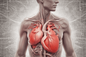
Cardiac Auscultation Overview
- Four heart sounds are possible, but usually only two are heard in normal small animals and the sounds are more audible in large animals.
- S1 is the sound of the closure of the Atrioventricular valves (left and right) which occurs at the start of ventricular systole.
- S2 is the sound of the closure of the semilunar valves (pulmonary and aortic) which occurs at the end of ventricular systole.
- S3 is the sound of rapid ventricular filling during diastole but is not normally auscultated in small animals.
- S3 can be associated with congestive heart failure and cardiomyopathy
- S4 is the sound of atrial systole, and is not normally auscultated in small animals.
- S4 can be associated with filling of non-compliant ventricles, or hypertrophic cardiomyopathy.
Describing Heart Murmurs
- Intensity is graded from I to VI, with Grade I being not distinct and Grade VI being very loud, not requiring a stethoscope.
- Position and Duration are described in relation to the cardiac cycle such as systolic, diastolic, pan-, holo-, early-, late, and continuous.
- Location matches the valve area which has the point of maximum intensity as Pulmonic, Aortic, Mitral, or Tricuspid.
- Quality can be described by the intensity profile such as plateau, crescendo, decrescendo, or diamond (crescendo-decrescendo).
- Character/Pitch/Frequency Profile is used to describe the sound as Blowing, musical, honking, harsh, or noisy.
Murmurs and Blood Flow
- Murmurs are the turbulent sound of blood flow and thus is classified as(ABNORMAL).
- Murmurs can be caused by an abnormal flow pattern such as Valvular insufficiency where the valve doesn't close completely.
- Murmurs can be caused by abnormal vessel diameter such as narrowing of the aorta (aortic stenosis).
- Murmurs can be caused by abnormal blood viscosity such as anemia causing low viscosity blood.
- Cardiac disease severity doesn't always link to the severity of the murmur.
Cardiac Output and Volume Regulation
- Cardiac pathology needs some physiology in order to understand it.
- The volume of blood moving in circulation is the responsibility of the heart along with the kidneys.
- Cardiac Output = Heart Rate X Stroke Volume.
- Stroke volume can be altered by contractility, and/or changes in blood volume.
Mitral Insufficiency Consequences
- Mitral insufficiency is where some of the blood leaks through the valve back into the left atrium.
- Cardiac output decreases as a result of mitral insufficiency.
- The body increases the heart rate in response to try and bring cardiac output bring it back to normal, but the disease progresses.
- The kidneys compensate by retaining water and electrolytes to increase blood volume and pressure, thus increasing pre-load (end diastolic volume).
- The kidneys short term compensation also increases the amount of blood regurgitated through the faulty valve.
- The heart must compensate to accommodate the increased volume and increased pressure, which results in dilated cardiomyopathy.
Mitral Insufficiency Final Result
- Without therapy, the end result of Mitral insufficiency is congestive heart failure.
- The left side of the heart can no longer enlarge and blood pressure continues to rise, causing more regurgitation and pressure in the lungs to increase eventually causing pulmonary edema.
- Myocardial failure can occur the heart muscle fails to pump fluid forward.
Mitral and Tricuspid Valve Insufficiency Effects
- Mitral Valve insufficiency eventually causes a reduction of flow to the left side of the heart
- An end result of Mitral valve insufficiency is pulmonary edema (fluid in the lungs), resulting in increased respiratory rate and effort/coughing or eventual exercise intolerance.
- Tricuspid valve insufficiency eventually causes a reduction in flow to the right side of the heart.
- Systemic circulation becomes congested because capillaries of the body cavity linings, abdomen, and chest manifest signs of disease first.
- Abdomen is lined by peritoneum, so the accumulation of fluid in the peritoneal space is called “Ascites” causing a distended abdomen.
- Chest is lined by pleura, so accumulation of fluid in the plural space is called "Plural effusion" causing respiratory effort, rate, and noise.
Tricuspid Insufficiency (Regurgitation)
- Tricuspid insufficiency is the result of a very similar disease process (pathophysiology) as mitral disease.
- There is right-sided heart congestion and pressures of the systemic circulation increases and starts “leaking”.
- The lining of the body cavities (thorax/abdomen) have tiny vessels that can become “leaky" under stress of high pressure.
- The abdominal cavity and organs are lined by the peritoneum.
- Excessive fluid in the peritoneal space is called "Ascites".
- The thoracic cavity and organs are lined with pleura.
- Excessive fluid in the plural space is called "Plural Effusion”.
Predisposed Breeds
- Mitral valve disease (acquired) predisposed breeds include Cavalier King Charles spaniel, dachshund, and any small dog breeds.
- Mitral valve disease (congenital) predisposed breeds include bull terriers and rottweilers.
- Myocardial failure predisposed breeds include boxers, cocker spaniels, Doberman pinschers, Great Danes, Irish wolfhounds, and Labrador retrievers.
- Patent ductus arteriosus predisposed breeds include cocker spaniels, English springer spaniels, German shepherds, Maltese terriers, and poodles.
- Pulmonary stenosis predisposed breeds include boxers, cocker spaniels, English bulldogs, mastiffs, miniature schnauzers, Samoyeds, and West Highland white terriers.
- Subvalvular aortic stenosis predisposed breeds include boxers, German shepherds, golden retrievers, Newfoundlands, and rottweilers.
- Tricuspid valve disease (congenital) predisposed breed is the Labrador retriever.
- Ventricular septal defect predisposed breed is the English springer spaniel.
Dilated Cardiomyopathy
- Dilated Cardiomyopathy occurs most frequently in boxers and Doberman pinchers due to a decrease in contractility of cardiac muscle because of genetics.
- Initially it is compensated by increasing heart rate and blood volume, but eventually the orientation of the mitral valve is affected resulting in mitral valve insufficiency ensues.
- Dilated cardiomyopathy is directly related to Taurine deficiency in a cat's diet.
- Dilated cardiomyopathy is very rare in household cats due to commercially available balanced nutrition.
Feline Hypertrophic Cardiomyopathy
- The exact cause of Feline Hypertrophic Cardiomyopathy is unknown, but there is a definitive genetic predisposition (Ragdoll, Maincoon).
- Ventricular walls thicken, pumps well, but cannot relax during diastole resulting in decreased Cardiac Output (CO).
- Can result in Malalignment of valves (especially the mitral).
Patent Ductus Arteriosus (PDA)
- Patent Ductus Arteriosus blood results in a a connection between the aorta vessels and the pulmonary artery.
Cardiac Silhouette on Radiographs
- Cardiac silhouette is NORMALLY about 60 to 65 percent of the thoracic width.
- VHS stands for Vertebral Heart Size
- VHS = L(ong) + S(hort)
- Normal ranges for the average dog is 8.5 - 10.9 Vertebra, and a normal range for the average cat is 6.8 - 8.1 Vertebra.
- To measure heart size on radiographs you measure the Long axis from the Carina to the Apex.
- To measure heart size on radiographs you measure the short axis from the widest part with in the cardiac silhouette.
- Measure both in cm/mm then with your measurement tool move the measurement length towards vertebrae starting at T4.
- This will give you a rough vertebral heart size
- Feline VD radiographs are more reliable than dog radiographs and can also be utilized because there is relatively little variation in cat chest shape (ratio of depth : width).
- Dogs have a large variation in shape between breeds.
Studying That Suits You
Use AI to generate personalized quizzes and flashcards to suit your learning preferences.