Podcast
Play an AI-generated podcast conversation about this lesson
Download our mobile app to listen on the go
Get App
Questions and Answers
What is a trial gallop?
What is a trial gallop?
An abnormal heart sound heard after S1 during late diastole.
What are the two layers that compose the pericardium?
What are the two layers that compose the pericardium?
- Fibrous and serous (correct)
- Muscular and fibrous
- Epithelial and muscular
- Serous and vascular
Which landmark corresponds to the mitral area?
Which landmark corresponds to the mitral area?
- Left 4th ICS
- Left 2nd ICS
- Right 2nd ICS
- Left 5th ICS, midclavicular (correct)
What is the function of the fibrous pericardium?
What is the function of the fibrous pericardium?
Signup and view all the answers
What are the layers of the serous pericardium?
What are the layers of the serous pericardium?
Signup and view all the answers
What is the amount of pericardial fluid normally present?
What is the amount of pericardial fluid normally present?
Signup and view all the answers
The right ventricle contains trabeculae carneae.
The right ventricle contains trabeculae carneae.
Signup and view all the answers
Match the following chambers with their descriptions:
Match the following chambers with their descriptions:
Signup and view all the answers
The anterior surface of the heart is also known as the ______.
The anterior surface of the heart is also known as the ______.
Signup and view all the answers
Flashcards are hidden until you start studying
Study Notes
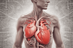
Heart Structure and Function
- Shape: Cone-shaped and similar in size to a closed fist.
- Orientation:
- Apex: Anteriorly directed, Positioned left and inferior (ALI).
- Base: Posteriorly directed, Positioned right and superior (PRS).
- Location: Situated in the mediastinum, above the diaphragm.
Pericardium
- Definition: The protective covering of the heart.
- Layers: Composed of two main layers:
- Fibrous Pericardium:
- Outermost layer, made of tough, inelastic, dense irregular connective tissue.
- Fuses with connective tissues of blood vessels entering and leaving the heart.
- Functions include preventing heart overstretching, providing protection, and anchoring the heart in the mediastinum.
- Serous Pericardium:
- Innermost layer, thinner and more delicate.
- Forms a double layer around the heart.
- Comprised of:
- Parietal Serous Pericardium (outermost layer).
- Visceral Serous Pericardium (innermost layer, also called epicardium).
- Fibrous Pericardium:
Pericardial Cavity and Fluid
- Pericardial Fluid (PF): Lubricating fluid between the parietal and visceral layers.
- Function: Reduces friction during heart movements.
- Typical volume: Ranges from 15 to 50 ml.
- Conditions:
- Decreased PF results in pericardial friction rub.
- Increased PF can lead to cardiac tamponade.
Heart Auscultation Landmarks
- Aortic Area: Right 2nd intercostal space (ICS).
- Pulmonic Area: Left 2nd ICS.
- Tricuspid Area: Left 4th ICS.
- Mitral Area: Left 5th ICS, at midclavicular line.
- Erb’s Point: Left 3rd ICS.
Chambers of the Heart
- Right Atrium:
- Receives blood from three veins: Superior vena cava (SVC), Inferior vena cava (IVC), and coronary sinus.
- Has a rough texture due to pectinate muscles.
- Left Atrium: Forms most of the heart's base.
- Right Ventricle: Contributes to the inferior surface, known as diaphragmatic surface.
- Left Ventricle: Also contributes to the inferior surface.
Surfaces of the Heart
- Anterior Surface: Known as the sternocostal surface.
- Posterior Surface: Referred to as the base surface, primarily consisting of the right and left atria.
- Inferior Surface: Also referred to as the diaphragmatic surface, includes right and left ventricles.
Studying That Suits You
Use AI to generate personalized quizzes and flashcards to suit your learning preferences.