Podcast
Questions and Answers
What is the primary source of Pulmonary Embolism?
What is the primary source of Pulmonary Embolism?
- Lower limb deep veins (correct)
- Pelvic veins
- Upper limb deep veins
- Abdominal veins
What is the path that an embolus takes to reach the pulmonary artery?
What is the path that an embolus takes to reach the pulmonary artery?
- Femoral artery → IVC → RA → LA → Pulmonary artery
- Femoral artery → IVC → RA → RV → Pulmonary artery
- Femoral vein → IVC → RA → RV → Pulmonary artery (correct)
- Femoral vein → IVC → LV → LA → Pulmonary artery
What is the primary component in the formation of a thrombus?
What is the primary component in the formation of a thrombus?
- Fibrin (correct)
- Platelets
- Red blood cells
- White blood cells
What is the term for a blood clot that forms within a blood vessel?
What is the term for a blood clot that forms within a blood vessel?
What is the term for a piece of a thrombus that breaks off and travels to another part of the body?
What is the term for a piece of a thrombus that breaks off and travels to another part of the body?
What is the main symptom of massive pulmonary embolism?
What is the main symptom of massive pulmonary embolism?
Why does hypotension occur in massive pulmonary embolism?
Why does hypotension occur in massive pulmonary embolism?
What is the characteristic ECG finding in massive pulmonary embolism?
What is the characteristic ECG finding in massive pulmonary embolism?
What is the classical ECG finding in massive pulmonary embolism?
What is the classical ECG finding in massive pulmonary embolism?
What is the most common abnormality seen on chest X-ray in massive pulmonary embolism?
What is the most common abnormality seen on chest X-ray in massive pulmonary embolism?
What is the initial investigation of choice in massive pulmonary embolism?
What is the initial investigation of choice in massive pulmonary embolism?
What is the approximate incidence of Deep Vein Thrombosis (DVT) per year?
What is the approximate incidence of Deep Vein Thrombosis (DVT) per year?
What percentage of Venous Thromboembolism (VTE) present with Pulmonary Embolism (PE) initially?
What percentage of Venous Thromboembolism (VTE) present with Pulmonary Embolism (PE) initially?
What is a strong risk factor for Deep Vein Thrombosis (DVT)?
What is a strong risk factor for Deep Vein Thrombosis (DVT)?
What is the characteristic of a proximal Deep Vein Thrombosis (DVT)?
What is the characteristic of a proximal Deep Vein Thrombosis (DVT)?
What is a characteristic of a distal Deep Vein Thrombosis (DVT)?
What is a characteristic of a distal Deep Vein Thrombosis (DVT)?
What is a significant percentage of Venous Thromboembolism (VTE) cases?
What is a significant percentage of Venous Thromboembolism (VTE) cases?
What is an indication for prophylactic anticoagulants?
What is an indication for prophylactic anticoagulants?
What is a characteristic of Pulmonary Embolism (PE)?
What is a characteristic of Pulmonary Embolism (PE)?
What is the primary reason for hearing localized crackles on auscultation in an area of pulmonary infarction?
What is the primary reason for hearing localized crackles on auscultation in an area of pulmonary infarction?
What is the significance of a pleural rub heard on auscultation in an area of pulmonary infarction?
What is the significance of a pleural rub heard on auscultation in an area of pulmonary infarction?
What is the most likely cause of localized crackles and a pleural rub over an area of pulmonary infarction?
What is the most likely cause of localized crackles and a pleural rub over an area of pulmonary infarction?
What is the importance of auscultation in diagnosing pulmonary infarction?
What is the importance of auscultation in diagnosing pulmonary infarction?
What is the relationship between localized crackles and a pleural rub in an area of pulmonary infarction?
What is the relationship between localized crackles and a pleural rub in an area of pulmonary infarction?
What is the primary modality of choice for imaging deep venous thrombosis?
What is the primary modality of choice for imaging deep venous thrombosis?
What is the gold standard for pulmonary embolism imaging?
What is the gold standard for pulmonary embolism imaging?
What is the primary indication for thrombolysis in pulmonary embolism?
What is the primary indication for thrombolysis in pulmonary embolism?
What is the primary advantage of direct oral anticoagulants (DOACs) over traditional anticoagulation?
What is the primary advantage of direct oral anticoagulants (DOACs) over traditional anticoagulation?
What is the primary difference between a DVT and a PE?
What is the primary difference between a DVT and a PE?
What is the Wells score used for?
What is the Wells score used for?
What is the primary reason for using compression ultrasound in DVT diagnosis?
What is the primary reason for using compression ultrasound in DVT diagnosis?
What is the primary indication for surgical intervention in PE?
What is the primary indication for surgical intervention in PE?
Flashcards are hidden until you start studying
Study Notes
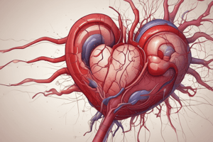
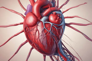
Venous Thromboembolic Disease
- Venous thromboembolic disease encompasses two diseases: deep vein thrombosis (DVT) and pulmonary embolism (PE)
- DVT occurs when a blood clot forms in the deep veins, usually in the lower limbs
- If the clot breaks loose and travels to the lungs, it's called a pulmonary embolism (PE)
Pathogenesis of Thrombosis
- Thrombosis is the formation of a blood clot within a blood vessel, mainly due to fibrin formation with contributions from platelets and other cells
- The more proximal the thrombosis, the higher the risk of PE
- Distal thromboses are less likely to cause PE and are harder to diagnose on ultrasound
Epidemiology and Risk Factors
- Venous thromboembolism is a common disease, affecting 1 in 1000 people per year
- Incidence increases with age, and 60% of cases present with DVT, while 40% present with PE
- A significant percentage of VTEs are subclinical
- Risk factors include major surgery, trauma, and absolute bed rest
Clinical Features
- Massive PE can cause central chest pain, syncope, hypotension, shock, and raised JVP and right ventricular heave
- Localized crackles and a pleural rub may be heard on auscultation
Diagnosis
- Initial investigations include ECG and chest X-ray, which may be normal or show unspecific abnormalities
- DVT imaging modalities include ultrasonography of the deep venous system and venography
- PE imaging modalities include computed tomographic pulmonary angiography (CTPA) and V/Q isotope scan
- Pulmonary artery angiography is the gold standard for diagnosing PE
Wells Score and D-Dimer
- Wells score is used to determine the likelihood of DVT
- D-dimer is a fibrin degradation product, and high levels indicate activation of the coagulation system
Treatment
- Anticoagulation with traditional anticoagulants or direct oral anticoagulants
- Thrombolysis may be indicated in selected patients with massive PE
- Surgical intervention may be necessary in some cases
Traditional Anticoagulation
- Low molecular weight heparin (LMWH) is used until sufficient coagulation with warfarin is achieved
- Warfarin is monitored using the international normalized ratio (INR)
Direct Oral Anticoagulants
- Direct factor Xa inhibitors (apixaban, rivaroxaban, and edoxaban) and direct thrombin inhibitor (dabigatran)
- Advantages: no need for monitoring, no drug/food interactions
Studying That Suits You
Use AI to generate personalized quizzes and flashcards to suit your learning preferences.