Podcast
Questions and Answers
Which of the following structures is responsible for carrying urine from the kidneys to the urinary bladder?
Which of the following structures is responsible for carrying urine from the kidneys to the urinary bladder?
- Renal artery
- Ureter (correct)
- Urethra
- Renal vein
What is the primary function of the detrusor muscle?
What is the primary function of the detrusor muscle?
- Filtering waste products from the blood
- Controlling voluntary urination
- Contracting to expel urine from the bladder (correct)
- Regulating blood flow to the kidneys
Which function does the spongy urethra perform in males, in addition to urine transport?
Which function does the spongy urethra perform in males, in addition to urine transport?
- Secreting prostate fluid
- Transporting sperm (correct)
- Producing seminal fluid
- Regulating testosterone
In the nephron, where does filtrate dilution primarily occur?
In the nephron, where does filtrate dilution primarily occur?
What is the role of ADH (antidiuretic hormone) in urine formation?
What is the role of ADH (antidiuretic hormone) in urine formation?
Which of the structures is the primary site for selective reabsorption in the nephron?
Which of the structures is the primary site for selective reabsorption in the nephron?
What type of cells are found in the walls of the afferent arterioles and secrete renin?
What type of cells are found in the walls of the afferent arterioles and secrete renin?
Which component of the juxtaglomerular complex is responsible for detecting NaCl concentration in the ascending limb of the loop of Henle?
Which component of the juxtaglomerular complex is responsible for detecting NaCl concentration in the ascending limb of the loop of Henle?
Which of the following is a function of the mesangial cells within the glomerulus?
Which of the following is a function of the mesangial cells within the glomerulus?
What structural adaptation of the ureters facilitates the movement of urine from the kidneys to the bladder?
What structural adaptation of the ureters facilitates the movement of urine from the kidneys to the bladder?
Which of the following lists the correct order of structures through which filtrate flows in the nephron?
Which of the following lists the correct order of structures through which filtrate flows in the nephron?
Which type of neurons are responsible for carrying impulses away from the central nervous system to effectors such as muscles or glands?
Which type of neurons are responsible for carrying impulses away from the central nervous system to effectors such as muscles or glands?
What is the primary function of oligodendrocytes?
What is the primary function of oligodendrocytes?
What is the functional significance of the decussation of pyramids in the medulla oblongata?
What is the functional significance of the decussation of pyramids in the medulla oblongata?
Damage to which area of the brain would most likely result in difficulties with language comprehension?
Damage to which area of the brain would most likely result in difficulties with language comprehension?
Which of the following is the correct order of meningeal layers from outermost to innermost?
Which of the following is the correct order of meningeal layers from outermost to innermost?
What is the role of the choroid plexus?
What is the role of the choroid plexus?
Which of the following is a characteristic of the white matter in the spinal cord?
Which of the following is a characteristic of the white matter in the spinal cord?
In a reflex arc, what is the role of the integration center?
In a reflex arc, what is the role of the integration center?
What does the Babinski sign (toes fanning out upon stroking the sole of the foot) indicate in adults?
What does the Babinski sign (toes fanning out upon stroking the sole of the foot) indicate in adults?
Flashcards
Kidneys
Kidneys
Organs that filter blood and produce urine.
Ureters
Ureters
Tubes that carry urine from the kidneys to the bladder using peristalsis.
Urinary Bladder
Urinary Bladder
The organ that stores urine temporarily.
Urethra
Urethra
Signup and view all the flashcards
Kidney Cortex
Kidney Cortex
Signup and view all the flashcards
Medullary Region
Medullary Region
Signup and view all the flashcards
Renal Pyramids
Renal Pyramids
Signup and view all the flashcards
Renal Papilla
Renal Papilla
Signup and view all the flashcards
Renal Columns
Renal Columns
Signup and view all the flashcards
Renal Pelvis
Renal Pelvis
Signup and view all the flashcards
Minor Calyces
Minor Calyces
Signup and view all the flashcards
Nephrons
Nephrons
Signup and view all the flashcards
Glomerular Capsule
Glomerular Capsule
Signup and view all the flashcards
Proximal Convoluted Tubule (PCT)
Proximal Convoluted Tubule (PCT)
Signup and view all the flashcards
Loop of Henle
Loop of Henle
Signup and view all the flashcards
Distal Convoluted Tubule (DCT)
Distal Convoluted Tubule (DCT)
Signup and view all the flashcards
Collecting Ducts
Collecting Ducts
Signup and view all the flashcards
Podocytes
Podocytes
Signup and view all the flashcards
Juxtaglomerular Complex
Juxtaglomerular Complex
Signup and view all the flashcards
Juxtaglomerular Complex cells
Juxtaglomerular Complex cells
Signup and view all the flashcards
Study Notes
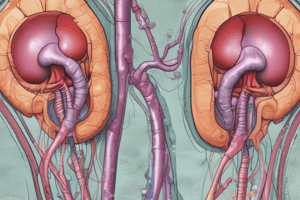
Anatomy of the Urinary System
- The gross anatomy consists of the kidneys, renal arteries and veins, ureters, urinary bladder, and urethra (male and female).
- Ureters use peristalsis
- The urinary bladder temporarily stores urine
Gross Anatomy: Male vs. Female
- The male urethra has three parts, namely the prostatic, membranous, and spongy urethra
- The penis has a dual function
- The female urethra is shorter at around 4cm
- The external urethral orifice is also different between males and females
Gross Internal Anatomy
- Key components of the internal Kidney include:
- Kidney cortex region
- Medullary region
- Renal pyramids and renal papilla
- Renal columns and renal pelvis
- Major and minor calyces
Microscopic Anatomy of Kidney
- Nephrons consist of the glomerulus with capsule which makes up the renal corpuscle
- Renal Tubule
- Glomerular (Bowman's) capsule connects to the Proximal Convoluted Tubule (PCT) via the podocytes
- Loop of Henle (descending and ascending limb) which connects to the
- Distal Convoluted Tubule (DCT)
- Collecting Ducts
Microscopic Anatomy of Kidney (cont.)
- Cortical nephrons are in the cortex
- Juxtamedullary nephrons start in the cortex, but the loop of Henle descends into medulla
- Two Capillary beds:
- Glomerulus and the Peritubular capillary bed
- Vasa recta in juxtamedullary nephrons
- Granular cells known as JG cells are mainly in the walls of afferent arterioles
- Macula densa cells line the wall of the cortical thick ascending limb at the transition to the DCT
- Mesangial cells influence surface area for filtration
Urine Formation
- Filtrate is diluted in the ascending loop of Henle
- Dilute urine is created allowing this filtrate to continue to the renal pelvis
- Collecting ducts remain impermeable to water in the absence of ADH, preventing further water reabsorption with a urine osmolality as low as 50 mOsm
- Concentrated urine is formed when ADH inhibits diuresis and increases urea reabsorption
- In the presence of ADH, 99% of the water in filtrate is reabsorbed
- ADH-dependent water reabsorption is the signal to produce concentrated urine when necessary
Urinary Bladder
- Micturition and incontinence are regulated by the internal and external urethral sphincters
- The Internal urethral sphincter is involuntary smooth muscle and relaxes during micturition when detrusor muscle contracts
- The External urethral sphincter is voluntary skeletal muscle and provides conscious control over urination
- Weakness or dysfunction leads to incontinence
- In both sexes, damage to the external sphincter or overactivity of the detrusor muscle can cause incontinence
Histology of the Nervous System
- Nervous Tissue can consists of Neuroglia (Glial cells) and Neurons
Neuroglia (Glial cells)
- Four types exist in the CNS:
- Astrocytes, Oligodendrocytes, Microglia, and Ependymal cells
- Two types exist in the PNS:
- Schwann cells, and Satellite cells
Neurons
- Neural cell body clusters are called nuclei in the CNS and ganglia in the PNS
- Components include:
- Neuroplasm (cytoplasm), Neurofibrils, Nissl bodies (chromatophilic substance, RER)
- Bundles of processes in the CNS are called tracts, while they are called nerves in the PNS
- Dendrites are the receptive regions
- Axons generate and conduct nerve impulses, also known as a nerve fiber
- Axon hillock is the initial segment with axon terminals (with synaptic knobs)
Synapse components
- neurotransmitters in synaptic vesicles
- presynaptic cell and postsynaptic cell
- synaptic cleft, which requires calcium
Classification of neurons By structure
- Unipolar: Almost all neurons that carry impulses toward CNS
- Bipolar: Part of the receptor apparatus of eye, ear, olfactory mucosa
- Multipolar: Most neurons in brain and spinal cord and those carrying impulses away from CNS
Classification of neurons By Function
- Sensory or afferent
- Motor or efferent
- Association or interneurons
Structure of a Nerve
- There are mixed, sensory or afferent, and motor or efferent types
- Its components are:
- Endoneurium surrounds the nerve fiber
- Perineurium surrounds the fascicle
- Epineurium surrounds the nerve
Anatomy of the Brain
- Nervous System:
- CNS: brain + spinal cord
- PNS: cranial + spinal nerves, ganglia, sensory receptors
- 4 Regions of the Brain:
- Cerebral Hemispheres
- Lobes:
- frontal, parietal, temporal, and occipital
External structures of the Brain
- Gyri are ridges
- The Postcentral gyrus location is the primary somatosensory cortex; the somatosensory association area Wernicke's area are at junction of parietal and temporal lobes
- The Precentral gyrus location is the primary motor area and Broca's area is at the base
- Sulci are indentations
- Central, lateral, parieto-occipital sulcus
- Fissures are deep indentations
- Longitudinal fissure and transverse cerebral fissure
Intenal structures of the Brain
- Cerebral cortex: composed of gray matter because of cell bodies (e.g. pyramidal cells)
- Cerebral white matter: composed of fiber tracts (axons of e.g. pyramidal cells)
- Association tracts
- Projection tracts, e.g. corona radiata (don't see in lab)
- Commissures, e.g. corpus callosum
- Basal nuclei: "islands” of gray matter within white matter involved in regulating voluntary motor activities
- Nuclei includes the Caudate, Lentiform, Putamen, and Globus Pallidus
Diencephalon
- Its External markers are NOT of it:
- Olfactory bulbs and tracts, optic nerves, optic chiasma and tracts, pituitary gland, mammillary bodies
- Contains internal structures:
- Thalamus (2 lobes)
- Interthalamic adhesion
- Location of interventricular foramen
- The hypothalamus connects to:
- Infundibulum connects to the pituitary gland
- Epithalamus
- Pineal body or gland choroid plexus
Brain stem
- Midbrain has external cerebral peduncles, with corpora quadrigemina
- A cerebral aqueduct travels through midbrain and connects 3rd and 4th ventricles
- Pons is dorsal to the 4th ventricle
- Medulla oblongata contains decussation of pyramids
Cerebellum
- It has two lateral hemispheres with 3 lobes anterior, posterior, flocculonodular connected by vermis
- Outer cortex and inner white matter called Arbor vitae
Ventricles of the Brain
- The ventricles are paired C-shaped lateral structures
- They are anteriorly separated by SEPTUM PELLUCIDUM
- They communicate with the 3rd ventricle via the INTERVENTRICULAR FORAMEN
- The 3rd ventricle is found in the diencephalon and the cerebral aqueduct btw the 3rd and 4th ventricle
- The 4th ventricle is found in the hindbrain dorsal to the pons
- There are three openings that mark the walls of the 4th ventricle:
- paired lateral apertures, a median aperture
Meninges of the Brain
- Outer: Dura mater (periosteal and meningeal layers)
- Includes the falx cerebri, falx cerebelli, tentorium cerebelli
- Subdural with a potential space
- Middle: Arachnoid mater with arachnoid space and arachnoid villi
- Inner: Pia mater
CSF: Choroid Plexus
- Falx cerebri folds into the longitudinal fissure of cerebrum
- Falx cerebelli separates the 2 cerebellar hemispheres
- Tentorium cerebelli separates cerebrum from cerebellum
- The plexus makes cerebrospinal fluid
Anatomy of the Spinal Cord
- Conus medullaris with
- filum terminale
- denticulate ligaments
- cauda equina
- cervical and lumbar enlargement
- Lumbar puncture below L3 and saddle block between L3 and L5
- Spinal nerves: 31 pairs, made from the fusion of dorsal+ventral root
Spinal Cord Grey and White Matter
- Gray Matter
- Has 2 Dorsal (posterior) horns, 2 ventral (anterior) horns, lateral horn (only found in thoracic and lumbar regions)
- The Gray commissure surrounds a central canal
- Has a Dorsal root with cell bodies of sensory fibers located in dorsal root ganglion
- Has a Ventral root with axons of motor neurons of somatic NS and axons of motor neurons of ANS
- White Matter (myelinated fibers)
- Dorsal median sulcus, ventral median fissure
- White columns: posterior, lateral and anterior funiculi
Studying That Suits You
Use AI to generate personalized quizzes and flashcards to suit your learning preferences.