Podcast
Questions and Answers
A patient presents with Subclavian Steal Syndrome. Where is the most likely location of the arterial occlusion or stenosis causing this condition?
A patient presents with Subclavian Steal Syndrome. Where is the most likely location of the arterial occlusion or stenosis causing this condition?
- Distal to the origin of the internal thoracic artery.
- Distal to the origin of the vertebral branch.
- Proximal to the origin of the thyrocervical trunk.
- Proximal to the origin of the vertebral branch. (correct)
During a surgical procedure in the axilla, a surgeon must carefully consider the anatomical relationships to avoid damaging critical structures. Which of the following is the correct spatial relationship between the axillary artery and vein?
During a surgical procedure in the axilla, a surgeon must carefully consider the anatomical relationships to avoid damaging critical structures. Which of the following is the correct spatial relationship between the axillary artery and vein?
- The axillary vein lies lateral to the axillary artery throughout its course.
- The axillary vein lies medial to the axillary artery throughout its course. (correct)
- The axillary vein crosses anterior to the axillary artery at the level of the teres major muscle.
- The axillary vein is posterior to the axillary artery in the second part of its course.
A trauma surgeon is treating a patient with a mid-shaft humeral fracture. If the profunda brachii artery is lacerated, which muscle compartment is most likely to suffer ischemia as a direct result?
A trauma surgeon is treating a patient with a mid-shaft humeral fracture. If the profunda brachii artery is lacerated, which muscle compartment is most likely to suffer ischemia as a direct result?
- Posterior compartment of the arm. (correct)
- Anterior compartment of the forearm.
- Anterior compartment of the arm.
- Posterior compartment of the forearm.
A patient requires a coronary artery bypass graft (CABG). Which artery, a branch of the subclavian artery, is often harvested for this procedure due to its reliable blood supply and proximity to the heart?
A patient requires a coronary artery bypass graft (CABG). Which artery, a branch of the subclavian artery, is often harvested for this procedure due to its reliable blood supply and proximity to the heart?
During a mastectomy, the lateral thoracic artery is inadvertently ligated. What is the MOST likely consequence of ligating the lateral thoracic artery?
During a mastectomy, the lateral thoracic artery is inadvertently ligated. What is the MOST likely consequence of ligating the lateral thoracic artery?
A patient is diagnosed with thoracic outlet syndrome (TOS) involving compression of the subclavian artery. Which anatomical structure is MOST likely implicated in causing this compression?
A patient is diagnosed with thoracic outlet syndrome (TOS) involving compression of the subclavian artery. Which anatomical structure is MOST likely implicated in causing this compression?
Following a stab wound to the cubital fossa, a patient exhibits decreased blood flow to the hand. If the ulnar artery remains intact, which arterial structure is MOST likely damaged?
Following a stab wound to the cubital fossa, a patient exhibits decreased blood flow to the hand. If the ulnar artery remains intact, which arterial structure is MOST likely damaged?
A patient presents with ischemia of the first two intercostal spaces. Which arterial branch is MOST likely compromised?
A patient presents with ischemia of the first two intercostal spaces. Which arterial branch is MOST likely compromised?
After a motorcycle accident, a patient has a compromised blood supply to the infraspinatus muscle. Which artery is MOST likely affected?
After a motorcycle accident, a patient has a compromised blood supply to the infraspinatus muscle. Which artery is MOST likely affected?
A surgeon is planning to perform a radial artery graft for coronary artery bypass. Prior to the procedure, which artery should be evaluated to ensure adequate collateral circulation to the hand in case the radial artery is harvested?
A surgeon is planning to perform a radial artery graft for coronary artery bypass. Prior to the procedure, which artery should be evaluated to ensure adequate collateral circulation to the hand in case the radial artery is harvested?
Flashcards
Subclavian Artery
Subclavian Artery
The major vessel supplying oxygenated blood to the upper limb.
Subclavian Artery Termination
Subclavian Artery Termination
Becomes the axillary artery at the lateral border of the first rib. Key vessel in the upper limb for blood.
Vertebral Artery
Vertebral Artery
First branch off the subclavian artery that ascends through cervical vertebrae to enter the cranial cavity.
Axillary Artery
Axillary Artery
Signup and view all the flashcards
Axillary Artery Parts
Axillary Artery Parts
Signup and view all the flashcards
Brachial Artery
Brachial Artery
Signup and view all the flashcards
Radial Artery
Radial Artery
Signup and view all the flashcards
Ulnar Artery
Ulnar Artery
Signup and view all the flashcards
Superficial Palmar Arch
Superficial Palmar Arch
Signup and view all the flashcards
Deep Palmar Arch
Deep Palmar Arch
Signup and view all the flashcards
Study Notes
- The vascular system of the upper limb contains arteries, veins, and lymphatic vessels
- The vascular system supplies blood and drains deoxygenated blood from the arm, forearm, and hand
- Arterial supply involves subclavian, axillary, brachial, radial, and ulnar arteries
- Venous drainage involves deep and superficial veins
- Lymphatic drainage comprises deep and superficial lymphatic vessels
- Arterial and venous systems are supported by lymphatic drainage for immune functions
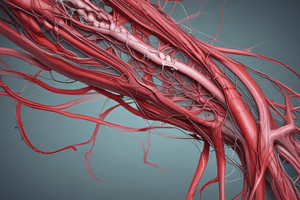
Arterial Supply of the Upper Limb
- Five main arteries: Subclavian, Axillary, Brachial, Radial, and Ulnar
Subclavian Artery - Origin
- Is the major vessel supplying oxygenated blood to the upper limb
- Arises and branches from the brachiocephalic trunk on the right side
- Arises directly from the aortic arch on the left side
Subclavian Artery - Course
- Travels laterally and inferior to the clavicle toward the axilla
- Passes posteriorly to the anterior scalene muscle
- Becomes the axillary artery at the lateral border of the first rib
Subclavian Artery - Branches
- Can be divided into three parts based on its anatomical relationship with the anterior scalene muscle
- The three parts are the First, Second, and Third parts
- Major branches across the 3 parts include vertebral, internal thoracic, thyrocervical, costocervical, and dorsal scapular arteries
Subclavian Artery - Course of Branches
- The first part (prescalene) extends from its origin to the medial border of the anterior scalene muscle
- The second part (retroscalene) is located posterior to the anterior scalene muscle.
- The third part (postscalene) extends from the lateral border of anterior scalene muscle to the lateral border of the first rib.
Subclavian Artery - Branches of the First Part
- Is further divided into three primary branches which are the vertebral artery, internal thoracic artery, and thyrocervical trunk (VIT)
Subclavian Artery - Vertebral Artery
- Originates as the first branch off the subclavian artery
- Ascends through foramina in cervical vertebrae (C6-C1) to enter the cranial cavity via the foramen magnum
- Merges with its counterpart from the opposite side at the pontomedullary junction to form the basilar artery
- Supplies the brain and spinal cord
- Important for maintaining adequate blood flow for balance and coordination
- Its compromise or occlusion can lead to neurological deficits
Subclavian Artery - Internal Thoracic Artery
- Originates as the second branch off the subclavian artery
- Descends along the inner surface of the anterior chest wall
- Terminates by dividing into superior epigastric, pericardiacophrenic, and musculophrenic arteries
- Supplies blood to parts of the chest wall and breast
- Used in coronary bypass surgeries due to robust blood supply and proximity to cardiac structures
Subclavian Artery - Thyrocervical Trunk
- Originates as the third branch off near the medial border of the anterior scalene muscle
- Ascends and quickly divides into branches
- Includes inferior thyroid, superficial/transverse cervical, and suprascapular arteries
- Terminates by giving rise to branches
- Includes inferior thyroid that supplies blood to the thyroid and parathyroid gland
- Includes the suprascapular which supplies muscles such as infraspinatus and supraspinatus, along with parts of sternocleidomastoid and subclavius muscles
- Includes the transverse/superficial cervical artery that supplies muscles in the neck region (trapezius and rhomboids)
- Includes the ascending cervical artery that supplies parts of the cervical spinal cord and surrounding muscles
- Essential for vascularizing various structures in both cervical regions
- Contributes significantly to collateral circulation around critical areas like the shoulder girdle
Subclavian Artery - Costocervical Trunk
- Originates as the second branch of the subclavian artery; located posterior to the anterior scalene muscle and posteriorly over the apex of the lung
- Descends within the neck before bifurcating into its terminal branches along the inner surface of the anterior chest wall
- Divides into the supreme intercostal artery and deep cervical artery
Subclavian Artery - Supreme Intercostal Artery
- Originates as the costocervical trunk
- Descends posteriorly and laterally along the upper border of the first two intercostal spaces
- Travels in a groove on the inner surface of the ribs
- Gives off branches that supply these intercostal spaces
- Supplies blood to the first two intercostal spaces and provides oxygenated blood to the muscles and tissues in this region
- Supplies parts of the serratus anterior muscle and overlying skin
- Terminates by dividing into smaller branches that anastomose with other arteries supplying adjacent areas, such as branches from the thoracic aorta
Subclavian Artery - Deep Cervical Artery
- Originates at the costocervical trunk
- Ascends along the neck's posterior aspect
- Travels deep to structures including muscles like splenius capitis and semispinalis capitis
- Runs towards the deep cervical fascia and reaches higher levels in the neck
- Supplies blood to muscles in the region involved in neck movement and stabilization
- Contributes to vascular supply for surrounding soft tissues
- Terminates by anastomosing with branches from the vertebral or occipital arteries for collateral circulation around critical neck structures
Subclavian Artery - Dorsal Scapular Artery
- Originates as the third branch of the subclavian artery
- Originates directly from the cervical trunk
- Descends along the medial border of the scapula
- Terminates by anastomosing with other arteries such as the suprascapular and circumflex scapular arteries
- Supplies blood to the back and shoulder (levator scapulae and rhomboids) causing injuries or functional variations in compromised key muscle groups
Subclavian Artery - Clinical Significance
- Subclavian Steal Syndrome occurs when there is occlusion or stenosis (narrowing) of a subclavian artery proximal to where it gives off its vertebral branch; blood flow may reverse through this branch, causing dizziness
- Thoracic Outlet Syndrome (TOS) means compression at this area vascular symptoms due to pressure on either or both subclavian arteries
- Aneurysms and Thrombosis creates susceptibility to aneurysms (localized dilation) and thrombosis (blockage), which can lead to ischemia in associated tissues
Axillary Artery - Introduction and Origin
- At the lateral border of the first rib, the subclavian artery becomes the axillary artery in the axilla region
- Runs alongside important structures, including the axillary vein that lies medial to it
- The cords of the brachial plexus surround it; cords are named according to their position relative to this artery (lateral, medial, and posterior)
- Runs deep to the pectoralis minor; the three parts are based on position relative to this muscle -First part: Proximal to pectoralis minor/suprapectoral -Second part: Posterior to pectoralis minor/ rectopectoral -Third part: Distal to pectoralis minor/ infrapectoral
Axillary Artery - Major Branches
- The major branches include the superior thoracic, thoracoacromial, lateral thoracic, subscapular, anterior circumflex humeral, and posterior circumflex humeral arteries (STLSAP)
- The three parts are based on their anatomical relationship with the pectoralis minor muscle -First part (Suprapectoral) branches to the superior thoracic artery -Second part (Retropectoral) branches to the thoracoacromial and lateral thoracic arteries -Third part (Infrapectoral) branches to the subscapular, anterior, and posterior circumflex arteries
Axillary Artery - Branches of the First Part
- First part (Suprapectoral) feeds the superior thoracic artery
- The superior thoracic artery arises from the axillary artery, located above the pectoralis minor muscle
- Travels anteriorly and inferiorly towards the thoracic wall branching once
- Terminates by anastomosing with branches of the internal thoracic artery and the superior intercostal arteries
Axillary Artery - Blood Supply, First Part Branches
- Primarily supplies blood to the pectoralis major and minor, subclavius, the superior aspect of serratus anterior muscle, and the muscles and skin of the first two intercostal spaces.
Axillary Artery - Branches of the Second Part
- Second part (Retropectoral) feeds the thoracoacromial artery
- The thoracoacromial artery branches off from the axillary artery
- It pierces the clavipectoral fascia and divides into four terminal branches which are clavicular, pectoral, acromial, and deltoid (CPAD)
Axillary Artery - Course and Termination, Branches of the Second Part
- The clavicular branch supplies the sternoclavicular joint and subclavius muscle
- The pectoral branch supplies both pectoralis major and minor muscles
- The acromial branch supplies blood to the acromion and deltoid muscle
- The deltoid branch travels in the deltopectoral groove to supply both deltoid and pectoralis major muscles
Axillary Artery - Lateral Thoracic Artery Branch of Second Part
- The lateral thoracic artery arises from the axillary artery
- It runs inferomedially along the inferior border of the pectoralis minor muscle
- Supplies blood to structures such as serratus anterior, both pectoralis muscles, subscapularis muscle, and mammary glands
Axillary Artery - Third Part Branches
- Third part (Infrapectoral) feeds the subscapular artery
- The subscapular artery is a large branch that originates from the axillary artery
- It travels caudally from the inferior border of pectoralis minor to the inferior border of teres major before bifurcating into two main branches which are the circumflex scapular artery and thoracodorsal artery
Axillary Artery - Third Part Branches
- The circumflex scapular artery courses around the lateral border of the scapula to enter the infraspinatus fossa
- The thoracodorsal artery runs inferiorly alongside the thoracodorsal nerve to supply the latissimus dorsi muscle
- The circumflex scapular artery contributes to scapular anastomosis
- The thoracodorsal artery supplies latissimus dorsi
Axillary Artery - Anterior Circumflex Humeral Artery (ACHA) Third Part Branches
- Originates as part of the axillary artery
- It travels horizontally towards the surgical neck of the humerus-deep head of the biceps brachii and coracobrachialis muscles
- The ACHA Supplies blood to shoulder joint via its ascending branch
Axillary Artery - Posterior Circumflex Humeral Artery (PCHA) Third Part Branches
- The PCHA also arises from the axillary artery
- It passes posteriorly alongside the axillary nerve through the quadrangular space before wrapping around the surgical neck of the humerus
- Supplies the shoulder joint and the deltoid muscle and anastomoses with ACHA
Axillary Artery - Clinical Correlates
- Injury risks can lead to hemorrhage or ischemia which can be caused by trauma or fractures around shoulder
- Aneurysms can compress surrounding structures such as nerves of the brachial plexus which can cause neurological symptoms
- Variations in branching pattern of subscapular or circumflex arteries vary significantly among individuals
- Anatomy knowledge is important for surgery, and preserving vascular integrity is essential for recovery
Brachial Artery - Introduction
- It originates as a continuation of the axillary artery at the inferior border of the teres major muscle-transition occurs in the upper arm with blood to structures arm, forearm, and hand
- It courses down the medial aspect of the humerus and runs medial to the biceps brachii muscle and anterior to the medial head of triceps brachii
- As it descends towards the elbow, it moves more anteriorly and enters the cubital fossa at the elbow joint where it bifurcates
Brachial Artery - Branches
- Gives rise to several branches which include the deep brachial artery (profunda brachii), superior ulnar collateral arteries, inferior ulnar collateral arteries
- Terminates by bifurcating into ulnar and radial arteries approximately 1 cm distal to the elbow joint
Brachial Artery - Deep Brachial Artery & Profunda Brachii Artery
- The deep brachial artery arises from the posterior aspect of the brachial artery just distal to the teres major
- It travels posteriorly through triangular intervals between the long and medial heads of triceps brachii
- Follows along with the radial nerve in a groove on the posterior surface of the humerus
- Supplies primarily to muscles in the posterior compartment of the arm.
Brachial Artery - Superior Ulnar Collateral Artery
- Branches off from the brachial artery slightly distal to mid-level of the arm
- Travales medially along with ulnar nerve through the intermuscular septum into the posterior compartment
- It anastomoses with branches around the medial epicondyle at the elbow
Brachial Artery - Inferior Ulnar Collateral Artery
- Originates near 5cm proximal to elbow joint
- Runs medially between brachialis muscle and median nerve
- Crosses intermuscular septum
- Forms anastomoses with both anterior and posterior ulnar recurrent arteries near olecranon fossa
Brachial Artery - Clinical Correlates
- The median nerve typically runs adjacent or deep; variation could lead to nerve compression syndromes, such as carpal tunnel syndrome
- Anatomical variations can occur, so knowledge in variations such as a superficial brachial artery or high bifurcation can complicate procedures like intravenous access or arterial catheterization
- In cases like supracondylar fractures in children, there is a high risk for vascular injuries
- Occlusion or laceration of this artery can lead to severe complications including ischemia or necrosis
Radial Artery
- Is one of two terminal branches that arise from the bifurcation of the brachial artery in the cubital fossa.
- It runs deep to the brachioradialis muscle and continues down the forearm
- Supplies blood to the posterolateral region of the forearm
- Contributes to arterial arches in hand
Ulnar Artery
- Is a terminal branch that arises alongside the radial artery from the bifurcation point in the cubital fossa
- Travels down the medial side of forearm beneath the flexor carpi ulnaris muscle
- Gives rise to the anterior interosseous artery and posterior interosseous arteries.
- Supplies blood to the anteromedial side of the forearm, wrist, hand, and fingers
- Common interosseous arteries provide blood also
Hand - Supply
- Radial and ulnar arteries form two vascular arches in the hand
- Superficial Palmar Arch is formed from the ulnar artery anteriorly to the flexor tendons in the hand, deep to the palmar aponeurosis
- Supplies digital arteries of the four fingers
- Deep Palmar Arch is formed mainly by the radial artery
- Located deep within the flexor of the hand
- Supplies the digits and wrist joint
Studying That Suits You
Use AI to generate personalized quizzes and flashcards to suit your learning preferences.