Podcast
Questions and Answers
Which frequency range is typically utilized in ultrasound imaging?
Which frequency range is typically utilized in ultrasound imaging?
- 10-15 MHz
- 5-25 MHz
- 1-3 MHz
- 2-20 MHz (correct)
A curvilinear probe offers more resolution but less penetration compared to a linear probe.
A curvilinear probe offers more resolution but less penetration compared to a linear probe.
False (B)
What is the typical velocity of sound in human tissue?
What is the typical velocity of sound in human tissue?
1540 m/s
The principle of ultrasound imaging relies on the ______ effect to generate and receive sound waves.
The principle of ultrasound imaging relies on the ______ effect to generate and receive sound waves.
Match the ultrasound probe type with its application:
Match the ultrasound probe type with its application:
What role does Dr. Khaleel Ahmed hold at the Arise Medical Academy?
What role does Dr. Khaleel Ahmed hold at the Arise Medical Academy?
Dr. Khaleel Ahmed has been an educator for less than five years.
Dr. Khaleel Ahmed has been an educator for less than five years.
What has Dr. Khaleel Ahmed contributed to the training of medical graduates?
What has Dr. Khaleel Ahmed contributed to the training of medical graduates?
Dr. Khaleel Ahmed is a nationally renowned faculty in the field of ______.
Dr. Khaleel Ahmed is a nationally renowned faculty in the field of ______.
Match the following titles with Dr. Khaleel Ahmed's roles:
Match the following titles with Dr. Khaleel Ahmed's roles:
A 30-year-old patient presents after a road traffic accident with blunt abdominal trauma. Initial FAST exam is positive. The patient's blood pressure is 130/80 mmHg and their heart rate is 72 bpm. What is the next appropriate step in management?
A 30-year-old patient presents after a road traffic accident with blunt abdominal trauma. Initial FAST exam is positive. The patient's blood pressure is 130/80 mmHg and their heart rate is 72 bpm. What is the next appropriate step in management?
Ultrasound (USG) is a reliable imaging modality for detecting gallbladder cancer and emphysematous cholecystitis.
Ultrasound (USG) is a reliable imaging modality for detecting gallbladder cancer and emphysematous cholecystitis.
The ______ mode of ultrasound is used to assess the motion of structures, particularly in echocardiography.
The ______ mode of ultrasound is used to assess the motion of structures, particularly in echocardiography.
What color coding in color Doppler ultrasound indicates flow towards the probe?
What color coding in color Doppler ultrasound indicates flow towards the probe?
Match the following ultrasound modalities with their respective applications:
Match the following ultrasound modalities with their respective applications:
Which of the following ultrasound findings is characteristically associated with renal calculi?
Which of the following ultrasound findings is characteristically associated with renal calculi?
Ultrasound is a reliable method for detecting and differentiating between a cystic mass and a solid mass.
Ultrasound is a reliable method for detecting and differentiating between a cystic mass and a solid mass.
What is the typical ultrasound appearance of a thyroid cancer?
What is the typical ultrasound appearance of a thyroid cancer?
What is the maximum radiation exposure limit for a pregnant occupational worker?
What is the maximum radiation exposure limit for a pregnant occupational worker?
Stochastic effects of radiation have a threshold dose.
Stochastic effects of radiation have a threshold dose.
What does ALARA stand for?
What does ALARA stand for?
The absorbed dose is measured in _____ and _____.
The absorbed dose is measured in _____ and _____.
Match the radiation syndrome with the corresponding dose level:
Match the radiation syndrome with the corresponding dose level:
Which imaging modality does NOT use ionizing radiation?
Which imaging modality does NOT use ionizing radiation?
A T-Score compares bone mineral density (BMD) of a patient with an age-matched individual.
A T-Score compares bone mineral density (BMD) of a patient with an age-matched individual.
What is the most sensitive view for detecting hemoperitoneum using FAST?
What is the most sensitive view for detecting hemoperitoneum using FAST?
Thermoluminescent dosimeters are used to measure radiation exposure in _____ workers.
Thermoluminescent dosimeters are used to measure radiation exposure in _____ workers.
Match the imaging term with its description:
Match the imaging term with its description:
What type of radiation effect is 'skin erythema' classified under?
What type of radiation effect is 'skin erythema' classified under?
The terms 'radiolucent' and 'hyperdense' both refer to areas where X-rays are absorbed more.
The terms 'radiolucent' and 'hyperdense' both refer to areas where X-rays are absorbed more.
What is the primary purpose of a DEXA scan?
What is the primary purpose of a DEXA scan?
In ultrasound, a linear probe is typically used for _____ imaging.
In ultrasound, a linear probe is typically used for _____ imaging.
What condition is indicated by the sign of air around the aorta, pulmonary artery, bronchus, and trachea?
What condition is indicated by the sign of air around the aorta, pulmonary artery, bronchus, and trachea?
Thymic Sail sign is indicative of abnormality in children.
Thymic Sail sign is indicative of abnormality in children.
What is the most common cause of pneumatocele in hospitalized children?
What is the most common cause of pneumatocele in hospitalized children?
The presence of _____ sign indicates a cavitating mass in the lung.
The presence of _____ sign indicates a cavitating mass in the lung.
Match the conditions with their typical radiographic signs:
Match the conditions with their typical radiographic signs:
Which clinical scenario is associated with a suspected foreign body in a child?
Which clinical scenario is associated with a suspected foreign body in a child?
Miliary mottling can be associated with mitral stenosis.
Miliary mottling can be associated with mitral stenosis.
What is the common appearance of cavalier lung abscesses on CXR?
What is the common appearance of cavalier lung abscesses on CXR?
_____ collection is characterized by a D-shape and does not change position.
_____ collection is characterized by a D-shape and does not change position.
What is the role of IV fluids and emergency laparotomy in treatment?
What is the role of IV fluids and emergency laparotomy in treatment?
Egg shell calcification is a hallmark sign of RMS (Rheumatological Management Syndrome).
Egg shell calcification is a hallmark sign of RMS (Rheumatological Management Syndrome).
What is the primary sign associated with the presence of a ruptured liver abscess?
What is the primary sign associated with the presence of a ruptured liver abscess?
The best indicator of ARDS on CT is an _____ gradient.
The best indicator of ARDS on CT is an _____ gradient.
Which organism is commonly responsible for causing red currant jelly sputum?
Which organism is commonly responsible for causing red currant jelly sputum?
Allergic Bronchopulmonary Aspergillosis (ABPA) typically presents with unilateral lung opacities.
Allergic Bronchopulmonary Aspergillosis (ABPA) typically presents with unilateral lung opacities.
What is the chief source of scatter radiation in diagnostic radiology?
What is the chief source of scatter radiation in diagnostic radiology?
Characteristic Radiation is the chief source of x-ray generation in mammography.
Characteristic Radiation is the chief source of x-ray generation in mammography.
What is measured in Hounsfield units in a CT scan?
What is measured in Hounsfield units in a CT scan?
The Hounsfield unit for air is ______.
The Hounsfield unit for air is ______.
Match the following types of CT scans with their primary use:
Match the following types of CT scans with their primary use:
Which structure is referred to as the chief site for blood pressure regulation in cases of hypertension?
Which structure is referred to as the chief site for blood pressure regulation in cases of hypertension?
The swallow tail sign is a normal appearance of the substantia nigra.
The swallow tail sign is a normal appearance of the substantia nigra.
The most common imaging modality for detecting acute aortic dissection is ______.
The most common imaging modality for detecting acute aortic dissection is ______.
Match the following Hounsfield unit ranges with their corresponding tissue types:
Match the following Hounsfield unit ranges with their corresponding tissue types:
Flashcards
How does ultrasound work?
How does ultrasound work?
Ultrasound uses the principle of pulse-echo, where sound waves are emitted and reflected back to the transducer. The time it takes for the sound to return determines the distance of the object.
What is ultrasound?
What is ultrasound?
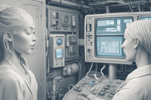
Ultrasound is a medical imaging technique that uses high-frequency sound waves to create real-time images of internal organs and tissues.
What is a transducer in ultrasound?
What is a transducer in ultrasound?
A transducer, often made of lead zirconate titanate (PZT), generates sound waves and receives the echoes. It's like a microphone and speaker combined.
What is the speed of sound in human tissue?
What is the speed of sound in human tissue?
Signup and view all the flashcards
What are different types of ultrasound probes?
What are different types of ultrasound probes?
Signup and view all the flashcards
Who is Dr. Khaleel Ahmed?
Who is Dr. Khaleel Ahmed?
Signup and view all the flashcards
What is Dr. Khaleel Ahmed's role in medical education?
What is Dr. Khaleel Ahmed's role in medical education?
Signup and view all the flashcards
What is Arise Medical Academy?
What is Arise Medical Academy?
Signup and view all the flashcards
How has Dr. Khaleel Ahmed contributed to the medical field?
How has Dr. Khaleel Ahmed contributed to the medical field?
Signup and view all the flashcards
What is the goal of Arise Medical Academy?
What is the goal of Arise Medical Academy?
Signup and view all the flashcards
When is USG the initial investigation of choice?
When is USG the initial investigation of choice?
Signup and view all the flashcards
What are the common scenarios where USG is used to detect fluid collections?
What are the common scenarios where USG is used to detect fluid collections?
Signup and view all the flashcards
What are the limitations of USG for Gallbladder conditions?
What are the limitations of USG for Gallbladder conditions?
Signup and view all the flashcards
What is Duplex USG and what are its applications?
What is Duplex USG and what are its applications?
Signup and view all the flashcards
How does Color Doppler work?
How does Color Doppler work?
Signup and view all the flashcards
What is a posterior acoustic shadow in USG?
What is a posterior acoustic shadow in USG?
Signup and view all the flashcards
What is Endoscopic Ultrasound (EUS)?
What is Endoscopic Ultrasound (EUS)?
Signup and view all the flashcards
Absorbed Dose
Absorbed Dose
Signup and view all the flashcards
Equivalent Dose
Equivalent Dose
Signup and view all the flashcards
Effective Dose
Effective Dose
Signup and view all the flashcards
Radioactivity
Radioactivity
Signup and view all the flashcards
Thermoluminescent dosimeter (TLD badge)
Thermoluminescent dosimeter (TLD badge)
Signup and view all the flashcards
ALARA
ALARA
Signup and view all the flashcards
DICOM
DICOM
Signup and view all the flashcards
PACS
PACS
Signup and view all the flashcards
POCUS
POCUS
Signup and view all the flashcards
RUSH
RUSH
Signup and view all the flashcards
Acute Radiation Syndrome
Acute Radiation Syndrome
Signup and view all the flashcards
Acute Hematopoietic Syndrome
Acute Hematopoietic Syndrome
Signup and view all the flashcards
Acute GI Syndrome
Acute GI Syndrome
Signup and view all the flashcards
Acute CNS Syndrome
Acute CNS Syndrome
Signup and view all the flashcards
DEXA Scan
DEXA Scan
Signup and view all the flashcards
What is the primary source of scatter radiation in diagnostic radiology?
What is the primary source of scatter radiation in diagnostic radiology?
Signup and view all the flashcards
What is the primary source of X-ray generation in mammography?
What is the primary source of X-ray generation in mammography?
Signup and view all the flashcards
How do CT scans work?
How do CT scans work?
Signup and view all the flashcards
What are Hounsfield Units (HU) in CT?
What are Hounsfield Units (HU) in CT?
Signup and view all the flashcards
How do cortical bones and fat appear on CT scans?
How do cortical bones and fat appear on CT scans?
Signup and view all the flashcards
What is CT urography?
What is CT urography?
Signup and view all the flashcards
What is MR urography?
What is MR urography?
Signup and view all the flashcards
What is the double density sign in CT pneumothorax?
What is the double density sign in CT pneumothorax?
Signup and view all the flashcards
What is the Holman-Miller sign?
What is the Holman-Miller sign?
Signup and view all the flashcards
What is CT angiography?
What is CT angiography?
Signup and view all the flashcards
Horse Shoe Kidney
Horse Shoe Kidney
Signup and view all the flashcards
Pelvic Kidney
Pelvic Kidney
Signup and view all the flashcards
Duplex Kidney
Duplex Kidney
Signup and view all the flashcards
Crossed Ectopic Kidney
Crossed Ectopic Kidney
Signup and view all the flashcards
Weigert-Meyer Rule
Weigert-Meyer Rule
Signup and view all the flashcards
Maiden Waist Deformity
Maiden Waist Deformity
Signup and view all the flashcards
Reverse J/Fish Hook Appearance
Reverse J/Fish Hook Appearance
Signup and view all the flashcards
Popcorn Calcification
Popcorn Calcification
Signup and view all the flashcards
Thimble Bladder
Thimble Bladder
Signup and view all the flashcards
Molar Tooth Sign
Molar Tooth Sign
Signup and view all the flashcards
Pneumomediastinum
Pneumomediastinum
Signup and view all the flashcards
Naclerios V sign
Naclerios V sign
Signup and view all the flashcards
Pneumoperitoneum
Pneumoperitoneum
Signup and view all the flashcards
Chilaiditi sign
Chilaiditi sign
Signup and view all the flashcards
Klebsiella pneumonia
Klebsiella pneumonia
Signup and view all the flashcards
Interstitial pneumonia
Interstitial pneumonia
Signup and view all the flashcards
Sarcoidosis
Sarcoidosis
Signup and view all the flashcards
Miliary mottling
Miliary mottling
Signup and view all the flashcards
Millet seed opacities
Millet seed opacities
Signup and view all the flashcards
Aspergillosis
Aspergillosis
Signup and view all the flashcards
Bronchiectasis
Bronchiectasis
Signup and view all the flashcards
Canon ball opacities
Canon ball opacities
Signup and view all the flashcards
Hydatid cyst
Hydatid cyst
Signup and view all the flashcards
Cavitating mass
Cavitating mass
Signup and view all the flashcards
Hypersensitivity pneumonitis
Hypersensitivity pneumonitis
Signup and view all the flashcards
Study Notes
Ultrasound Principles
- Ultrasound is a real-time imaging technique, operator-dependent
- It uses the piezo electric effect, converting electrical energy into sound waves, and vice versa.
- Ultrasound transducers/ probes are made of lead zirconate titanate.
- Ultrasound uses a sound frequency range of 2 - 20 MHz, and the velocity of sound in human tissue is 1540 m/s
Types of Ultrasound Probes/Transducers
- Linear Probe: Has a frequency range of 10-12 MHz, providing less penetration but higher resolution, good for superficial structures like thyroid, carotid, and breast.
- Curvilinear Probe: Has a frequency range of 3-5 MHz, offering more penetration and less resolution, useful for deeper structures like the abdomen and OB/GYN applications.
- Endocavitary Probe: Features a 7-10 MHz frequency for endocavitary locations, used in procedures such as transrectal ultrasound (TRUS) for prostate and transvaginal ultrasound for detecting ectopic pregnancies.
Additional Ultrasound Applications
- Phase Array Probe: Small footprint. Used for echocardiography and cardiovascular imaging.
- Intravascular Ultrasound: Highly sensitive. Used for evaluating plaques in blood vessels.
- Ultrasound Biomicroscopy: Used for evaluating the anterior chamber of the eye. Tissue harmonic imaging enhances the quality of ultrasound images by reducing noise and improving image quality.
Studying That Suits You
Use AI to generate personalized quizzes and flashcards to suit your learning preferences.