Podcast
Questions and Answers
What determines the choice of an ultrasound?
What determines the choice of an ultrasound?
- Neither good resolution nor deep penetration
- Only good resolution
- Good resolution and deep penetration (correct)
- Only deep penetration
What type of reflection originates the echo signal?
What type of reflection originates the echo signal?
- Parallel reflection
- Non-perpendicular reflection
- Perpendicular reflection (correct)
- Oblique reflection
What happens to the echo signal when the surface is rough?
What happens to the echo signal when the surface is rough?
- The echo signal remains the same
- The echo signal increases
- The echo signal decreases (correct)
- The echo signal is inverted
What does a smooth surface result in?
What does a smooth surface result in?
What is the mode of ultrasound imaging used to obtain diagnostic information about the depth of a structure?
What is the mode of ultrasound imaging used to obtain diagnostic information about the depth of a structure?
What is the formula to calculate the depth of an interface?
What is the formula to calculate the depth of an interface?
What is the average speed of sound in soft tissue?
What is the average speed of sound in soft tissue?
What is the application of A-Mode scan in ophthalmology?
What is the application of A-Mode scan in ophthalmology?
What is the shift in the middle structure considered abnormal in an adult?
What is the shift in the middle structure considered abnormal in an adult?
What is the application of A-Mode scan in echo encephalography?
What is the application of A-Mode scan in echo encephalography?
What is the advantage of using high frequency ultrasound in the eye?
What is the advantage of using high frequency ultrasound in the eye?
What type of ultrasound mode is used to study motion such as heart and heart valves?
What type of ultrasound mode is used to study motion such as heart and heart valves?
What is the effect of continued ultrasound at an intensity of 1 W/cm²?
What is the effect of continued ultrasound at an intensity of 1 W/cm²?
What is the purpose of B-mode ultrasound?
What is the purpose of B-mode ultrasound?
What is the effect of focused ultrasound at an intensity of 10³ W/cm²?
What is the effect of focused ultrasound at an intensity of 10³ W/cm²?
What is the advantage of using ultrasound frequencies of up to 20 MHz in the eye?
What is the advantage of using ultrasound frequencies of up to 20 MHz in the eye?
What type of ultrasound mode is used to obtain 3D images with motion?
What type of ultrasound mode is used to obtain 3D images with motion?
What is the effect of continued ultrasound at an intensity of 0.01 W/cm²?
What is the effect of continued ultrasound at an intensity of 0.01 W/cm²?
What is the purpose of A-mode ultrasound?
What is the purpose of A-mode ultrasound?
What is the effect of continued ultrasound at an intensity of 35 W/cm²?
What is the effect of continued ultrasound at an intensity of 35 W/cm²?
What is the frequency range of ultrasound waves used in medical applications?
What is the frequency range of ultrasound waves used in medical applications?
What is the principle behind the generation and detection of ultrasound signals?
What is the principle behind the generation and detection of ultrasound signals?
What is the purpose of using water or a jelly paste in medical ultrasound diagnosis?
What is the purpose of using water or a jelly paste in medical ultrasound diagnosis?
What is the term for the device that uses ultrasound waves to generate an image of a particular soft tissue structure in the body?
What is the term for the device that uses ultrasound waves to generate an image of a particular soft tissue structure in the body?
What is the focal zone related to in ultrasound image production?
What is the focal zone related to in ultrasound image production?
What is the purpose of the transducer in medical ultrasound diagnosis?
What is the purpose of the transducer in medical ultrasound diagnosis?
What is the effect of applying an electric potential difference between the faces of a piezoelectric crystal?
What is the effect of applying an electric potential difference between the faces of a piezoelectric crystal?
What is the term for the conversion of electrical energy to mechanical energy and vice versa?
What is the term for the conversion of electrical energy to mechanical energy and vice versa?
What is affected by the acoustic impedance in ultrasound image production?
What is affected by the acoustic impedance in ultrasound image production?
What is the result of the push-pull action of the transducer in medical ultrasound diagnosis?
What is the result of the push-pull action of the transducer in medical ultrasound diagnosis?
What happens when an ultrasound wave encounters a boundary between two tissues with different acoustic impedance values?
What happens when an ultrasound wave encounters a boundary between two tissues with different acoustic impedance values?
Why is a thick liquid (jelly) used between the transducer and the patient's skin?
Why is a thick liquid (jelly) used between the transducer and the patient's skin?
What is refraction in the context of ultrasound imaging?
What is refraction in the context of ultrasound imaging?
What determines the quality of ultrasound imaging?
What determines the quality of ultrasound imaging?
What is the main cause of attenuation of the ultrasound beam as it propagates through tissue?
What is the main cause of attenuation of the ultrasound beam as it propagates through tissue?
What is the characteristic of the attenuation of the ultrasound beam as it propagates through tissue?
What is the characteristic of the attenuation of the ultrasound beam as it propagates through tissue?
What is the purpose of aligning the US transducer perpendicular to the interface between two media?
What is the purpose of aligning the US transducer perpendicular to the interface between two media?
What is spatial resolution in the context of ultrasound imaging?
What is spatial resolution in the context of ultrasound imaging?
What happens to the intensity of the ultrasound wave as it passes through tissue?
What happens to the intensity of the ultrasound wave as it passes through tissue?
What is the effect of refraction on the ultrasound image?
What is the effect of refraction on the ultrasound image?
Flashcards are hidden until you start studying
Study Notes
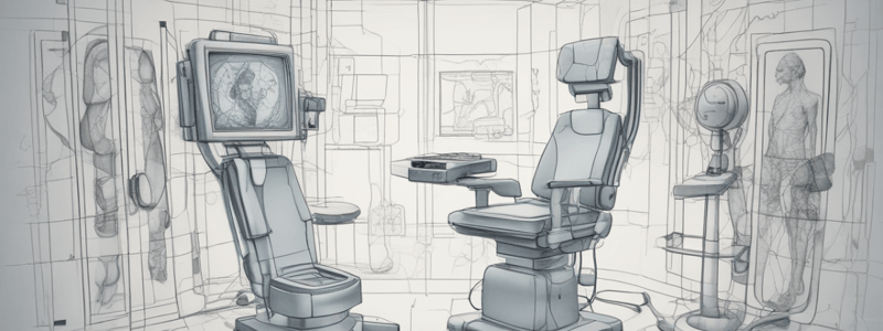
Ultrasound Imaging
- Ultrasound image quality is determined by a compromise between good resolution and deep penetration.
Reflection
- Perpendicular reflection originates the echo signal, while non-perpendicular reflection causes an intensity loss in the echo signal.
- Smooth surface → low scattering → good image
- Rough surface → high scattering → bad image
A-Mode (1D)
- Used to obtain diagnostic information about the depth of structures (image with 1-dimension).
- Sends US waves into the body and measures the time required to receive the reflected sound (echoes) from the interface between different tissues.
- Depth = Velocity x time
- Used to detect brain tumors and eye diseases.
Applications of A-Mode Scan
- Echo Encephalography: • Used in the detection of brain tumors. • Pulses of ultrasound are sent to a thin region of the skull above the ear, and the echoes from different structures are displayed on the oscilloscope. • Compare the echoes from the left side of the head to the right side, and find the shift in the middle structure. • If the shift > 3 mm for an adult (abnormal) or > 2 mm for a child (abnormal).
- Ophthalmology: • Used in diagnosis of eye diseases. • Used in biometry (measurement of distance in the eye, such as lens thickness, depth from cornea to lens, distance to the retina, and thickness of the vitreous tumor).
US Image Modes
-
B-Mode (2D): • Used to obtain 2D images of the body. • Principle is the same as in A-mode, except that the transducer is moving. • Provides information about the internal structure of the body, such as size, location, and change with time of the eye, liver, breast, heart, and fetus.
-
M-Mode (2D + motion): • Used to study motion, such as that of the heart and heart valves (image with 2D + motion). • Combines features of A- and B-mode. • Used in diagnostic information about the heart (mitral valve) and detection of pericardial effusion.
-
D-Mode (3D + motion; or 4D): • Takes 3-dimensional US images and adds the element of time to the process (image with 3D with motion).
Physiological Effects of Ultrasound
- Various physiological and chemical effects occur when ultrasonic waves pass through the body, and they can cause physiological effects.
- The magnitude of physiological effects depends on the frequency and amplitude of the sound.
Low Intensity US (~ 0.01 W/cm²)
- No harmful effects are observed → used for diagnostic work (as in sonar).
Continues US (~1 W/cm²)
- Deep heating effect (diathermy) → temperature rise due to the absorption of acoustic energy in the tissue.
Continues US (1-10 W/cm²)
- Sound moves through → region of compression and rarefactions → pressure differences in adjacent regions of tissues (micromassage).
Continues US (~ 35 W/cm²)
- Tissue destroying effect → rupture DNA molecules.
Continues and focused US (~ 10³ W/cm²)
- Selective destroying of deep tissue using a focused ultrasound beam.
Ultrasound Generation
- Ultrasound is sound with a frequency of 20 kHz to 1 GHz (for medical applications).
- It is greater than the upper limit of human hearing.
Piezoelectric Principle
- The ultrasound signal is generated and detected by the sensor.
- The transducer is based on the piezoelectric principle.
- Many crystals can be used so that AC voltage (electrical energy) across the crystal will produce a vibration of the crystal (mechanical energy), thus generating an ultrasound wave and vice versa.
SONAR
- It is a device that uses an US wave to generate an image of a particular soft tissue structure in the body.
Image Production
- Three concepts that affect US image production: • Focal zone • Acoustic impedance • Refraction
Studying That Suits You
Use AI to generate personalized quizzes and flashcards to suit your learning preferences.