Podcast
Questions and Answers
What is the primary function of the arm in a microscope?
What is the primary function of the arm in a microscope?
- To provide stability to the microscope
- To enhance the magnification of the lenses
- To connect the base to the body tube and support optical components (correct)
- To adjust the coarse focus
What does magnification in a microscope primarily refer to?
What does magnification in a microscope primarily refer to?
- The process of enlarging the appearance of an object (correct)
- The clarity of the image produced
- The ability to distinguish fine details
- The maximum distance between two resolvable points
Which of the following lenses typically has a magnification of 10x?
Which of the following lenses typically has a magnification of 10x?
- Fine focus lens
- Eyepiece (Ocular lens) (correct)
- Objective lens
- Coarse focus lens
What is the definition of resolution in the context of microscopy?
What is the definition of resolution in the context of microscopy?
How does the resolving power of a microscope relate to the minimum resolvable distance?
How does the resolving power of a microscope relate to the minimum resolvable distance?
What happens to the field of view as magnification increases?
What happens to the field of view as magnification increases?
Which range of magnification provides a larger field of view in microscopy?
Which range of magnification provides a larger field of view in microscopy?
What role does the body tube play in a microscope?
What role does the body tube play in a microscope?
What type of microscope provides a 3D view of the specimen?
What type of microscope provides a 3D view of the specimen?
Which component controls the amount of light passing through the specimen on a light microscope?
Which component controls the amount of light passing through the specimen on a light microscope?
What is the primary purpose of electron microscopes?
What is the primary purpose of electron microscopes?
What is the primary purpose of using medium magnification in microscopy?
What is the primary purpose of using medium magnification in microscopy?
Which type of lens provides the initial magnification in a light microscope?
Which type of lens provides the initial magnification in a light microscope?
What is the role of the revolving nosepiece in a light microscope?
What is the role of the revolving nosepiece in a light microscope?
Which type of microscope passes electrons through a thin specimen?
Which type of microscope passes electrons through a thin specimen?
Which knob should be used exclusively when focusing specimens under high power?
Which knob should be used exclusively when focusing specimens under high power?
How do scanning probe microscopes operate?
How do scanning probe microscopes operate?
What happens to the letter 'e' when viewed under a microscope?
What happens to the letter 'e' when viewed under a microscope?
What type of microscope is best suited for observing large, three-dimensional specimens?
What type of microscope is best suited for observing large, three-dimensional specimens?
Why should backpacks and materials be kept out of the aisles when using a microscope?
Why should backpacks and materials be kept out of the aisles when using a microscope?
What could occur if the coarse knob is used while looking through a microscope on high power?
What could occur if the coarse knob is used while looking through a microscope on high power?
What is the typical field of view diameter when using high magnification?
What is the typical field of view diameter when using high magnification?
What is a wet mount in microscopy?
What is a wet mount in microscopy?
What should be done after using the coarse knob on low power?
What should be done after using the coarse knob on low power?
Which of the following is a cautionary measure when using methylene blue?
Which of the following is a cautionary measure when using methylene blue?
What is the purpose of staining specimens?
What is the purpose of staining specimens?
Which technique helps avoid air bubbles when applying a cover slip?
Which technique helps avoid air bubbles when applying a cover slip?
What is the initial step in preparing a wet mount slide?
What is the initial step in preparing a wet mount slide?
Which of the following components is necessary for making a wet mount slide?
Which of the following components is necessary for making a wet mount slide?
What is the appropriate way to find the specimen under the microscope?
What is the appropriate way to find the specimen under the microscope?
Which step is NOT part of preparing a wet mount slide?
Which step is NOT part of preparing a wet mount slide?
To achieve the total magnification of a microscope with an ocular lens of 10x and a high-power objective of 50x, what is the total magnification?
To achieve the total magnification of a microscope with an ocular lens of 10x and a high-power objective of 50x, what is the total magnification?
What should be done after placing the specimen in the drop of liquid?
What should be done after placing the specimen in the drop of liquid?
Which of the following statements is true regarding microscope storage?
Which of the following statements is true regarding microscope storage?
When using the high-power objective, which knob should be used for focusing?
When using the high-power objective, which knob should be used for focusing?
What is the first step in the wet mount technique for observing onion cells?
What is the first step in the wet mount technique for observing onion cells?
Which part of the microscope should be adjusted to control the amount of light hitting the slide?
Which part of the microscope should be adjusted to control the amount of light hitting the slide?
Flashcards are hidden until you start studying
Study Notes
Types of Microscopes
- Optical Microscopes: Use light and lenses for magnification
- Compound Microscopes: Utilize multiple lenses for high magnification
- Stereo Microscopes: Provide a 3D view and used for lower magnification
- Electron Microscopes: Employ electron beams instead of light, achieving significantly higher magnifications
- Transmission Electron Microscopes (TEM): Pass electrons through thin specimens
- Scanning Electron Microscopes (SEM): Scan the surface of specimens with electron beams
- Scanning Probe Microscopes: Utilize physical probes to scan surfaces on an atomic level
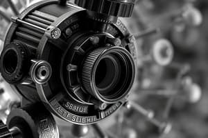
Anatomy of a Light Microscope
- Eyepiece (Ocular Lens): The lens you look through, typically providing 10x or 15x magnification
- Objective Lenses: Multiple lenses on a rotating nosepiece, each with different magnification levels (e.g., 4x, 10x, 40x, 100x)
- Revolving Nosepiece: Holds and allows selection of different objective lenses
- Stage: Platform where the slides are placed, often with clips or a holder to secure the slide.
- Stage Clips: Secure the slide to the stage.
- Condenser: Lens system below the stage focusing light onto the specimen.
- Diaphragm (Aperture Iris): Controls the amount of light passing through the specimen, located beneath the condenser.
- Illuminator (Light Source): Provides the light needed for imaging, usually a built-in lamp or LED.
- Base: Bottom part providing stability and housing the illuminator.
- Arm: Connects the base to the body tube, supporting optical components.
- Coarse Focus: Adjusts focus by moving the stage or lenses.
- Fine Focus: Allows precise fine-tuning of the focus.
- Body Tube: Connects the eyepiece to objective lenses, maintaining alignment.
Magnification
- Magnification: Enlarging the appearance of an object for detailed examination
- Objective Lenses: Primary lenses near the specimen, providing initial magnification (common magnifications: 4x, 10x, 40x, 100x)
- Eyepiece (Ocular Lens): Further magnifies the image from the objective lenses (typical magnifications: 10x or 15x)
Resolution
- Resolution: Ability to distinguish two closely spaced points as separate entities
- Resolving Power: A microscope's ability to distinguish fine details, inversely related to the minimum resolvable distance (higher resolving power = seeing finer details)
Field of View (FoV)
- Field of View: Diameter of the visible area through the eyepiece
- Relationship between Magnification and Field of View: As magnification increases, the field of view decreases.
- Typical Field of View Sizes: Larger view with lower magnification (scanning), smaller view with higher magnification (high-power)
Using a Microscope
- Begin with the scanning objective lens
- Place the specimen in the center of the field of view
- Focus using the coarse knob
- Fine-tune focus with the fine knob
- Switch to low power and repeat focusing steps 3-4
- Switch to high power and use ONLY the fine adjustment knob for focusing
- Always start with the scanning objective
- Store the microscope with the scanning objective in place and cord wrapped around
- Carry with both hands, base and arm
Wet Mounts
- Wet Mount Technique: Prepares a specimen for viewing under a microscope by placing it in a drop of liquid on a slide and covering with a cover slip.
- Purpose: Observing living organisms or specimens in their hydrated state.
- Steps:
- Clean slide
- Drop of liquid (water, saline, staining solution)
- Add specimen (small piece or crushed if large)
- Apply cover slip at 45 degrees to avoid air bubbles
- Spread evenly
- Examine under microscope, starting with low-power lens
Staining Specimens
- Purpose: Improving visibility of cell parts
- Common Stain: Iodine
- Steps:
- Drop of Stain on slide
- Specimen centered on stain
- Cover slip applied
Onion Cell Wet Mount
- Steps:
- Obtain clean slide and cover slip
- Cut small piece of onion
- Peel inner surface (tissue-like)
- Place onion on slide
- Add drop of iodine
- Cover with cover slip
- Observe under microscope
- Describe observations
Cheek Cell Wet Mount
- Steps:
- Clean Slide, coverslip, and toothpick
- Gently scrape inside cheek with toothpick
- Place scrapings on slide
- Add drop of stain (methylene blue)
- Coverslip applied
- Observe under microscope
Microscope Quiz
- Start with the scanning objective.
- Use fine adjustment knob when using high power objective.
- Type used in most science classes is the compound microscope.
- The diaphragm controls the amount of light.
- Carry by the base and arm.
- Objectives are attached to the revolving nosepiece.
- Always store with the scanning objective in place.
- Total magnification is the ocular magnification multiplied by the objective lens magnification.
Studying That Suits You
Use AI to generate personalized quizzes and flashcards to suit your learning preferences.