Podcast
Questions and Answers
What is the primary function of mucous connective tissue?
What is the primary function of mucous connective tissue?
- To provide structural support to bones
- To store energy in fat cells
- To give rise to all other connective tissue types (correct)
- To allow for elasticity in blood vessels
Which of the following features is characteristic of dense connective tissue?
Which of the following features is characteristic of dense connective tissue?
- High cellularity with numerous fibroblasts
- Abundant amorphous intercellular material
- Presence of elastic lamina in blood vessels
- Close packing of fibers and few cells (correct)
Where is dense white collagen connective tissue found?
Where is dense white collagen connective tissue found?
- In the dermis of the skin (correct)
- In the respiratory tract
- In the lining of arteries
- In the brain tissue
What distinguishes regular dense white collagenous connective tissue from irregular dense white collagenous connective tissue?
What distinguishes regular dense white collagenous connective tissue from irregular dense white collagenous connective tissue?
What type of tissue is Wharton's jelly classified as?
What type of tissue is Wharton's jelly classified as?
Which statement correctly describes the umbilical vein in comparison to the arteries?
Which statement correctly describes the umbilical vein in comparison to the arteries?
What is the main distinguishing characteristic of irregular dense white collagenous connective tissue?
What is the main distinguishing characteristic of irregular dense white collagenous connective tissue?
Which tissue type is primarily involved in resisting stretching from multiple directions?
Which tissue type is primarily involved in resisting stretching from multiple directions?
What is the primary function of the fibrocytes in tendons?
What is the primary function of the fibrocytes in tendons?
What characterizes dense yellow elastic connective tissue?
What characterizes dense yellow elastic connective tissue?
Which cells develop into chondrocytes once matured?
Which cells develop into chondrocytes once matured?
Which type of loose connective tissue primarily serves as the packing and anchoring material for various structures in the body?
Which type of loose connective tissue primarily serves as the packing and anchoring material for various structures in the body?
Where are chondrocytes primarily located?
Where are chondrocytes primarily located?
What distinguishes brown adipose connective tissue from white adipose connective tissue?
What distinguishes brown adipose connective tissue from white adipose connective tissue?
What distinguishes immature chondrocytes from mature ones?
What distinguishes immature chondrocytes from mature ones?
Where is reticular connective tissue predominantly found?
Where is reticular connective tissue predominantly found?
What is the primary function of cartilage?
What is the primary function of cartilage?
Which of the following tissues contains dense yellow elastic connective tissue?
Which of the following tissues contains dense yellow elastic connective tissue?
Which type of connective tissue is characterized by the presence of undifferentiated mesenchymal cells?
Which type of connective tissue is characterized by the presence of undifferentiated mesenchymal cells?
Chondroblasts are known for their role in which process?
Chondroblasts are known for their role in which process?
What pigment gives white adipose connective tissue its yellow appearance?
What pigment gives white adipose connective tissue its yellow appearance?
Which of the following functions is NOT associated with loose areolar connective tissue?
Which of the following functions is NOT associated with loose areolar connective tissue?
Which characteristic is true of white adipose tissue?
Which characteristic is true of white adipose tissue?
The main cell types found in loose areolar connective tissue include:
The main cell types found in loose areolar connective tissue include:
What type of collagen fibers is primarily found in the matrix of elastic cartilage?
What type of collagen fibers is primarily found in the matrix of elastic cartilage?
Which of the following locations is elastic cartilage NOT found?
Which of the following locations is elastic cartilage NOT found?
What characteristic distinguishes fibrocartilage from elastic cartilage?
What characteristic distinguishes fibrocartilage from elastic cartilage?
What is the primary function of fibrocartilage?
What is the primary function of fibrocartilage?
Which component primarily nourishes cartilage tissue?
Which component primarily nourishes cartilage tissue?
What is the structure surrounding the nucleus pulposus in the intervertebral disc?
What is the structure surrounding the nucleus pulposus in the intervertebral disc?
How does cartilage grow by appositional growth?
How does cartilage grow by appositional growth?
In which condition does the nucleus pulposus herniate through the annulus fibrosus?
In which condition does the nucleus pulposus herniate through the annulus fibrosus?
What is the primary function of osteogenic cells in bone tissue?
What is the primary function of osteogenic cells in bone tissue?
Which component constitutes the majority of the organic part of bone matrix?
Which component constitutes the majority of the organic part of bone matrix?
What is the mineral primarily responsible for the hardness of bone?
What is the mineral primarily responsible for the hardness of bone?
Which layer of the periosteum is primarily composed of collagen fibers and fibroblasts?
Which layer of the periosteum is primarily composed of collagen fibers and fibroblasts?
What is the function of osteoblasts in bone development?
What is the function of osteoblasts in bone development?
What is the composition of the inorganic matrix of bone primarily formed of?
What is the composition of the inorganic matrix of bone primarily formed of?
Where are osteogenic cells located within the bone structure?
Where are osteogenic cells located within the bone structure?
How do osteogenic cells respond to a highly vascular environment?
How do osteogenic cells respond to a highly vascular environment?
What characterizes the appearance of osteoblasts at the EM level?
What characterizes the appearance of osteoblasts at the EM level?
What happens to osteoblasts when they become trapped in lacunae?
What happens to osteoblasts when they become trapped in lacunae?
Which component is synthesized by osteoblasts in the bone matrix?
Which component is synthesized by osteoblasts in the bone matrix?
What is the main function of osteocytes in mature bone?
What is the main function of osteocytes in mature bone?
What distinguishes osteoclasts from osteoblasts at the LM level?
What distinguishes osteoclasts from osteoblasts at the LM level?
Which structure is unique to osteoclasts that aids in their function?
Which structure is unique to osteoclasts that aids in their function?
Where can osteoclasts typically be found?
Where can osteoclasts typically be found?
What distinguishes the nucleus of osteocytes?
What distinguishes the nucleus of osteocytes?
Flashcards
Loose Connective Tissue
Loose Connective Tissue
A type of connective tissue with a loose arrangement of cells, fibers, and ground substance.
Areolar Connective Tissue
Areolar Connective Tissue
The most common type of loose connective tissue, containing fibroblasts, macrophages, and collagen fibers.
Adipose Tissue
Adipose Tissue
Loose connective tissue dominated by fat cells, with white and brown types.
White Adipose Tissue
White Adipose Tissue
Signup and view all the flashcards
Brown Adipose Tissue
Brown Adipose Tissue
Signup and view all the flashcards
Reticular Connective Tissue
Reticular Connective Tissue
Signup and view all the flashcards
Embryonic Mesenchymal Connective Tissue
Embryonic Mesenchymal Connective Tissue
Signup and view all the flashcards
Tendons, Aponeuroses, and Ligaments
Tendons, Aponeuroses, and Ligaments
Signup and view all the flashcards
Fibrocytes (Tendons)
Fibrocytes (Tendons)
Signup and view all the flashcards
Dense Yellow Elastic Connective Tissue
Dense Yellow Elastic Connective Tissue
Signup and view all the flashcards
Yellow Elastic Ligaments
Yellow Elastic Ligaments
Signup and view all the flashcards
Ligamentum Nuchae
Ligamentum Nuchae
Signup and view all the flashcards
Ligamentum Flavum
Ligamentum Flavum
Signup and view all the flashcards
Cartilage
Cartilage
Signup and view all the flashcards
Chondroblasts
Chondroblasts
Signup and view all the flashcards
Chondrocytes
Chondrocytes
Signup and view all the flashcards
Lacunae
Lacunae
Signup and view all the flashcards
Matrix
Matrix
Signup and view all the flashcards
Mucous Connective Tissue
Mucous Connective Tissue
Signup and view all the flashcards
Wharton's Jelly
Wharton's Jelly
Signup and view all the flashcards
Dense Connective Tissue
Dense Connective Tissue
Signup and view all the flashcards
Dense White Collagenous Connective Tissue
Dense White Collagenous Connective Tissue
Signup and view all the flashcards
Irregular Dense White Collagenous C.T.
Irregular Dense White Collagenous C.T.
Signup and view all the flashcards
Regular Dense White Collagenous C.T.
Regular Dense White Collagenous C.T.
Signup and view all the flashcards
Tendons
Tendons
Signup and view all the flashcards
Ligaments
Ligaments
Signup and view all the flashcards
Elastic Cartilage
Elastic Cartilage
Signup and view all the flashcards
Perichondrium
Perichondrium
Signup and view all the flashcards
Fibrocartilage
Fibrocartilage
Signup and view all the flashcards
Collagen fibers in fibrocartilage
Collagen fibers in fibrocartilage
Signup and view all the flashcards
Intervertebral Disc structure
Intervertebral Disc structure
Signup and view all the flashcards
Avascular Cartilage
Avascular Cartilage
Signup and view all the flashcards
Appositional Growth (Cartilage)
Appositional Growth (Cartilage)
Signup and view all the flashcards
Interstitial Growth
Interstitial Growth
Signup and view all the flashcards
Bone Matrix
Bone Matrix
Signup and view all the flashcards
Osteoid
Osteoid
Signup and view all the flashcards
Bone Mineral
Bone Mineral
Signup and view all the flashcards
Osteogenic Cells
Osteogenic Cells
Signup and view all the flashcards
Periosteum
Periosteum
Signup and view all the flashcards
Endosteum
Endosteum
Signup and view all the flashcards
Osteoblasts
Osteoblasts
Signup and view all the flashcards
Osteoblast Function
Osteoblast Function
Signup and view all the flashcards
Osteoblast Structure (LM)
Osteoblast Structure (LM)
Signup and view all the flashcards
Osteoblast Structure (EM)
Osteoblast Structure (EM)
Signup and view all the flashcards
Osteocyte Function
Osteocyte Function
Signup and view all the flashcards
Osteocyte Structure (LM)
Osteocyte Structure (LM)
Signup and view all the flashcards
Osteocyte Structure (EM)
Osteocyte Structure (EM)
Signup and view all the flashcards
Osteoclast Function
Osteoclast Function
Signup and view all the flashcards
Osteoclast Structure (LM)
Osteoclast Structure (LM)
Signup and view all the flashcards
Osteoclast Structure (EM)
Osteoclast Structure (EM)
Signup and view all the flashcards
Study Notes
Types of Connective Tissue Proper
- Connective tissue proper is classified according to matrix density and cell/fiber types.
- Loose connective tissue includes:
- Loose areolar connective tissue
- Adipose connective tissue
- Reticular connective tissue
- Embryonic mesenchymal connective tissue
- Mucous connective tissue
- Dense connective tissue includes:
- Dense white collagen connective tissue (irregular and regular)
- Dense yellow elastic connective tissue
Loose Areolar Connective Tissue
- Composed of cells, fibers, and ground substance
- Predominantly white collagenous fibers
- Functions as packing and anchoring material, embedding medium for nerves/blood vessels, and binding other tissues/organs.
- Found throughout the body.
Adipose Connective Tissue
- Two types: white and brown
- White adipose tissue: Contains one large fat droplet per cell, appearing yellow due to carotene. Primarily for fat metabolism/storage. Found throughout the body
- Brown adipose tissue: Stores fat in multiple droplets; higher vascularity gives it a brown color. Crucial for heat production in newborns. Found in smaller amounts in adults.
Reticular Connective Tissue
- Primitive connective tissue with a network of reticular fibers (associated with reticular cells).
- Creates the framework for many organs (lymph nodes, bone marrow, liver).
Embryonic Mesenchymal Connective Tissue
- Unspecialized tissue in early embryonic development.
- Composed of undifferentiated mesenchymal cells, ground substance, and fine reticular fibers.
- Develops into other connective tissue types.
Mucous Connective Tissue
- Found in the umbilical cord (Wharton's jelly)
- Composed of stellate fibroblasts, abundant ground substance (mucin-rich), and fine collagenous fibers.
- Supports umbilical blood vessels and acts as a gelatinous substance.
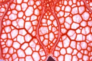
Dense Connective Tissue
- Characterized by the close packing of fibers, few cells, and little ground substance.
- Classified based on fiber type (collagen or elastic).
Dense White Collagen Connective Tissue
- Irregular: Collagen fibers in various directions. Found in dermis, capsules, and around organs.
- Regular: Collagen fibers in parallel arrangement. Found in tendons and ligaments.
Dense Yellow Elastic Connective Tissue
- Predominantly composed of elastic fibers, providing elasticity/ability to return to original shape.
- Located in ligaments, like ligamentum nuchae and flavum, and walls of large arteries.
Cartilage
- Firm, flexible, and strong connective tissue
- Unlike bone, it lacks blood vessels so nutrients are delivered through diffusion.
- Made up of cells, fibers, and extracellular matrix.
- Cells: Chondroblasts and chondrocytes
- Fibers: Collagen fibers (mostly type II)
- Matrix: Ground substance (cartilage matrix), chondroitin sulfate, glycoproteins, water
Chondroblasts
- Active cartilage-forming cells.
- Synthesize collagen fibers and the matrix, producing new cartilage matrix.
- Located in the inner portion of the perichondrium.
- Once enclosed in lacunae they are called chondrocytes.
Chondrocytes
- Mature cartilage cells.
- Inside cartilage lacunae.
- Maintain the cartilage matrix.
- Can divide, creating cell nests of two, four, or more cells.
Cartilage Fibers
- Collagen fibers (primarily type II)
- Elastic fibers (present in elastic cartilage only)
Cartilage Matrix
- Ground substance rich in water & proteins (proteoglycans, chondromucoprotein, chondroitin sulfate).
- Provides resilience and support.
Types of Cartilage
- Hyaline: Most common type; smooth surface for joint articulation; found in articular surfaces, respiratory tracts.
- Elastic: Flexible; ear, epiglottis
- Fibrocartilage: Strongest; intervertebral discs, pubic symphysis.
Bone
- Strong, hard, rigid connective tissue. Matrix composed of organic components (osteoid) and inorganic components (bone mineral).
- Matrix has collagen fibers, ground substance, water
Osteogenic Cells
- Mesenchymal stem cells
- Located in periosteum & endosteum
- Can differentiate into osteoblasts and chondroblasts.
Osteoblasts
- Bone-forming cells
- Synthesize and secrete organic components (osteoid)
- When trapped in matrix, differentiate into osteocytes (mature bone cells)
Osteocytes
- Mature bone cells residing in lacunae within the bone matrix.
- Maintain bone matrix and communicate with other osteocytes.
Osteoclasts
- Bone-resorbing cells
- Responsible for breaking down and remodeling bone tissue.
Cartilage Nutrition
- Cartilage is avascular; nutrients, oxygen diffuse through matrix from perichondrium.
Cartilage Growth
- Appositional: New layers added from perichondrium surface.
- Interstitial: Growth from within the tissue, due to chondrocyte division.
Clinical Note - Herniated Intervertebral Disc
- Prolapsed disc: Nucleus pulposus (disc's soft core) herniates through annulus fibrosus (outer ring).
- Causes inflammation and pain; potentially compresses spinal nerves.
Studying That Suits You
Use AI to generate personalized quizzes and flashcards to suit your learning preferences.