Podcast
Questions and Answers
What is the primary function of the articular disc in the TMJ?
What is the primary function of the articular disc in the TMJ?
- Divides the TMJ into superior and inferior compartments (correct)
- Serves as a lubricant for the joint
- Acts as a shock absorber during jaw movement
- Provides structural stability to the TMJ
Which component of the TMJ provides attachment for the retrodiscal pad?
Which component of the TMJ provides attachment for the retrodiscal pad?
- Posterior band of the articular disc
- Superior retrodiscal lamina (correct)
- Anterior band of the articular disc
- Inferior retrodiscal lamina
Which of the following describes the location of the glenoid fossa in the TMJ?
Which of the following describes the location of the glenoid fossa in the TMJ?
- Located lateral to the mandibular condyles
- A projection on the zygomatic arch
- A depression sitting anterior to the tympanic plate
- A depression into which the condyle is located (correct)
What type of tissue primarily composes the articular disc?
What type of tissue primarily composes the articular disc?
Which part of the TMJ capsule prevents posterior displacement of the condyle?
Which part of the TMJ capsule prevents posterior displacement of the condyle?
What is the volume of the superior compartment of the TMJ?
What is the volume of the superior compartment of the TMJ?
Which characteristic is true regarding the mandibular condyles?
Which characteristic is true regarding the mandibular condyles?
What is the main purpose of synovial fluid in the TMJ?
What is the main purpose of synovial fluid in the TMJ?
Which muscle is considered the most powerful muscle of mastication?
Which muscle is considered the most powerful muscle of mastication?
What is the primary action of the superficial head of the masseter muscle?
What is the primary action of the superficial head of the masseter muscle?
What movement involves shifting the mandible to one side?
What movement involves shifting the mandible to one side?
Which muscles are involved in the process of mastication?
Which muscles are involved in the process of mastication?
Which artery is NOT part of the arterial supply to the TMJ?
Which artery is NOT part of the arterial supply to the TMJ?
Which of the following nerves provides sensory innervation to the TMJ?
Which of the following nerves provides sensory innervation to the TMJ?
Where does the deep head of the medial pterygoid muscle originate?
Where does the deep head of the medial pterygoid muscle originate?
What type of motion is retraction in the context of TMJ movements?
What type of motion is retraction in the context of TMJ movements?
Flashcards are hidden until you start studying
Study Notes
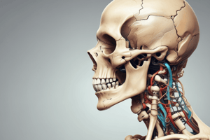
TMJ Structural Components
- Located on each side of the head; movements on one side affect the other side.
- Articulates between the squamous portion of the temporal bone and the condyle of the mandible (Md).
- Key components include:
- Squamous portion of the temporal bone
- Articular disc
- Condyle of the mandible
- Capsule
- Ligaments
Key Anatomical Features
- Articular Eminence: Strong bony prominence on the base of the zygomatic arch.
- Articular Tubercle: Located on the lateral part of the articular eminence; serves as an attachment point for the capsule and lateral TMJ ligament.
- Glenoid (Mandibular) Fossa: Depression accommodating the condyle.
- Tympanic Plate: Vertical plate anterior to the external acoustic meatus.
- Postglenoid Tubercle: Posterior aspect of the glenoid fossa; attachment for the capsule and retrodiscal pad.
Mandibular Condyles
- Shape resembles footballs.
- Dimensions: Mediolateral 20mm, Anteroposterior 10mm.
- Surface consists of avascular fibrous connective tissue with primary load-bearing areas on the lateral aspects.
Articular Disc
- Made of dense fibrous connective tissue; avascular and aneural in the center.
- Peripherally attaches to the capsule, divided into three bands:
- Anterior: Thick
- Intermediate: Thinnest, biconcave
- Posterior: Thickest
TMJ Capsule and Ligaments
- Completely encloses the TMJ; made of fibrous connective tissue.
- Lined by a highly vascular synovial membrane, which is highly innervated.
- Temporomandibular (Lateral) Ligament: Thickened lateral aspect of the capsule, preventing lateral and posterior displacement of the condyle.
TMJ Compartments
- Division by the articular disc into superior and inferior compartments:
- Superior: Larger (1.2 ml); allows translational movement.
- Inferior: Smaller (0.9 ml); allows rotational movement.
- Internal surface lined by the synovial membrane that produces synovial fluid for lubrication and nutrient supply.
Posterior Attachment Complex
- Bilaminar Zone: Attaches the disc to the bone and provides nourishment and innervation.
- Superior Retrodiscal Lamina: Elastic; attaches the disc to the temporal bone, allowing the disc to stretch during joint movement.
- Retrodiscal Pad: Loose vascular tissue that nourishes the joint, fills with blood upon forward mandible movement.
- Inferior Retrodiscal Lamina: Comprising collagen fibers; attaches the disc to the condyle and prevents displacement.
Innervation and Vascularization
- Arterial Supply:
- External Carotid a.
- Superficial temporal a.
- Maxillary a.
- Deep auricular a.
- Anterior tympanic a.
- Venous Drainage:
- Superficial temporal v.
- Maxillary v.
- Sensory Innervation:
- V3 (trigeminal nerve branch)
- Auriculotemporal n.
- Masseteric n.
- Posterior deep temporal n.
Movement of the TMJ
- Functions include:
- Protrusion: Bringing the mandible forward.
- Retraction: Bringing the mandible backward.
- Depression: Lowering the mandible.
- Elevation: Raising the mandible.
- Lateral Deviation: Shifting the mandible to one side.
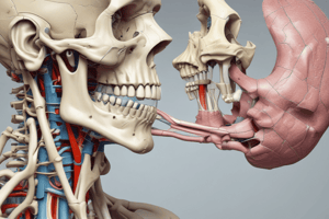
Mastication
- The process of chewing food for swallowing and digestion.
- Major muscles originate from the skull and insert onto the mandible, including:
- Temporalis
- Masseter
- Lateral pterygoid
- Medial pterygoid
- Additional muscles involved include those of the tongue, facial expression (buccinator, orbicularis oris), and suprahyoid/infrahyoid muscles.
Temporalis Muscle
- Powerful muscle of mastication; shape is quadrilateral with superficial and deep heads.
- Origin: Zygomatic arch; Insertion: Coronoid process and lateral surface of mandible.
- Actions include elevation and protrusion (superficial head) and elevation and retrusion (deep head).
Masseter Muscle
- Fan-shaped muscle located on the lateral surface of the skull.
- Origin: Temporal fossa; Insertion: Coronoid process and anterior border of ramus.
- Actions: Elevates and retrudes the mandible.
Medial Pterygoid Muscle
- The deepest muscle of mastication with two heads (superficial and deep).
- Location: Medial to the ramus of the mandible (infratemporal fossa).
- Actions: Elevation, protrusion, and lateral excursion of the mandible.
- Deep Head: Originates from medial surface of lateral pterygoid plate, inserts on medial surface of ramus.
- Superficial Head: Originates from maxillary tuberosity and pyramidal process of palatine bone, inserts on medial surface of ramus.
Studying That Suits You
Use AI to generate personalized quizzes and flashcards to suit your learning preferences.