Podcast
Questions and Answers
What role do cadherins specifically play in adherens junctions?
What role do cadherins specifically play in adherens junctions?
- They facilitate intercellular communication.
- They bind to each other in the presence of Ca²⁺. (correct)
- They create hydrophilic pores in cell membranes.
- They form connections with intermediate filaments.
What distinguishes desmosomes from adherens junctions in terms of the proteins involved?
What distinguishes desmosomes from adherens junctions in terms of the proteins involved?
- Adherens junctions bind to intermediate filaments rather than actin.
- Adherens junctions use desmocollins for adhesion.
- Desmosomes feature desmogleins which connect to plakoglobins. (correct)
- Desmosomes utilize connexins for junction formation.
What is the primary function of gap junctions?
What is the primary function of gap junctions?
- Mediating intercellular communication. (correct)
- Anchoring cells to the basement membrane.
- Forming tight seals between neighboring cells.
- Providing structural strength to epithelial tissues.
How do connexins contribute to the structure of gap junctions?
How do connexins contribute to the structure of gap junctions?
Which statement about adherens junctions is incorrect?
Which statement about adherens junctions is incorrect?
What is the primary function of epithelial tissues?
What is the primary function of epithelial tissues?
Which of the following best describes the composition of epithelial tissues?
Which of the following best describes the composition of epithelial tissues?
Which cell type is specialized for absorption within epithelial tissues?
Which cell type is specialized for absorption within epithelial tissues?
What characterizes connective tissue compared to other tissue types?
What characterizes connective tissue compared to other tissue types?
What determines the shape of epithelial cell nuclei?
What determines the shape of epithelial cell nuclei?
In which of the following tissues are myoepithelial cells primarily found?
In which of the following tissues are myoepithelial cells primarily found?
What is the main structural difference between parenchyma and stroma in organs?
What is the main structural difference between parenchyma and stroma in organs?
Which characteristic is NOT associated with epithelial tissues?
Which characteristic is NOT associated with epithelial tissues?
What primarily indicates the shape and density of epithelial cells in microscopic examination?
What primarily indicates the shape and density of epithelial cells in microscopic examination?
Which structure is primarily responsible for facilitating increased contact between epithelial tissues and connective tissues?
Which structure is primarily responsible for facilitating increased contact between epithelial tissues and connective tissues?
What characterizes the apical pole of epithelial cells?
What characterizes the apical pole of epithelial cells?
What is a primary function of the basement membrane in epithelial tissues?
What is a primary function of the basement membrane in epithelial tissues?
What differentiates the basal lamina from the reticular lamina in the basement membrane?
What differentiates the basal lamina from the reticular lamina in the basement membrane?
Which statement about epithelial tissue is inaccurate?
Which statement about epithelial tissue is inaccurate?
Epithelial cells that are cuboidal or columnar typically have what feature?
Epithelial cells that are cuboidal or columnar typically have what feature?
Which of the following substances is not typically found in the basement membrane?
Which of the following substances is not typically found in the basement membrane?
What feature allows epithelial cells to effectively connect to one another?
What feature allows epithelial cells to effectively connect to one another?
The term 'basal lamina' specifically refers to which of the following?
The term 'basal lamina' specifically refers to which of the following?
What role does type IV collagen play in the structure of the basal lamina?
What role does type IV collagen play in the structure of the basal lamina?
Which protein is responsible for binding to integrin proteins in the basal cell membrane?
Which protein is responsible for binding to integrin proteins in the basal cell membrane?
What is one of the functions of the basal lamina in relation to epithelial cells?
What is one of the functions of the basal lamina in relation to epithelial cells?
Which type of collagen is found in the reticular lamina?
Which type of collagen is found in the reticular lamina?
How do nidogen and perlecan contribute to the basal lamina?
How do nidogen and perlecan contribute to the basal lamina?
Which type of intercellular junction is responsible for preventing the passage of molecules between adjacent cells?
Which type of intercellular junction is responsible for preventing the passage of molecules between adjacent cells?
What are external laminae primarily associated with?
What are external laminae primarily associated with?
What is the primary role of adherent or anchoring junctions in epithelial cells?
What is the primary role of adherent or anchoring junctions in epithelial cells?
Which statement best defines the role of basement membranes?
Which statement best defines the role of basement membranes?
In which order are tight junctions arranged in most epithelia?
In which order are tight junctions arranged in most epithelia?
What types of proteins primarily create tight junctions?
What types of proteins primarily create tight junctions?
What structural feature distinguishes the basal lamina from the reticular lamina?
What structural feature distinguishes the basal lamina from the reticular lamina?
What characteristic does the basement membrane confer to the epithelium regarding cellular migration?
What characteristic does the basement membrane confer to the epithelium regarding cellular migration?
Which pathway do molecules primarily take when crossing an epithelium due to the presence of tight junctions?
Which pathway do molecules primarily take when crossing an epithelium due to the presence of tight junctions?
Which of the following correctly describes the composition source of the basal lamina and reticular lamina?
Which of the following correctly describes the composition source of the basal lamina and reticular lamina?
How does the permeability of epithelia change based on the number of fused sealing strands in tight junctions?
How does the permeability of epithelia change based on the number of fused sealing strands in tight junctions?
What characteristic of tight junctions allows them to act as fences within cell membranes?
What characteristic of tight junctions allows them to act as fences within cell membranes?
What is true about gap junctions in epithelial tissues?
What is true about gap junctions in epithelial tissues?
What effect does the arrangement of tight junctions have on the function of epithelial cells?
What effect does the arrangement of tight junctions have on the function of epithelial cells?
Which statement accurately reflects the role of epithelial tight junctions?
Which statement accurately reflects the role of epithelial tight junctions?
Flashcards
What are the four basic tissue types in the human body?
What are the four basic tissue types in the human body?
Four basic tissue types found in the human body, each with specialized cells and functions.
What is the defining characteristic of connective tissue?
What is the defining characteristic of connective tissue?
A type of tissue characterized by cells producing an abundant extracellular matrix (ECM), which provides support and structure.
What is parenchyma?
What is parenchyma?
Cells in an organ responsible for its specific function.
What is stroma?
What is stroma?
Signup and view all the flashcards
What is the composition of epithelial tissue?
What is the composition of epithelial tissue?
Signup and view all the flashcards
Where can epithelial tissue be found in the body?
Where can epithelial tissue be found in the body?
Signup and view all the flashcards
What are the principal functions of epithelial tissues?
What are the principal functions of epithelial tissues?
Signup and view all the flashcards
How does the shape of an epithelial cell nucleus relate to its function?
How does the shape of an epithelial cell nucleus relate to its function?
Signup and view all the flashcards
Nuclei as indicators of cell shape and density
Nuclei as indicators of cell shape and density
Signup and view all the flashcards
Determining number of cell layers in epithelia
Determining number of cell layers in epithelia
Signup and view all the flashcards
Epithelial dependence on connective tissue
Epithelial dependence on connective tissue
Signup and view all the flashcards
Lamina propria's role in epithelial tissues
Lamina propria's role in epithelial tissues
Signup and view all the flashcards
Function of papillae in epithelia
Function of papillae in epithelia
Signup and view all the flashcards
Polarity in epithelial cells
Polarity in epithelial cells
Signup and view all the flashcards
Basement membrane in epithelial tissues
Basement membrane in epithelial tissues
Signup and view all the flashcards
Structure of the basement membrane
Structure of the basement membrane
Signup and view all the flashcards
Basal lamina: Ultrastructural level
Basal lamina: Ultrastructural level
Signup and view all the flashcards
Terminology: Basement membrane vs. basal lamina
Terminology: Basement membrane vs. basal lamina
Signup and view all the flashcards
What is the basement membrane?
What is the basement membrane?
Signup and view all the flashcards
What is the basal lamina?
What is the basal lamina?
Signup and view all the flashcards
What is type IV collagen?
What is type IV collagen?
Signup and view all the flashcards
What is laminin?
What is laminin?
Signup and view all the flashcards
What are nidogen and perlecan and their functions?
What are nidogen and perlecan and their functions?
Signup and view all the flashcards
What are basal laminae, also known as external laminae?
What are basal laminae, also known as external laminae?
Signup and view all the flashcards
What is the reticular lamina?
What is the reticular lamina?
Signup and view all the flashcards
What are the functions of basement membranes?
What are the functions of basement membranes?
Signup and view all the flashcards
What are intercellular adhesion and junctions in epithelial cells?
What are intercellular adhesion and junctions in epithelial cells?
Signup and view all the flashcards
Why are intercellular junctions important?
Why are intercellular junctions important?
Signup and view all the flashcards
What is an adherens junction?
What is an adherens junction?
Signup and view all the flashcards
What is a desmosome?
What is a desmosome?
Signup and view all the flashcards
What are gap junctions?
What are gap junctions?
Signup and view all the flashcards
What are connexins?
What are connexins?
Signup and view all the flashcards
What is a terminal web?
What is a terminal web?
Signup and view all the flashcards
What are intercellular junctions?
What are intercellular junctions?
Signup and view all the flashcards
What are tight junctions?
What are tight junctions?
Signup and view all the flashcards
What are adhering junctions?
What are adhering junctions?
Signup and view all the flashcards
What is the order of intercellular junctions?
What is the order of intercellular junctions?
Signup and view all the flashcards
What are zonulae occludens?
What are zonulae occludens?
Signup and view all the flashcards
What is occludin?
What is occludin?
Signup and view all the flashcards
What is the transcellular pathway?
What is the transcellular pathway?
Signup and view all the flashcards
What is the paracellular pathway?
What is the paracellular pathway?
Signup and view all the flashcards
How do tight junctions act as membrane fences?
How do tight junctions act as membrane fences?
Signup and view all the flashcards
Study Notes
Basic Tissue Types
- The human body's organs are composed of four basic tissue types: epithelial, connective, muscular, and nervous tissues.
- Each tissue type is a group of specialized cells performing specific functions, containing extracellular matrix (ECM) as well as cells.
- These tissues associate in varying proportions and morphologies characteristic of each organ.
Connective, Muscle, and Nervous Tissues
- Connective tissue is characterized by cells producing an abundant ECM.
- Muscle tissue is composed of elongated cells specialized for contraction and movement.
- Nervous tissue comprises cells with long, fine processes specialized for receiving, generating, and transmitting nerve impulses.
Parenchyma and Stroma
- Organs are divided into parenchyma and stroma.
- Parenchyma consists of cells responsible for the organ's specialized functions.
- Stroma, primarily composed of connective tissue (except in the brain and spinal cord), supports the parenchyma.
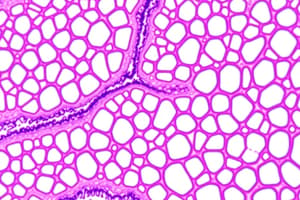
Epithelial Tissues
- Epithelial tissues are composed of closely grouped polyhedral cells adhering strongly to one another with a thin layer of ECM.
- They form cellular sheets that line the cavities of organs and cover the body's surface, lining all internal and external surfaces.
- All substances entering or leaving an organ must cross this tissue type.
Principal Functions of Epithelial Tissues
- Covering, lining, and protecting surfaces (e.g., epidermis of skin).
- Absorption (e.g., intestinal lining).
- Secretion (e.g., parenchymal cells of glands).
- Certain epithelial cells may be contractile (myoepithelial cells) or specialized as sensory cells (e.g., taste buds or olfactory epithelium).
Characteristics of Epithelial Cells
- Epithelial cells vary in shape and dimensions, ranging from tall columnar to low squamous cells.
- Cell shape is generally dictated by function.
- Cell nuclei shapes correspond to the cell shape: columnar cells have elongated nuclei, squamous cells have flattened nuclei, and cuboidal or pyramidal cells have more spherical nuclei.
Epithelial Tissue Adjacency to Connective Tissue
- Most epithelia are adjacent to connective tissue containing blood vessels, supplying nutrients and oxygen to the epithelial cells.
- Even thick epithelia typically do not contain their own blood vessels.
- The connective tissue under the epithelia in the digestive, respiratory, and urinary systems is known as the lamina propria.
- Epithelial-connective tissue contact is facilitated by papillae, small evaginations.
Polarity and Structural Aspects of Epithelial Cells
- Epithelial cells display polarity, having organelles and membrane proteins distributed unevenly across the cell.
- Basal pole contacts the ECM and connective tissue, while the apical pole faces a space.
- Cuboidal or columnar cells have lateral surfaces adjoining neighboring cells with membrane folds increasing surface area and functional capacity.
Basement Membranes
- The basal surface of epithelia rests on a basement membrane consisting of macromolecules.
- The basement membrane acts as a semipermeable filter for substances reaching epithelial cells from below.
- It contains glycoproteins and other components, often visible with a light microscope.
Basal Lamina and Reticular Lamina
- The basement membrane consists of the basal lamina and the reticular lamina.
- Basal Lamina is a thin sheet-like layer of fibrils close to epithelial cells.
- Reticular Lamina is a more diffuse layer beneath the basal lamina.
Components of the Basal Lamina
- Type IV collagen forms a two-dimensional network structure similar to a window screen.
- Laminin is a large glycoprotein that attaches to integrins.
- Nidogen and perlecan contribute to basal lamina structure and are proteins and proteoglycans, respectively.
Intercellular Adhesion and Junctions
- Epithelial cells adhere strongly to neighboring cells and basal lamina, especially in areas subject to friction.
- Lateral surfaces of epithelial cells feature intercellular junctions with distinct functions.
- Tight junctions create a seal between adjacent cells.
- Adherent junctions anchor cells together.
- Gap junctions form channels for communication.
Desmosomes
- Desmosomes (or macula adherens) are disc-shaped anchoring junctions resembling spot-welds, rather than belts.
- Desmosomes consist of cadherins which link to plakoglobins (catenin-like proteins) that are linked to desmoplakins.
Gap Junctions
- Gap junctions connect cells mediating intercellular communication, and are common in nearly all mammalian tissues.
- Cryofracture preparations show transmembrane protein complexes that form circular patches in the plasma membrane known as connexons.
Hemidesmosomes
- Hemidesmosomes are anchoring junctions on the basal surface of epithelial cells that link to laminin of the basal lamina.
Focal Adhesions
- Focal adhesions are basal anchoring junctions that link integrins to bundled actin filaments, rather than intermediate filaments.
- Focal adhesions are involved in epithelial repair and reorganization.
- Integrins in focal adhesions connect to paxillin and focal adhesion kinase (FAK), a signaling protein that affects cell mobility and gene expression.
Specializations of Apical Surface
- The apical ends of many columnar and cuboidal epithelial cells feature specialized structures like microvilli and stereocilia for enhancing absorption or moving substances along the surface, observed in some absorptive epithelial cells.
Microvilli
- Microvilli are cytoplasmic projections formed by bundled actin filaments, found in large numbers in epithelial cells specialized for absorption.
- They dramatically enhance the cell's surface area.
Stereocilia
- Stereocilia are even longer microvilli found in the male reproductive system for absorption.
Cilia
- Cilia are long, motile structures containing internal microtubules.
- Cilia are often found in epithelial cells lining the upper respiratory tract or in other specialized epithelial cells (e.g., those of the inner ear).
- The rhythmic beating patterns of cilia move fluid along the epithelial surface.
Basal Bodies and Axoneme Continuity
- The microtubules of cilia are continuous with basal bodies, which are similar to centrioles.
Types of Epithelia
- Epithelia are classified as covering/lining or secretory (glandular).
Covering Epithelia
- Covering epithelia are single-layered or multiple-layered cells that cover surfaces or line cavities of organs.
- They are classified based on the number and shape of cell layers.
- Simple (single layer) — squamous, cuboidal, columnar
- Stratified (multiple layers) — squamous, cuboidal, columnar
Stratified Squamous Epithelia
- These epithelia have thin surface cells and can be either keratinized or nonkeratinized
- Keratinized (epidermis)
- Nonkeratinized (moist areas like mouth, esophagus)
Transitional Epithelium
- Also known as urothelium and lines the urinary tract.
- It can distend as the urinary bladder fills, a unique morphological feature.
Secretory Epithelia
- Secretory epithelia are primarily involved in producing and releasing macromolecules.
- They are also part of specialized organs called glands.
- Cells in glands synthesize, store, and release either proteins (e.g., pancreas), lipids (e.g., adrenal glands), or carbohydrate-protein complexes, (e.g., salivary glands).
Mechanisms of Secretion
- Merocrine: Secretion via exocytosis (most common).
- Holocrine: Secretory product released with cell debris.
- Apocrine: Product released with cell membrane.
Serous and Mucous Cells (Merocrine Secretion)
- Serous cells synthesize protein secretions typically non-glycosylated (e.g., digestive enzymes), with a well-developed endoplasmic and Golgi apparatus, filled with secretory granules, stained with acidophilic stains.
- Mucous cells synthesize and secrete protective secretions that become mucous upon hydration (e.g., goblet cells).
Myoepithelial Cells
- Myoepithelial cells are contractile cells found in many secretory glands (sweat, lacrimal, salivary, mammary)
Endocrine Glands
- Endocrine glands lose their connection to the original epithelium (ductless) and release hormones directly into the blood, lacking myoepithelial cells.
- Protein hormones are released by exocytosis, whereas lipophilic steroid hormones diffuse through the cell membrane.
Ion Transport and Transcellular Transport
- Active ion transport moves ions against concentration/electrical gradients (e.g., Na+/K+-ATPase).
- Transcellular transport involves ion pumps and water channels, through aquaporins, to cross the epithelium.
- Tight junctions prevent paracellular diffusion between cells.
Renewal of Epithelial Tissues
- Epithelial tissues renew constantly via mitotic activity and stem cells, with rates varying across tissues.
Repair and Replacement in Epithelial Tissues
- Epithelial cells have a high capacity for rapid repair and replacement, with cell division in target areas after significant damage.
- Stem cells may facilitate epithelial regeneration in specific locations.
Studying That Suits You
Use AI to generate personalized quizzes and flashcards to suit your learning preferences.