Podcast
Questions and Answers
What is the name of the area where the axon is connected to the cell body?
What is the name of the area where the axon is connected to the cell body?
- Dendrite
- Node of Ranvier
- Myelin sheath
- Axon hillock (correct)
What is the function of the myelin sheath in the axon?
What is the function of the myelin sheath in the axon?
- To speed up action potential propagation by stopping ion exchange (correct)
- To slow down action potential propagation
- To receive signals
- To synthesize proteins
What is the process called when the action potential jumps from gap to gap in the myelin sheath?
What is the process called when the action potential jumps from gap to gap in the myelin sheath?
- Neurotransmission
- Ionic conduction
- Electric conduction
- Saltatory conduction (correct)
What is the purpose of Na+/K+ ATPases in maintaining resting potential?
What is the purpose of Na+/K+ ATPases in maintaining resting potential?
What is the voltage range of the peak of the action potential in neurons?
What is the voltage range of the peak of the action potential in neurons?
What is the term for when the membrane potential becomes more negative than the normal resting potential?
What is the term for when the membrane potential becomes more negative than the normal resting potential?
What is the purpose of K+ leak channels in maintaining resting potential?
What is the purpose of K+ leak channels in maintaining resting potential?
What happens during the refractory period?
What happens during the refractory period?
What is the main reason why another action potential cannot be fired during the absolute refractory period?
What is the main reason why another action potential cannot be fired during the absolute refractory period?
What is the role of Ca2+ ions in the steps of synaptic transmission?
What is the role of Ca2+ ions in the steps of synaptic transmission?
What type of potential is produced when excitatory neurotransmitters bind to ligand-gated ion channels?
What type of potential is produced when excitatory neurotransmitters bind to ligand-gated ion channels?
What is the function of microglial cells in the nervous system?
What is the function of microglial cells in the nervous system?
What happens to the membrane potential during an inhibitory postsynaptic potential (IPSP)?
What happens to the membrane potential during an inhibitory postsynaptic potential (IPSP)?
What is the purpose of the synapse?
What is the purpose of the synapse?
During which period can a stronger than normal stimulus cause another action potential to be fired?
During which period can a stronger than normal stimulus cause another action potential to be fired?
What type of ions flow into the cell during an excitatory postsynaptic potential (EPSP)?
What type of ions flow into the cell during an excitatory postsynaptic potential (EPSP)?
Flashcards are hidden until you start studying
Study Notes
The Neuron
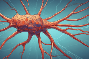
- The neuron is the most basic unit of the nervous system, composed of three parts: soma (cell body), dendrites (extensions that receive signals), and the axon (sends signals out).
The Axon
- The axon has three key components: axon hillock, myelin sheath, and nodes of Ranvier.
- The axon hillock is responsible for the summation of graded potentials.
- The myelin sheath is a fatty insulation formed by oligodendrocytes (in the central nervous system) and Schwann cells (in the peripheral nervous system) that speeds up action potential propagation by stopping ion exchange.
- Nodes of Ranvier are gaps between myelin sheaths where ion exchange occurs, and propagation of the action potential occurs through saltatory conduction.
Action Potential
- At resting potential, the membrane potential of the neuron is around -70mV, maintained by Na+/K+ ATPases and K+ leak channels.
- The steps of an action potential are:
- Depolarization: threshold potential reached (around -55mV), voltage-gated Na+ channels open, letting Na+ in, and the neuron depolarizes (reaches +30mV to +40mV).
- Repolarization: voltage-gated K+ channels open, letting K+ out, and the membrane potential becomes less positive.
- Hyperpolarization: the membrane potential becomes more negative than the normal resting potential, establishing a refractory period.
- Return to normal resting potential: through the pumping of Na+/K+ ATPases and K+ leak channels.
Refractory Period
- The absolute refractory period is the period after the initiation of the action potential during which another action potential cannot be fired, no matter how powerful the stimulus is, due to the inactivation of voltage-gated Na+ channels.
- The relative refractory period is the period after the action potential fires during which a stronger than normal stimulus could cause another action potential to be fired.
Synaptic Transmission
- The synapse is the space between two neurons, where the presynaptic neuron sends the signal and releases neurotransmitters, and the postsynaptic neuron receives the signal by interacting with the released neurotransmitters.
- The steps of synaptic transmission are:
- Action potential reaches the end of the presynaptic axon, causing voltage-gated calcium channels to open and letting Ca2+ ions into the neuron.
- Ca2+ ions cause synaptic vesicles to fuse and undergo exocytosis, releasing neurotransmitters into the synapse.
- Neurotransmitters bind to ligand-gated ion channels on the postsynaptic neuron, producing graded potentials (depolarizations or hyperpolarizations of the membrane).
- Graded potentials summate at the axon hillock, and an action will fire if the summation of graded potentials is higher than the threshold potential of neurons.
Types of Graded Potentials
- Excitatory postsynaptic potential (EPSP): graded potential that depolarizes the membrane, caused by excitatory neurotransmitters that open Na+ ion gates and let Na+ ions flow into the cell.
- Inhibitory postsynaptic potential (IPSP): graded potential that hyperpolarizes the membrane, caused by inhibitory neurotransmitters that open K+ ion gates and let K+ ions flow out of the cell, or allow influx of Cl- ions.
Glial Cells
- Glial cells are non-neuronal cells in the nervous system that help support and surround neurons.
- They are divided into microglial cells (macrophages that protect the CNS) and macroglial cells.
Studying That Suits You
Use AI to generate personalized quizzes and flashcards to suit your learning preferences.


