Podcast
Questions and Answers
Which division of the human skeleton contains more bones?
Which division of the human skeleton contains more bones?
- Appendicular skeleton (correct)
- Both divisions have the same number of bones
- Axial skeleton
- The skeleton is made up of only one group
The adult human skeleton contains 206 bones overall.
The adult human skeleton contains 206 bones overall.
True (A)
What type of bone is mostly composed of spongy bone tissue surrounded by a thin layer of compact bone?
What type of bone is mostly composed of spongy bone tissue surrounded by a thin layer of compact bone?
Short bones
The two major types of bone surface markings are _____ and processes.
The two major types of bone surface markings are _____ and processes.
Match the following types of bones with their descriptions:
Match the following types of bones with their descriptions:
What treatment options are available for unequal leg length?
What treatment options are available for unequal leg length?
Folic acid supplementation during pregnancy can reduce the risk of spina bifida.
Folic acid supplementation during pregnancy can reduce the risk of spina bifida.
What is the term for the protrusion of the spinal cord in spina bifida?
What is the term for the protrusion of the spinal cord in spina bifida?
The axial skeleton includes the bones of the _____ and the vertebral column.
The axial skeleton includes the bones of the _____ and the vertebral column.
Match the following conditions with their descriptions:
Match the following conditions with their descriptions:
What is one of the functions of the skull?
What is one of the functions of the skull?
The mandible is the only immovable bone of the skull.
The mandible is the only immovable bone of the skull.
What are the three auditory ossicles called?
What are the three auditory ossicles called?
The passage for the optic nerve is found in the _______ bone.
The passage for the optic nerve is found in the _______ bone.
Match the cranial bone to its description:
Match the cranial bone to its description:
Which bone is known as the 'keystone' of the cranial floor?
Which bone is known as the 'keystone' of the cranial floor?
The zygomatic bone forms the upper jaw of the skull.
The zygomatic bone forms the upper jaw of the skull.
How many total bones are there in the adult human skull?
How many total bones are there in the adult human skull?
The _________ forms the inferior portion of the nasal septum.
The _________ forms the inferior portion of the nasal septum.
What is the function of fontanels in an infant's skull?
What is the function of fontanels in an infant's skull?
The maxilla bones are comprised of only one bone in the skull.
The maxilla bones are comprised of only one bone in the skull.
What are the nasal bones primarily responsible for?
What are the nasal bones primarily responsible for?
The _______ bones complete the posterior portion of the hard palate.
The _______ bones complete the posterior portion of the hard palate.
Match the facial bones with their characteristics:
Match the facial bones with their characteristics:
What is the primary function of the intervertebral discs?
What is the primary function of the intervertebral discs?
The hyoid bone is part of the vertebral column.
The hyoid bone is part of the vertebral column.
What are the two special vertebrae in the cervical region and their names?
What are the two special vertebrae in the cervical region and their names?
The ____ processes of the cervical vertebrae are bifid.
The ____ processes of the cervical vertebrae are bifid.
Match the following vertebrae with their characteristics:
Match the following vertebrae with their characteristics:
Which of the following statements about the vertebral column is false?
Which of the following statements about the vertebral column is false?
The thoracic vertebrae have demifacets for rib articulation.
The thoracic vertebrae have demifacets for rib articulation.
What is the term for the tiny inferior bone of the sternum?
What is the term for the tiny inferior bone of the sternum?
The spinal cord passes through the ____ foramen of the vertebrae.
The spinal cord passes through the ____ foramen of the vertebrae.
Match the ribs with their types:
Match the ribs with their types:
Which part of the vertebrae bears weight?
Which part of the vertebrae bears weight?
The cervical curve is acquired after learning to walk.
The cervical curve is acquired after learning to walk.
What is scoliosis?
What is scoliosis?
The ____ vertebrae contain transverse foramina for the vertebral artery.
The ____ vertebrae contain transverse foramina for the vertebral artery.
What type of cartilage are the costal cartilages made of?
What type of cartilage are the costal cartilages made of?
Flashcards are hidden until you start studying
Study Notes
The Human Skeleton
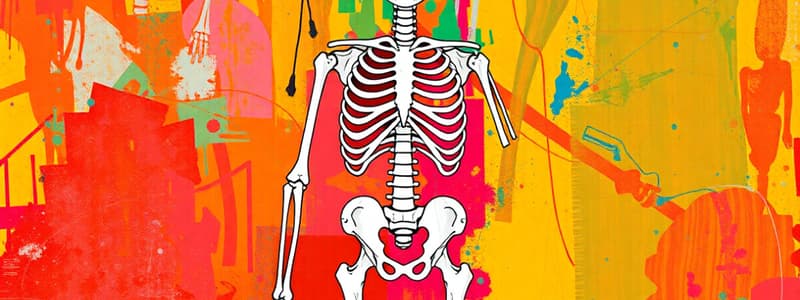
- The adult human skeleton contains 206 bones.
- The skeleton can be divided into two main groups:
- Axial Skeleton
- Contains 80 bones.
- Runs from the head to the bottom of the vertebral column.
- Appendicular Skeleton
- Contains 126 bones.
- Includes bones of the limbs and the girdles that attach the limbs to the axial skeleton.
- Axial Skeleton
- The musculoskeletal system includes both the muscular and skeletal systems, which are interdependent and allow for movement.
Types of Bones
- There are five main types of bones:
- Long Bones:
- Longer than they are wide.
- Curved to absorb shock evenly.
- Spongy bone tissue is located in the epiphyses (ends of the bone) while compact bone tissue is found in the diaphysis (shaft).
- Short Bones:
- Nearly as long as they are wide.
- Consist mostly of spongy bone tissue surrounded by a thin layer of compact bone.
- Flat Bones:
- Thin plates of compact bone.
- Contain spongy bone interiors.
- Sesamoid Bones:
- Thin, small bones that develop in areas of high mechanical stress.
- Protect tendons by modulating tension applied during movement.
- Irregular Bones:
- Have an irregular shape or distribution.
- Long Bones:
Surface Markings of Bones
- Bones have surface markings that serve physiological functions.
- Depressions and Openings:
- Provide passage for blood vessels and nerves.
- Types include foramina, fossae, and meati.
- Processes, Projections, or Outgrowths:
- Attachment points for ligaments and tendons.
- Types include condyles, facets, heads, crests, and processes.
- Depressions and Openings:
The Human Skull
- The skull performs many important functions:
- Protects the brain.
- Serves as a point of attachment for facial muscles.
- Forms portions of the orbits, nasal, and oral cavities.
- Includes the auditory ossicles, enabling hearing.
Cranial Bones
- There are 22 bones in the skull, some of which are paired.
- Frontal Bone:
- Forms the forehead at the anterior of the skull.
- Contains the supraorbital foramen for the supraorbital artery and nerve.
- Ethmoid Bone:
- Forms the medial portion of the orbits.
- Forms the anterior portion of the cranial floor
- Contains the crista galli, a triangular process where the membrane dividing the two halves of the brain attaches.
- The cribriform plate contains the olfactory foramina for sensory structures involved in smell.
- The perpendicular plate forms the superior part of the nasal septum.
- Sphenoid Bone:
- “Keystone” of the cranial floor
- Contains the optic foramen for the ophthalmic artery and optic nerve.
- Temporal Bones:
- Form the lateral and inferior portions of the cranium.
- The zygomatic arch forms the lateral part of the “cheekbone.”
- The mandibular fossa forms a cavity that accommodates the mandibular condyle, forming the temporomandibular joint.
- The styloid process is an attachment point for neck and tongue muscles.
- The mastoid process is an attachment point for neck muscles.
- The external auditory meatus forms the ear canal and directs sound waves to the auditory ossicles.
- Auditory Ossicles:
- Sound waves hit the tympanic membrane, vibrating the auditory ossicles, and transmitting vibrations to auditory sensory structures.
- Malleus (hammer)
- Incus (anvil)
- Stapes (stirrup)
- Sound waves hit the tympanic membrane, vibrating the auditory ossicles, and transmitting vibrations to auditory sensory structures.
- Occipital Bone:
- Forms the posterior and inferior portion of the skull.
- The occipital condyles form joints with the first cervical vertebra (atlas), creating the atlantooccipital joint.
- The foramen magnum provides passage for the spinal cord to connect with the brain.
- Parietal Bones:
- Form the superior and lateral portions of the skull.
- No significant surface markings.
Facial Bones
- There are 14 facial bones forming the anterior portion of the skull, including the only movable bone: the mandible.
- Mandible:
- Forms the lower jaw.
- The largest and strongest bone of the skull.
- The condylar process articulates with the mandibular fossa of the temporal bone.
- Maxillae:
- Forms the upper jaw.
- Two bones (left and right) fused in adults.
- The palatine process forms most of the hard palate.
- The palatine process provides passage for the infraorbital blood vessels and nerves (fifth cranial nerve).
- Palatine Bones
- L-shaped bones that complete the posterior portion of the hard palate.
- Zygomatic Bones:
- Form the anterior portion of the “cheekbones.”
- Parts of the inferior and lateral walls of the orbits.
- Vomer:
- Forms the inferior portion of the nasal septum.
- Inferior Nasal Conchae:
- Form the lateral walls of the nasal cavity.
- Their curled shape swirls air around the nasal passages, increasing the chance of trapping airborne invaders.
- Nasal Bones:
- Form the bridge of the nose where glasses sit.
- Lacrimal Bones:
- The smallest of the facial bones.
- Located near the tear ducts (lacrimal ducts).
Review:
- Bones of the Cranium:
- Occipital bone
- Right/left parietal bone
- Right/left sphenoid bone
- Right/left temporal bone
- Frontal bone
- Bones of the Face:
- Left/right zygomatic
- Left/right maxilla
- Vomer
- Left/right Nasal bone
- Mandible
- Left/right palatine bone
- Left/right lacrimal
- Left/right inferior nasal concha
Special Features of the Skull
- Orbits:
- Contain the eyes.
- Form from seven bones:
- Frontal bone
- Lacrimal
- Ethmoid
- Zygomatic
- Sphenoid
- Maxilla
- Palatine
- Sutures:
- At birth, the skeleton is incompletely ossified, leaving "soft spots" called fontanels.
- Mesenchymal tissue persists and becomes dense connective tissue.
- Fontanels allow for brain growth and facilitate passage through the birth canal.
- Coronal and Sagittal Sutures:
- Coronal Suture: Connects the frontal and parietal bones (coronal means crown).
- Sagittal Suture: Connects the parietal bones superiorly.
- Other Sutures:
- Lambdoid Suture: Connects the parietal bone and occipital bone.
- Squamous Suture: Connects the parietal and temporal bone.
- Zygomatic Arch:
- The prominent bony portion that runs laterally and posteriorly from the zygomatic bone.
- Forms from the zygomatic bone and temporal bone.
- Sinuses:
- Cavities lined with mucous membranes, found in the facial bones.
- They help trap invaders and make the skull lighter.
- Paranasal Sinuses:
- Important to be familiar with for laboratory study.
The Hyoid Bone
- A unique bone that does not articulate with any other bone.
- It floats on tendon and ligaments.
- Muscles of the tongue attach to the hyoid bone.
- The hyoid bone is not the Adam's apple (thyroid cartilage).
The Vertebral Column
- The vertebral column serves many important functions:
- Supports and moves the skull.
- Protects the spinal cord.
- Provides attachment points for muscles of the back and abdomen.
- Contains intervertebral discs to cushion the vertebrae from shock.
- Protects nerves.
Properties of the Vertebral Column
- Subregions: Divided into specific regions.
- Curves: Curved to improve shock absorption.
- The normal curves of the spine are the four curves of healthy spine development.
- The cervical vertebrae acquire their curve after holding up one's head.
- The lumbar vertebrae acquire their curve after learning to walk.
- The normal curves of the spine are the four curves of healthy spine development.
Intervertebral Discs
- Discs of fibrocartilage located between vertebrae.
- They compress throughout the day due to dehydration.
- Individuals are tallest immediately after waking because the vertebrae are not under the force of gravity while lying down.
Vertebral Anatomy
- Body (Vertebral Body): Bears weight and contains nutrient foramina.
- Vertebral Foramen: Provides passage for the spinal cord.
- Superior Articular Processes: Articulate with the inferior articular processes of the vertebrae above.
- Facets: The surfaces on bones where articulations occur at joints.
Cervical Vertebrae
- Most superior vertebrae, forming the neck.
- Numbered C1-C7.
- C1 and C2 have special names:
- C1 (Atlas):
- Has no body or spinous process.
- Has a distinct anterior arch.
- A large vertebral foramen that accommodates the dens of the axis (C2).
- C2 (Axis):
- Contains the dens, a large superior and anterior projection that passes through the vertebral foramen of the atlas.
- The articulation of C1 and C2 forms the atlantoaxial joint, permitting head rotation.
- C1 (Atlas):
Special Properties of Cervical Vertebrae
- Spinous Processes: Bifid (two heads).
- Transverse Processes: Contain the transverse foramen, providing passage for the vertebral artery, a major posterior blood vessel servicing the head.
Thoracic Vertebrae
- Numbered T1-T12.
- Have large transverse processes for articulations with ribs.
- Unique features of the thoracic vertebrae:
- Demifacets: Facets where articulations occur.
- Connect the tubercle of the ribs with the vertebral column.
- The head of a rib connects with demifacets on two vertebrae.
- Demifacets: Facets where articulations occur.
Anatomy of a Rib
- Head: Articulates with demifacets on two vertebral bodies.
- Neck: Narrowed region adjacent to the head.
- Tubercle: Posterior and lateral projection that articulates with the facets on the transverse processes of the thoracic vertebrae.
Lumbar Vertebrae
- Numbered L1-L5.
- Have short and thick spinous processes, serving as attachment points for back muscles.
Sacrum
- Five fused vertebrae.
- Articulates with the pelvic girdle (hip bones) at the sacroiliac joints.
- The vertebral canal becomes the sacral canal within the sacral vertebrae.
- Ends at the sacral hiatus, the inferior opening.
Coccyx
- The tailbone.
- Consists of four fused coccygeal vertebrae (Co1-Co4).
Thoracic Cage
- Forms the ribcage and breastbone.
- Sternum:
- The breastbone.
- The medial bone to which ribs attach.
- Divided into three parts:
- Manubrium: The most superior part.
- Body: The long intermediate portion.
- Xiphoid process: The tiny inferior bone.
- Contains the suprasternal notch, a medial depression between the clavicular notches.
- Contains the clavicular notches where the sternum articulates with the clavicles.
Types of Ribs
- True Ribs: The first seven ribs (superior to inferior).
- Articulate with the thoracic vertebrae and the sternum.
- False Ribs: Five ribs that articulate with the thoracic vertebrae but not with the sternum.
- Ribs 8-10 articulate anteriorly with the costal cartilages of the 7th rib.
- Costal cartilages are made of hyaline cartilage.
- Floating Ribs: Ribs 11-12.
- Do not articulate with any bones anteriorly.
Disorders of the Axial Skeleton
-
Scoliosis:
- Lateral bending of the vertebral column.
- Abnormal curve of the vertebral column, usually in the thoracic vertebrae.
- Can be inherited or compensatory (e.g., in response to unequal leg length).
- Signs include uneven shoulders and/or waist, and difficulty breathing in severe cases.
- Symptoms include chronic back pain and arthritis.
- Treatment includes bracing, physical therapy, and surgery.
-
Spina Bifida:
- Incomplete closing of the vertebral column during fetal development.
- The spinal cord might protrude (meningocele).
- Folic acid supplementation during pregnancy reduces the risk of spina bifida and other neural tube defects.
- Treatment includes physical therapy and surgery.
Summary
-
The axial skeleton includes the bones of the:
- Cranium.
- Face.
- Vertebral column.
- Thoracic cage.
-
These bones are a source of red bone marrow.
-
They protect internal organs, including the brain, spinal cord, and viscera of the thoracic cavity.
Studying That Suits You
Use AI to generate personalized quizzes and flashcards to suit your learning preferences.


