Podcast
Questions and Answers
What critical structural adaptation within arterioles enables them to regulate blood flow effectively?
What critical structural adaptation within arterioles enables them to regulate blood flow effectively?
- A high concentration of elastin allows for significant stretching.
- A single layer of endothelial cells facilitates rapid diffusion.
- The presence of valves ensures unidirectional flow.
- The vast amount of smooth muscle allows contraction and relaxation. (correct)
Under what condition would the macula densa cells trigger the release of nitric oxide?
Under what condition would the macula densa cells trigger the release of nitric oxide?
- When blood pressure increases, leading to increased GFR.
- When adenosine is released, causing vasoconstriction.
- When GFR decreases due to a drop in blood pressure. (correct)
- When sodium concentration in the ascending loop of Henle is excessively high.
Why is the maintenance of blood volume and ion balance critically dependent on filtration in the kidneys?
Why is the maintenance of blood volume and ion balance critically dependent on filtration in the kidneys?
- It determines the rate of protein synthesis by liver cells.
- It directly regulates the production of red and white blood cells.
- It enables the kidneys to filter blood and selectively reabsorb or excrete substances. (correct)
- It facilitates the removal of metabolic waste products from the interstitial fluid.
In the cardiac cycle, what is the primary determinant of the transition from isovolumetric ventricular contraction to ventricular ejection?
In the cardiac cycle, what is the primary determinant of the transition from isovolumetric ventricular contraction to ventricular ejection?
How does the vestibulo-ocular reflex (VOR) achieve clear vision during head movements?
How does the vestibulo-ocular reflex (VOR) achieve clear vision during head movements?
What is the functional significance of the differing ionic compositions of perilymph and endolymph within the cochlea?
What is the functional significance of the differing ionic compositions of perilymph and endolymph within the cochlea?
What is the primary role of the spiral organ (organ of Corti) in the auditory system?
What is the primary role of the spiral organ (organ of Corti) in the auditory system?
During the cardiac cycle, how do ventricular cardiomyocytes ensure coordinated contraction?
During the cardiac cycle, how do ventricular cardiomyocytes ensure coordinated contraction?
What is the key significance of the Frank-Starling mechanism in cardiac physiology?
What is the key significance of the Frank-Starling mechanism in cardiac physiology?
If the colloid osmotic pressure in Bowman's capsule (πBC) were to increase significantly, what direct effect would this have on net filtration pressure and GFR?
If the colloid osmotic pressure in Bowman's capsule (πBC) were to increase significantly, what direct effect would this have on net filtration pressure and GFR?
How do the narrow intercellular clefts influence the transport properties of continuous capillaries?
How do the narrow intercellular clefts influence the transport properties of continuous capillaries?
Which mechanism primarily enables the thin ascending limb of the loop of Henle to contribute to the concentration of urine?
Which mechanism primarily enables the thin ascending limb of the loop of Henle to contribute to the concentration of urine?
How do the specialized cells of the juxtaglomerular apparatus (JGA) respond to decreased sodium chloride concentration in the distal tubule?
How do the specialized cells of the juxtaglomerular apparatus (JGA) respond to decreased sodium chloride concentration in the distal tubule?
What is the physiological consequence of increased antidiuretic hormone (ADH) secretion on the collecting ducts?
What is the physiological consequence of increased antidiuretic hormone (ADH) secretion on the collecting ducts?
What structural feature is critical for increasing surface area for reabsorption within the proximal tubule cells?
What structural feature is critical for increasing surface area for reabsorption within the proximal tubule cells?
How does increased sympathetic nervous system activity affect glomerular filtration rate (GFR)?
How does increased sympathetic nervous system activity affect glomerular filtration rate (GFR)?
How does the composition of the filtrate in Bowman's capsule differ from that of plasma in the glomerular capillaries?
How does the composition of the filtrate in Bowman's capsule differ from that of plasma in the glomerular capillaries?
What is the primary driving force behind the reabsorption of water in the descending limb of the loop of Henle?
What is the primary driving force behind the reabsorption of water in the descending limb of the loop of Henle?
In the absence of ADH, what would be the primary effect on fluid reabsorption in the collecting ducts??
In the absence of ADH, what would be the primary effect on fluid reabsorption in the collecting ducts??
How does the myogenic mechanism serve to maintain constant glomerular filtration rate (GFR) despite fluctuations in systemic blood pressure?
How does the myogenic mechanism serve to maintain constant glomerular filtration rate (GFR) despite fluctuations in systemic blood pressure?
Flashcards
Outer Ear
Outer Ear
Collects and amplifies sound, directing it into the ear canal.
Ear Canal
Ear Canal
Amplifies certain frequencies and protects the eardrum.
Auditory Ossicles
Auditory Ossicles
Transmit sound vibrations from the eardrum to the inner ear.
Semicircular Canals
Semicircular Canals
Signup and view all the flashcards
Cochlea
Cochlea
Signup and view all the flashcards
Auditory Tube
Auditory Tube
Signup and view all the flashcards
Tympanic Membrane
Tympanic Membrane
Signup and view all the flashcards
Round Window
Round Window
Signup and view all the flashcards
Oval Window
Oval Window
Signup and view all the flashcards
Interventricular septum
Interventricular septum
Signup and view all the flashcards
Apex of Heart
Apex of Heart
Signup and view all the flashcards
Left Ventricular Myocardium
Left Ventricular Myocardium
Signup and view all the flashcards
Heart Valves
Heart Valves
Signup and view all the flashcards
End Systolic Volume (ESV)
End Systolic Volume (ESV)
Signup and view all the flashcards
End Diastolic Volume (EDV)
End Diastolic Volume (EDV)
Signup and view all the flashcards
Stroke Volume (SV)
Stroke Volume (SV)
Signup and view all the flashcards
Arteries
Arteries
Signup and view all the flashcards
Venules
Venules
Signup and view all the flashcards
Veins
Veins
Signup and view all the flashcards
Nephron
Nephron
Signup and view all the flashcards
Study Notes
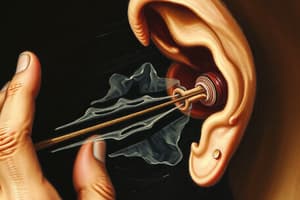
Auditory System
- Outer ear collects and amplifies sound, directs sound into the ear canal, and determines the direction and distance of sounds
- The S-shaped ear canal amplifies certain frequencies and protects the eardrum; glands produce cerumen to trap foreign substances
- The incus, malleus, and stapes (auditory ossicles) transmit sound vibrations from the eardrum to the inner ear
- Semicircular canals are three loop-shaped structures that maintain balance and spatial orientation in different planes
- The fluid-filled cochlea converts sound vibrations into neural signals
- The auditory tube (Eustachian tube) connects the middle ear to the nasopharynx, equalizing air pressure and draining fluid
- The tympanic membrane (eardrum) separates the external and middle ear
- Sound waves vibrate the tympanic membrane, which then vibrates the malleus
- The round window is were waves dissipate through
- At the Oval window the stapes vibrates, waves are detected by hair cells, which then transmit information to nerve cells transmit info to nerve cells, carry AP to auditory centre of brain.
- Cochlea Divisions:
- Upper scala vestibuli (vestibular duct)
- Middle cochlear duct
- Lower scala tympani
- Scala vestibuli and tympani are filled with perilymph fluid, which is high in Na and low in K concentrations
- The cochlear duct contains endolymph, which is high in K and low in Na
- The basilar membrane separates the cochlear duct and tympanic duct and contains the spiral organ
- The Organ of Corti is where sound waves are converted to action potentials by hair cells
- Hair cells, called stereocilia (one long kinocilia) move based on waves in endolymph fluid in ampullae (base of semi circular canals)
- Hair cells are embedded in a gelatinous structure called cupula
- Hairs normal = base line neurotransmitter release
- Hair movement = stimulate more or less release
- Towards kinocilia = more
- Head Movement Detection:
- Anterior: detects forward and backward head movement
- Posterior: detects head tilts towards shoulders
- Lateral: detects horizontal head movement (head shake)
- Vestibulo-Ocular Reflex (VOR): Semicircular canals play a key role, stabilizes vision by producing eye movements that counteract head movements
Cardiovascular System
- This system has three components: the heart, vessels, and blood
- The heart pumps to move blood, vessels transport blood, and blood carries gases
- The interventricular septum stops blood from mixing between ventricles
- The apex of the heart is where contractions start and move upwards
- The left ventricle wall is thicker to move blood forcefully
- Heart valves prevent backflow
- Heart Sounds:
- "Lub": AV valves closing (contraction)
- "Dub": aortic and pulmonary valves closing (relaxation)
- CUSPS prevent backflow by filling with blood
- Cardiac muscle cells (cardiomyocytes) allow for contraction and relaxation
- Types of Cells:
- Contractile cells are striated due to myofilament formatting
- Needs calcium to release from SR/ECF, ATP required
- Nodal/conducting cells behave like neurons
- Don't contract, take AP through heart, self excitable (create own AP)
- Action Potential:
- Moves through gap junctions and intercalated discs lock cells together
- AP influx on triggers depolarization, K channels cause repolarization
- Occurs every 0.8 seconds (70 beats/min)
- AP created by SA node moves through gap junctions to contractile cells in the atria
- The AV node slows the AP to allow atrial cardiomyocytes to finish contracting
- The AP goes to the atrioventricular bundle before spreading to both ventricles
- Subendocardial branches excite ventricular cardiomyocytes to contract from the bottom up
- Max HR equation: 220 - your age in years
- PSNS releases Ach to decrease heart rate below 100, decreasing Na and Ca permeability, and increasing K permeability
- SNS releases norepinephrine to increase HR, increasing Na and Ca permeability, and decreasing K permeability
- ECG:
- P wave indicates atrial depolarization
- QRS wave indicates ventricular depolarization
- T wave indicates ventricular repolarization
Cardiac Cycle
- Systole: cardiomyocytes contracting
- Diastole: cardiomyocytes relaxing
- Cardiac cycle refers to series of events that occurs with every heartbeat
- 5 Phases of the Cardiac Cycle:
- Isovolumetric ventricular systole; ventricles contract without volume change
- Ventricular systole; blood moves into the aorta and pulmonary arteries
- Isovolumetric ventricular diastole; ventricles relax without volume change
- Late ventricular diastole; ventricles relax and fill with blood
- Atrial systole; atria contract, moving blood into ventricles
- Volumes:
- End Systolic Volume (ESV): amount of blood remaining in the ventricles after systole
- End Diastolic Volume (EDV): amount of blood in ventricles before ventricular contraction
- Stroke Volume (SV): amount of blood pumped out with each heartbeat
- SV = EDV - ESV
- Ventricular systole and diastole have two phases, the first phase has no volume change, the second phase blood refills vesicles
Blood Vessels
- The aorta is the largest artery
- Blood flows through arteries into arterioles, then capillaries, and finally venules
- Total blood volume is 4-6 L
- The pulmonary circuit makes up 15% of vessels with the systemic having 85%
- Vessels transport; oxygenated and deoxygenated blood, and are involved in gas exchange
- Anatomy of a Blood Vessel:
- Tunica Externa: fibrous connective tissue, protects the vessels
- Tunica Media: smooth muscle and elastin which allows vessel to stretch
- Tunica Interna: endothelial cells, aka Endothelial cells
- Blood pressure is highest in arteries and lowest in veins
- Vessel Types:
- Arteries carry blood away from the heart, have large diameters, and contain lots of elastin maintaining higher pressure during systole and lower during diastole
- Arterioles (resistance vessels) have a lot of smooth muscle that can contract or relax to stimuli
- Capillaries (smallest blood vessels) act as exchange vessels (O2 leaves into the systemic circuit), hormones leave and bind to receptors
- Venules comes next, pressure is lower than capillaries to allow for flow
- Veins are capacitance vessels allowing back blood flow, containing valves to allow in ONE direction
Blood Flow
- Arterioles control the amount of blood flow to tissues
- Blood flow increases bloody supply to active tissues and decrease it to inactive tissues, maintains blood pressure and decrease heat loss
- Blood flow equation: Blood flow = pressure gradient/resistance
- Factors affecting resistance: viscosity, vessel length, lumen radius
- Capillary walls are a single cell thick and used for exchange.
- Have luminal and basolateral membranes
- Substances use trans cellular transport (enters and exits cell), simple diffusion and Diffusion using channels down a gradient
- Active transport in a gradient and facilitated diffusion proteins are also used
- Small ions and substances move in intercellular clefts in paracellular transport
- Continuous capillaries are less permeable, fenestrated capillaries more
- Intercellular cleft permeability is related to proteins holding endothelial cells together
- During bulk flow, fluid moves out of a capillary through filtration
- Starling forces promote return of fluid back in capillary
Vasoconstriction and Vasodilation
- Regulatory systems control flow.
- Local regulation involves changes within organs or tissues.
- Stimuli change blood flow as needed.
- Humoral regulation involves blood substances
- Substances change vessel radius by binding to receptors.
- Some cause vasoconstriction; others, vasodilation.
- Extrinsic mechanisms: regulation or hormones are produced elsewhere.
- Neural regulation is when neurons from the sympathetic nervous system innervate smooth muscle cells in tunica media.
- Sympathetic releases norepinephrine.
- Extrinsic mechanisms: Neurons originate outside the tissue/organ.
- Local Regulation:
- Myogenic theory causes Rise in BP (vasoconstriction) and Drop in BP (vasodilation)
- Metabolic theory causes Not enough vasodilator metabolites (promotes vasoconstriction or arterioles)
- Increases metabolism (causes vasodilation)
- Negative feedback loop using baroreceptor reflex maintains BP homeostasis.
- Detect changes in BP
- Send to the medulla oblongata of your brainstem.
- Initiates negative feedback loop to adjust heart rate, stroke volume, and vessel diameter to stabilize MAP.
- Mean Arterial Pressure (MAP): diastolic pressure + 1/3 (systolic pressure-diastolic pressure)
- Hypertension: high BP damaging vessels leads to heart diseases and stroke
- Hypotension: Low BP and causes inadequate blood flow to organs
Kidney
-
Kidney functions: Removing nonessential substances, and recovering essential substances
-
Kidney Location:
- Posterior to abdomen, outside abdominal cavity
- Sandwiched between member and that line abdomen
-
Precipitation and crystal action of higher than usual concentrations of minerals and ions
-
Nephron:
- Functional unit of kidney
- Made of two basic structures - renal corpuscle, and tubule
-
Bowman's capsule located outside of renal corpuscle
-
Nephrons ability carefully select item to be excreted as urine
-
Nephron positioning*:
- Cortical nephron 80%
- Juxtamedullary 20%
-
Blood: The kidneys receive almost 20% of total cardiac output which are filtered by nephrons, maintaining blood volume and ion balance
-
Plasma: Water with dissolves macromolecules (proteins, amino acids, hormones and glucose).
-
Blood Filtration refers to movement of fluid from glomerulus into bowman's capsule.
-
Reabsorption refers to movement of filtrate back into capillary bed
-
Secretion is putting items dissolved in blood, added to filtrate.
-
To renal vein, to bladder and outside the body
Filtration
- Glomerulus contains: many fenestrations (pores), making it leaky
- The Filtration Barriers are that the:
- Size of fenestrations, spaces and podocytes are small
- Endothelial cells have Basal lamina.
- Mostly affected by amount of blood floe into kidney and BP
- If total renal blood flow of BP increases so would net filtration.
- Net filtration pressure = (PGC+πBC) - (PBC + πGC)
- Filtration coefficient: influenced by surface area of glomerular capillaries and permeability
Glomerulus Filtration Rate
-
In tubuloglomerular feedback:
- GFR INCREASES - BP increases
-
Macula densa cells in the ascending loop of henle detect sodium levels and fast filtration
-
Cells will release adenosine and cause vasoconstriction
-
Calculations
-
Substances are processed in a different way ###Tubal Transport
-
Tubal Transport
-
Different channels and transporters are used.
-
Channels are small protein lined pores, passive that use and electrochemical gradient
- Uniporters permits movement of a single molecule through membrane, protein carriers
-
Symporters two or more molecules at the same time in the same direction co transport- Antiporters two molecules at the same time in the opposite direction, one must move to make other move
- Primary active transport needs ATP, against gradient
-
Proximal Tubule:
- 3 Na interstitial
- 2 k tubule o glucose,
-
Descending Limb:
- Responds to hormones
-
Ascending Limb:
-
Reabsorbs NA, Cl, K paracellular
-
Distal Convoluted - reabsorbs, Ca and Na
-
Collecting Ducts:
- They are A intercenated -Type B- respond to plasma PH
-
Most of the reabsorption and filtering is done in the proximal tubule
Studying That Suits You
Use AI to generate personalized quizzes and flashcards to suit your learning preferences.