Podcast
Questions and Answers
What is the capacity of the gallbladder?
What is the capacity of the gallbladder?
- 40-60 cc (correct)
- 70-90 cc
- 20-30 cc
- 100-120 cc
What is the length of the gallbladder?
What is the length of the gallbladder?
- 7-8 inches
- 3-4 inches (correct)
- 5-6 inches
- 2-3 inches
What is the function of the gallbladder?
What is the function of the gallbladder?
- Secretes digestive enzymes
- Stores and concentrates bile (correct)
- Produces bile
- Absorbs nutrients
What is the branch of the right hepatic artery that supplies the gallbladder?
What is the branch of the right hepatic artery that supplies the gallbladder?
What is the vein that drains the gallbladder?
What is the vein that drains the gallbladder?
What is the nerve plexus that supplies sympathetic fibers to the gallbladder?
What is the nerve plexus that supplies sympathetic fibers to the gallbladder?
What is the duct that forms by the union of the right and left hepatic ducts?
What is the duct that forms by the union of the right and left hepatic ducts?
What is the part of the common bile duct that lies behind the superior part of the duodenum?
What is the part of the common bile duct that lies behind the superior part of the duodenum?
What is the structure that the common bile duct descends in?
What is the structure that the common bile duct descends in?
What is the structure that the supraduodenal part of the common bile duct lies in front of?
What is the structure that the supraduodenal part of the common bile duct lies in front of?
Flashcards are hidden until you start studying
Study Notes
Liver Anatomy
- Location: occupies the whole right hypochondrium, a part of epigastrium, and a part of left hypochondrium
- Weight: 1/50 of body weight in adults (1500gm) and 1/20 of body weight in infants
- Functions:
- Exocrine (bile) and endocrine (albumin, prothrombin, and fibrinogen) gland
- Secretion of bile and bile salts
- Metabolism of carbohydrates, fats, and proteins
- Formation of heparin and anticoagulant substances
- Detoxification
- Storage of glycogen and vitamins
- Activation of vitamin D
Surface Anatomy of the Liver
- Upper border: presented by a line joining the following points:
- Left 5th intercostal space below the left nipple at the left midclavicular line
- Point at the xiphisternal junction
- Right 5th costal cartilage at the midclavicular line
- Right 7th rib at the midaxillary line
- Right border: convex line joining two points:
- Point at the 7th rib at the midaxillary line
- Point at the 11th rib at the midaxillary line
- Lower border: presented by a line joining the following points:
- Left 5th intercostal space below the left nipple at the left midclavicular line
- Left costal margin at the tip of the left 8th costal cartilage
- Point on the midline at the transpyloric plane
- Right 11th rib at the midaxillary line
- Shape: wedge shape with its base to the right
- Surfaces: five surfaces, including the base or right, superior, anterior, posterior, and inferior or visceral surfaces
- Borders: ill-defined except for the inferior border
Surfaces, Relations, and Impressions
- Anterior surface relations:
- Diaphragm separating it from the right and left pleura and lung
- Right costal cartilage
- Xiphoid process
- Anterior abdominal wall
- Superior surface relations:
- Diaphragm separating it from the pericardium and heart (in the middle) and pleurae and lungs (on each side)
- Right lateral surface (base) relations:
- Diaphragm separating it from the right lung and pleura and ribs from 7-11 of the right side
- Posterior surface relations:
- Bare area: triangular area related to the diaphragm (devoid of peritoneum)
- Groove for the IVC
- Caudate lobe: related to the diaphragm, projecting downwards to form the quadrate process separating the IVC from the porta hepatis
- Fissure for the ligamentum venosum (obliterated ductus venosus in embryology)
- Esophageal notch
Inferior Surface of the Liver (Visceral Surface)
- Relations:
- Right kidney
- Right suprarenal gland
- Right colic flexure
- 2nd part of the duodenum
- Gallbladder fossa
- Quadrate lobe: related to the pyloroduodenal junction and transverse colon
- Fissure for the ligamentum teres (obliterated left umbilical vein)
- Tuber omentale: related to the lesser omentum
Lobes of the Liver
- Anatomical division:
- The liver is divided into a smaller left and a larger right lobe by:
- Falciform ligament: anteriorly
- Fissure for the ligamentum venosum: posteriorly
- Fissure for the ligamental teres: inferiorly
- The liver is divided into a smaller left and a larger right lobe by:
- The right lobe contains two additional lobes: the caudate and quadrate lobes
- Functional division:
- Caudate lobe:
- Located in the posterior surface of the right lobe
- Relations: inferior to the porta hepatis, right to the fossa for the inferior vena cava, and left to the fossa for the ligamentum venosum
- Quadrate lobe:
- Located on the inferior surface of the right lobe
- Relations: anterior to the inferior margin of the liver, superior to the porta hepatis, right to the fossa for the gallbladder, and left to the fissure for the ligamentum teres
- Caudate lobe:
Porta Hepatis
- It is the hilum of the liver
- Located on the postero-inferior surface of the liver
- Lies between the caudate and quadrate lobes
- Structures passing through it:
- DAV (from before backwards): right and left hepatic ducts, right and left branches of the hepatic artery, and portal vein
- Lymphatics, lymph nodes, and autonomic fibers
Peritoneum of the Liver
- The liver is covered by peritoneum (intraperitoneal organ) except at the bare area
- Inferior surface covered with peritoneum of the greater sac except for the porta hepatis, gallbladder, and ligamentum teres fissure
- Right lateral surface covered with peritoneum, related to the diaphragm, which separates it from the right pleura, lung, and ribs from 7-11
Ligaments of the Liver
- Falciform ligament of the liver
- Ligamentum teres hepatis
- Coronary ligament
- Right triangular ligament
- Left triangular ligament
- Hepatogastric ligament
- Hepatoduodenal ligament
- Ligamentum venosum
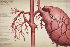
Gallbladder and Biliary System
- Parts:
- Fundus: projects below the inferior margin of the liver, lies opposite the tip of the right 9th costal cartilage, related anteriorly to the anterior abdominal wall and posteriorly to the transverse colon
- Body: related anteriorly to the liver and posteriorly to the 1st part of the duodenum
- Neck: continuous with the cystic duct, has the same relation as the body
- Peritoneal covering:
- Fundus: completely covered by peritoneum
- Body and neck: only covered posteriorly
- Function: stores and concentrates bile
- Artery: cystic branch of the right hepatic artery
- Vein: cystic vein that drains into the right branch of the portal vein
- Lymph drainage: cystic lymph nodes, then to hepatic and finally to celiac lymph nodes
- Nerves: sympathetic from the celiac nerve plexus (T7-9), parasympathetic through the vagus nerves, and pain through the right phrenic nerve with referral to the stomach, inferior angle of the right scapula, and right shoulder
Biliary Ducts
- Right and left hepatic ducts:
- Emerge from the porta hepatis
- Drain bile from the right and left lobes of the liver
- Unite to form the common hepatic duct
- Common hepatic duct:
- One and a half inches in length
- Formed by the union of the two hepatic ducts
- Unites with the cystic duct to form the common bile duct
- Common bile duct:
- Formation: formed below the porta hepatis by the union of the cystic and common hepatic ducts
- Descends in the free edge of the lesser omentum and divides into three parts:
- Supra duodenal part
- Retroduodenal part
- Infra duodenal part
Studying That Suits You
Use AI to generate personalized quizzes and flashcards to suit your learning preferences.