Podcast
Questions and Answers
Which of these nematode species are associated with Parasitic Gastro-Enteritis (PGE)?
Which of these nematode species are associated with Parasitic Gastro-Enteritis (PGE)?
- Haemonchus (correct)
- Ascaris
- Trichostrongylus (correct)
- Oesophagostomum (correct)
What percentage of prevalence does Haemonchus have?
What percentage of prevalence does Haemonchus have?
90%
What are the clinical signs of PGE?
What are the clinical signs of PGE?
Thin, no appetite, emaciated
The prepatent period for Haemonchus contortus is 5-6 weeks.
The prepatent period for Haemonchus contortus is 5-6 weeks.
Where do Trichostrongylus and Cooperia reside in ruminants?
Where do Trichostrongylus and Cooperia reside in ruminants?
Haemonchus is also known as the '__________' worm.
Haemonchus is also known as the '__________' worm.
What is the main route of infection for Haemonchus contortus?
What is the main route of infection for Haemonchus contortus?
What characteristic features are associated with Trichostrongylus?
What characteristic features are associated with Trichostrongylus?
Match the following types of strongyles with their characteristics:
Match the following types of strongyles with their characteristics:
Flashcards
Ruminant Strongyles
Ruminant Strongyles
A group of parasitic nematodes that infect the digestive tracts of ruminant animals, causing parasitic gastro-enteritis (PGE).
Life Cycle of Ruminant Strongyles
Life Cycle of Ruminant Strongyles
The process by which ruminant strongyles develop from a larval stage (L1) to an adult stage, involves multiple stages, including L2, L3, and L4.
Parasitic Gastro-Enteritis (PGE)
Parasitic Gastro-Enteritis (PGE)
A disease complex caused by multiple nematode species (mostly strongyles), characterized by diarrhea and weight loss (clinical), or reduced production (subclinical).
Haemonchus
Haemonchus
Signup and view all the flashcards
Trichostrongylus
Trichostrongylus
Signup and view all the flashcards
Oesophagostomum
Oesophagostomum
Signup and view all the flashcards
Cooperia
Cooperia
Signup and view all the flashcards
Diagnosis of PGE
Diagnosis of PGE
Signup and view all the flashcards
Clinical Signs (PGE)
Clinical Signs (PGE)
Signup and view all the flashcards
Post-mortem Findings
Post-mortem Findings
Signup and view all the flashcards
Fecal Egg Count
Fecal Egg Count
Signup and view all the flashcards
Haematocrit
Haematocrit
Signup and view all the flashcards
Prevalence (Strongyles)
Prevalence (Strongyles)
Signup and view all the flashcards
Prepatent period
Prepatent period
Signup and view all the flashcards
Epidemiology
Epidemiology
Signup and view all the flashcards
Study Notes
Strongylida
- Strongylida is a group of parasitic nematodes.
- Ruminant strongyles are a type of strongyle affecting ruminant animals.
- The life cycle of ruminant strongyles involves parasitic stages (L3-L4-Adult) and pre-parasitic stages (L1, L2).
- The parasitic stage lasts for several weeks.
- The pre-parasitic stage, typically takes seven days.
Ruminant Strongyles
- Strongylida can affect ruminants.
- Ruminant Strongyles include the following:
- Haemonchus contortus (90% prevalence in ruminants)
- Trichostrongylus (60% prevalence in ruminants)
- Oesophagostomum (30% prevalence in ruminants)
- Cooperia (infects the small intestine)
- Parasitic Gastro-Enteritis (PGE) is a disease complex associated with numerous nematode species (mostly strongyles) potentially occurring singly or in combination
Epidemiology and Diagnosis of PGE
- PGE is primarily diagnosed by diarrhea and weight loss (clinical disease) or suboptimal production (subclinical disease).
- Risk factors for PGE include: number of infected animals, age (typically younger than 6 months, but older animals can be affected if stressed), high stocking density, and anthelmintic drenching practices (type, method, dose, potency, and expiry date).
- Clinical signs include loss of protein and cellular components leading to thinness, lack of appetite, and emaciation.
Life Cycle of Ruminant Strongyles
- Strongyles develop from larval stages (L1, L2, L3, L4, L5)
- Infection occurs via ingestion of contaminated grass or herbage.
- Exsheathed L3 invade the host.
- L2 hatches after 7 days and transforms into L3.
- L3 transforms into the adult stage.
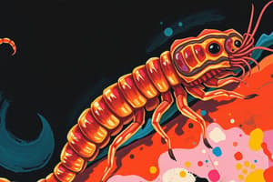
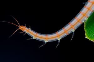
Haemonchus contortus
- This worm is also known as the "barber's pole" worm, reaching 2-3 cm in length.
- Found predominantly in the abomasum of sheep and cattle.
- Infection occurs by ingestion of infected grasses.
- A pre-patent period of 3-4 weeks exists.
- Adults suck blood, causing anemia and hypoalbuminemia.
Trichostrongylus and Cooperia
- These nematodes dwell in the small intestine of ruminants (0.5 cm in length).
- L3 infects by burrowing into the epithelium, progressing to L4 emerging into the lumen, causing enteritis, villi erosion, and protein loss.
Oesophagostomum
- This species of worm (1.2 cm long) inhabits the large intestine of ruminants.
- Infection occurs by ingestion of infected grasses.
- L3 larvae penetrate the mucosa causing inflammation and nodule formation.
- Subsequently, L4 causes ulceration.
Diagnosis of PGE
- Post-mortem findings: Bite marks, pinpoint hemorrhage (e.g., in Haemonchus contortus), and pale organs (anemia) with watery blood in fresh carcasses.
- Total worm counts: Assessment of parasite burden after post-mortem examination.
- Worm identification:
- Feces: McMaster technique to count eggs (Clinical PGE: FEC >1500 epg; Subclinical PGE: FEC 500-1500 epg).
- Blood: Haematocrit Centrifugation Technique (HCT) to determine packed cell volume (PCV) for anemia and hypoproteinemia.
Pathogenesis of Haemonchus contortus
- Anemia: High worm burden leads to substantial blood loss, reducing hemoglobin and iron absorption.
- Malabsorption/Maldigestion: Disruption of gastric function (low HCl, pH increase), which impairs protein digestion and nutrient intake. The cells producing HCl are lost thus producing inadequate amounts of this substance causing protein indigestion
Pathogenesis of Trichostrongylus
- Malabsorption: Villus damage and atrophy caused by larval migration and penetration disrupt nutrient absorption. Increased shedding from the villi and inadequate mitosis contribute to the problem
Pathogenesis of Oesophagostomum
- Inflammation and nodules: L3 larvae penetrate into the intestinal mucosa, leading to inflammation, nodule formation, and hemorrhages.
- Larval migration: L3 are initially found in the esophagus but migrate into the large intestine.
- Reduced tissue quality: The body's response to L3 larvae is inflammation in this instance, but it is possible for other conditions to adversely effect this as well.
General Nematode Characteristics
- Unisexual, females larger than males.
- Five larval stages (L1–L5).
- Majority reside in the gastrointestinal tract.
- Classified into Rhabditida, Strongylida, Enoplida, Ascaridida, Spirurida, and Filarida.
- Life cycles vary significantly based on the specific group.
Studying That Suits You
Use AI to generate personalized quizzes and flashcards to suit your learning preferences.