Podcast
Questions and Answers
What occurs during the patellar reflex?
What occurs during the patellar reflex?
- Sensory neurons synapse directly onto alpha motor neurons. (correct)
- The muscle spindle remains inactive during reflexive actions.
- The quadriceps muscle relaxes and the hamstrings contract.
- Sensory neurons activate spinal interneurons to inhibit motor neurons.
Which motor neurons are responsible for contracting extrafusal muscle fibers?
Which motor neurons are responsible for contracting extrafusal muscle fibers?
- Gamma motor neurons
- Alpha motor neurons (correct)
- Type Ia sensory fibers
- Interneurons
How does reciprocal inhibition function during muscle contraction?
How does reciprocal inhibition function during muscle contraction?
- Both muscle groups contract equally to maintain balance.
- Motor neurons of the agonist and antagonist simultaneously activate.
- The muscle spindle prevents contraction of both muscle groups.
- Antagonist muscles are inhibited to allow for agonist contraction. (correct)
What is the result of damage to the corticospinal tracts?
What is the result of damage to the corticospinal tracts?
Which type of reflex involves only one synapse?
Which type of reflex involves only one synapse?
What is the primary role of muscle spindles?
What is the primary role of muscle spindles?
What occurs during alpha-gamma coactivation?
What occurs during alpha-gamma coactivation?
Which sensory fibers are activated when quadriceps are stretched?
Which sensory fibers are activated when quadriceps are stretched?
What effect does contraction of extrafusal fibers without adjusting intrafusal fibers have?
What effect does contraction of extrafusal fibers without adjusting intrafusal fibers have?
What does hypertonia indicate in terms of motor neuron activity?
What does hypertonia indicate in terms of motor neuron activity?
What is the primary function of extrafusal muscle fibers?
What is the primary function of extrafusal muscle fibers?
Which type of fiber is primarily responsive to the degree of muscle stretch?
Which type of fiber is primarily responsive to the degree of muscle stretch?
Which of the following illustrates a characteristic of gamma motor neurons?
Which of the following illustrates a characteristic of gamma motor neurons?
What mechanism triggers depolarization in the stretch reflex pathway?
What mechanism triggers depolarization in the stretch reflex pathway?
What type of sensory fiber responds to both stretch and the velocity of that stretch?
What type of sensory fiber responds to both stretch and the velocity of that stretch?
What is the main role of intrafusal fibers in muscle spindles?
What is the main role of intrafusal fibers in muscle spindles?
Which type of intrafusal fiber detects both the degree of stretch and the speed of that stretch?
Which type of intrafusal fiber detects both the degree of stretch and the speed of that stretch?
How do sensory neurons respond when mechanically gated ion channels open?
How do sensory neurons respond when mechanically gated ion channels open?
What type of muscle fibers are responsible for muscle contractions during physical activities?
What type of muscle fibers are responsible for muscle contractions during physical activities?
Which part of the intrafusal fiber innervation maintains the sensitivity during muscle activation?
Which part of the intrafusal fiber innervation maintains the sensitivity during muscle activation?
What is the purpose of alpha-gamma coactivation during muscle movement?
What is the purpose of alpha-gamma coactivation during muscle movement?
Which statement accurately describes the relationship between antagonistic muscles and reciprocal inhibition?
Which statement accurately describes the relationship between antagonistic muscles and reciprocal inhibition?
What is a consequence of damage to the corticospinal tracts?
What is a consequence of damage to the corticospinal tracts?
During the patellar reflex, what is the role of type Ia and type II sensory fibers?
During the patellar reflex, what is the role of type Ia and type II sensory fibers?
What happens to sensory neuron activity when intrafusal fibers are slack?
What happens to sensory neuron activity when intrafusal fibers are slack?
How do alpha motor neurons differ from gamma motor neurons in muscle function?
How do alpha motor neurons differ from gamma motor neurons in muscle function?
What triggers the patellar reflex and the subsequent response of the quadriceps?
What triggers the patellar reflex and the subsequent response of the quadriceps?
What is the primary role of gamma motor neurons in muscle spindles?
What is the primary role of gamma motor neurons in muscle spindles?
Which aspect of muscle spindle function is most critical during physical activity?
Which aspect of muscle spindle function is most critical during physical activity?
What is the primary function of gamma motor neurons in relation to muscle spindles?
What is the primary function of gamma motor neurons in relation to muscle spindles?
Which type of sensory fiber primarily conveys information about the degree of muscle stretch?
Which type of sensory fiber primarily conveys information about the degree of muscle stretch?
How do intrafusal fibers contribute to proprioception?
How do intrafusal fibers contribute to proprioception?
What is the main structural difference between nuclear bag fibers and nuclear chain fibers?
What is the main structural difference between nuclear bag fibers and nuclear chain fibers?
What initiates depolarization in sensory neurons during the stretch reflex?
What initiates depolarization in sensory neurons during the stretch reflex?
Which of the following accurately describes extrafusal muscle fibers?
Which of the following accurately describes extrafusal muscle fibers?
What is the role of Type Ia sensory fibers in muscle spindles?
What is the role of Type Ia sensory fibers in muscle spindles?
What is a key function of intrafusal fibers in muscle spindles?
What is a key function of intrafusal fibers in muscle spindles?
Which characteristic distinguishes nuclear bag fibers from other intrafusal fibers?
Which characteristic distinguishes nuclear bag fibers from other intrafusal fibers?
What happens when mechanically gated ion channels open in sensory neurons?
What happens when mechanically gated ion channels open in sensory neurons?
What is the primary function of nuclear chain fibers compared to other types of intrafusal fibers?
What is the primary function of nuclear chain fibers compared to other types of intrafusal fibers?
What mechanism leads to depolarization in sensory neurons during a muscle stretch?
What mechanism leads to depolarization in sensory neurons during a muscle stretch?
What is the role of gamma motor neurons in relation to intrafusal fibers?
What is the role of gamma motor neurons in relation to intrafusal fibers?
Which type of sensory fiber primarily responds to the degree of muscle stretch?
Which type of sensory fiber primarily responds to the degree of muscle stretch?
In the context of the stretch reflex, what role do type Ia sensory fibers play?
In the context of the stretch reflex, what role do type Ia sensory fibers play?
How does the structure of intrafusal fibers differ from extrafusal fibers?
How does the structure of intrafusal fibers differ from extrafusal fibers?
What type of muscle fiber primarily mediates the proprioceptive input from muscle spindles?
What type of muscle fiber primarily mediates the proprioceptive input from muscle spindles?
What is the function of the polar ends of intrafusal fibers?
What is the function of the polar ends of intrafusal fibers?
Which groups of muscle fibers are responsible for generating movement by pulling on tendons?
Which groups of muscle fibers are responsible for generating movement by pulling on tendons?
Which characteristic best describes nuclear bag fibers?
Which characteristic best describes nuclear bag fibers?
What is the primary consequence of alpha-gamma coactivation during muscle movement?
What is the primary consequence of alpha-gamma coactivation during muscle movement?
What is the primary function of inhibitory interneurons in the reflex arc?
What is the primary function of inhibitory interneurons in the reflex arc?
What happens to muscle spindle function when intrafusal fibers are too slack?
What happens to muscle spindle function when intrafusal fibers are too slack?
What differentiates the activity of gamma motor neurons from alpha motor neurons?
What differentiates the activity of gamma motor neurons from alpha motor neurons?
Which mechanism is primarily responsible for muscle relaxation of antagonistic muscles during contraction?
Which mechanism is primarily responsible for muscle relaxation of antagonistic muscles during contraction?
How does damage to the corticospinal tracts affect gamma motor neuron activity?
How does damage to the corticospinal tracts affect gamma motor neuron activity?
Which characteristic is crucial for the effective performance of the patellar reflex?
Which characteristic is crucial for the effective performance of the patellar reflex?
What is the role of type Ia sensory fibers in the stretch reflex mechanism?
What is the role of type Ia sensory fibers in the stretch reflex mechanism?
During muscle contraction, what effect does activation of alpha motor neurons have on antagonistic muscle groups?
During muscle contraction, what effect does activation of alpha motor neurons have on antagonistic muscle groups?
What is primarily responsible for ensuring precise control in muscle movements?
What is primarily responsible for ensuring precise control in muscle movements?
What is the primary role of type Ia and type II sensory fibers during the patellar reflex?
What is the primary role of type Ia and type II sensory fibers during the patellar reflex?
What is a distinct effect of reciprocal inhibition during muscle contraction?
What is a distinct effect of reciprocal inhibition during muscle contraction?
What occurs as a result of coactivation of alpha and gamma motor neurons?
What occurs as a result of coactivation of alpha and gamma motor neurons?
Which statement best describes the implications of upper motor neuron lesions?
Which statement best describes the implications of upper motor neuron lesions?
What role do gamma motor neurons play during muscle contraction?
What role do gamma motor neurons play during muscle contraction?
Which of the following describes the outcome when extrafusal fibers contract without concurrent adjustments to intrafusal fibers?
Which of the following describes the outcome when extrafusal fibers contract without concurrent adjustments to intrafusal fibers?
What is the main function of inhibitory interneurons within the reflex arc?
What is the main function of inhibitory interneurons within the reflex arc?
How does the sensitivity of muscle spindles change during cocontraction of muscles?
How does the sensitivity of muscle spindles change during cocontraction of muscles?
In the context of muscle spindle activation, what is the primary function of the intrafusal fibers?
In the context of muscle spindle activation, what is the primary function of the intrafusal fibers?
Which type of intrafusal fiber is specifically designed to detect both the degree of muscle stretch and the rate of that stretch?
Which type of intrafusal fiber is specifically designed to detect both the degree of muscle stretch and the rate of that stretch?
How do Type Ia and Type II sensory fibers differ in their response to muscle stretch?
How do Type Ia and Type II sensory fibers differ in their response to muscle stretch?
What is the primary structural characteristic that differentiates nuclear bag fibers from nuclear chain fibers?
What is the primary structural characteristic that differentiates nuclear bag fibers from nuclear chain fibers?
What function do gamma motor neurons serve in relation to intrafusal fibers?
What function do gamma motor neurons serve in relation to intrafusal fibers?
When a muscle is stretched, what initial event occurs at the level of sensory neurons?
When a muscle is stretched, what initial event occurs at the level of sensory neurons?
What distinguishes extrafusal muscle fibers from intrafusal muscle fibers in terms of function?
What distinguishes extrafusal muscle fibers from intrafusal muscle fibers in terms of function?
In the context of muscle activity, what would happen if intrafusal fibers were not contracted by gamma motor neurons?
In the context of muscle activity, what would happen if intrafusal fibers were not contracted by gamma motor neurons?
What triggers action potentials to travel through sensory fibers to the spinal cord during the stretch reflex?
What triggers action potentials to travel through sensory fibers to the spinal cord during the stretch reflex?
Which of the following best describes the role of muscle spindles in movement?
Which of the following best describes the role of muscle spindles in movement?
What motivates the activation of the stretch reflex when a muscle is rapidly stretched?
What motivates the activation of the stretch reflex when a muscle is rapidly stretched?
What is the primary significance of reciprocal inhibition during muscle contraction?
What is the primary significance of reciprocal inhibition during muscle contraction?
What is the primary consequence of gamma motor neuron hyperactivity due to upper motor neuron lesions?
What is the primary consequence of gamma motor neuron hyperactivity due to upper motor neuron lesions?
Which best describes alpha-gamma coactivation?
Which best describes alpha-gamma coactivation?
What triggers the generation of action potentials in sensory neurons during stretching?
What triggers the generation of action potentials in sensory neurons during stretching?
In the context of the stretch reflex, which aspect of alpha motor neurons is essential for reflexive movement?
In the context of the stretch reflex, which aspect of alpha motor neurons is essential for reflexive movement?
Which characteristic of the patellar reflex distinguishes it from polysynaptic reflexes?
Which characteristic of the patellar reflex distinguishes it from polysynaptic reflexes?
What role do intracortical inhibitory interneurons play in motor neuron modulation?
What role do intracortical inhibitory interneurons play in motor neuron modulation?
How does damage to corticospinal tracts affect muscle spindle sensitivity?
How does damage to corticospinal tracts affect muscle spindle sensitivity?
In which manner do type Ia sensory fibers primarily contribute to the stretch reflex?
In which manner do type Ia sensory fibers primarily contribute to the stretch reflex?
What is the main function of intrafusal fibers in the context of muscle spindle activity?
What is the main function of intrafusal fibers in the context of muscle spindle activity?
Which statement is true regarding the roles of nuclear chain fibers compared to nuclear bag fibers?
Which statement is true regarding the roles of nuclear chain fibers compared to nuclear bag fibers?
How do gamma motor neurons contribute to the functionality of muscle spindles during contraction?
How do gamma motor neurons contribute to the functionality of muscle spindles during contraction?
What differentiates Type Ia sensory fibers from Type II fibers in terms of their sensitivity?
What differentiates Type Ia sensory fibers from Type II fibers in terms of their sensitivity?
Which component of muscle spindles is involved in detecting the speed of muscle stretch?
Which component of muscle spindles is involved in detecting the speed of muscle stretch?
What is the primary function of the mechanically gated ion channels on sensory neurons during the stretch reflex?
What is the primary function of the mechanically gated ion channels on sensory neurons during the stretch reflex?
Which aspect of intrafusal fibers enhances the brain's awareness of muscle positioning?
Which aspect of intrafusal fibers enhances the brain's awareness of muscle positioning?
What is a characteristic function of extrafusal fibers in human movement?
What is a characteristic function of extrafusal fibers in human movement?
Which type of muscle fiber is encased in a connective tissue capsule and functions as a proprioceptor?
Which type of muscle fiber is encased in a connective tissue capsule and functions as a proprioceptor?
What type of muscle fiber provides feedback about both the degree and speed of muscle stretch?
What type of muscle fiber provides feedback about both the degree and speed of muscle stretch?
How does the depolarization of sensory neurons occur in response to muscle stretch?
How does the depolarization of sensory neurons occur in response to muscle stretch?
Which type of intrafusal fiber is primarily responsible for detecting the speed of muscle stretch?
Which type of intrafusal fiber is primarily responsible for detecting the speed of muscle stretch?
What is the role of gamma motor neurons in relation to muscle spindles?
What is the role of gamma motor neurons in relation to muscle spindles?
How do Type Ia and Type II sensory fibers differ in their response to muscle stretching?
How do Type Ia and Type II sensory fibers differ in their response to muscle stretching?
What initiates the depolarization of sensory neurons during the stretch reflex?
What initiates the depolarization of sensory neurons during the stretch reflex?
Which characteristics differentiate extrafusal fibers from intrafusal fibers?
Which characteristics differentiate extrafusal fibers from intrafusal fibers?
Flashcards are hidden until you start studying
Study Notes
Stretch Reflex and Muscle Spindles
- The stretch reflex, also known as the muscle spindle reflex, involves proprioceptors that detect muscle stretch and tension.
- Two main types of skeletal muscle fibers exist: extrafusal and intrafusal muscle fibers.
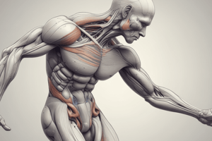
Extrafusal Muscle Fibers
- Extrafusal fibers are red, striated muscles attached to tendons; they generate movement by pulling on tendons that connect muscles to bones.
- They are responsible for muscle contractions and movement during activities.
Intrafusal Muscle Fibers
- Encased in a connective tissue capsule, intrafusal fibers are found within muscle spindles and serve as proprioceptors.
- They do not connect to tendons but inform the brain about the position and movement of muscles and joints in three-dimensional space.
Types of Intrafusal Fibers
- Intrafusal fibers include nuclear bag fibers (larger, with nuclei centrally located) and nuclear chain fibers (nuclei arranged in a chain).
- Nuclear bag fibers detect both the degree of muscle stretch and the speed (velocity) of that stretch, while nuclear chain fibers primarily detect just the degree of stretch.
Sensory and Motor Innervation
- Type Ia sensory fibers (annulo-spiral endings) and Type II fibers (flower spray endings) mediate proprioceptive input from intrafusal fibers.
- Type Ia fibers are sensitive to both stretch and velocity, while Type II fibers primarily respond to the degree of stretch.
- Gamma motor neurons are responsible for contracting intrafusal fibers at the polar ends, which maintains the sensitivity of muscle spindles during muscle activation.
Mechanism of the Stretch Reflex
- Stretching a muscle activates intrafusal fibers, leading to the opening of mechanically gated ion channels on sensory neurons, causing depolarization.
- This depolarization triggers action potentials that travel through the sensory fibers to the spinal cord.
- The reflex arc can involve monosynaptic pathways, where the sensory neuron directly synapses onto a motor neuron, causing muscle contraction.
Clinical Relevance
- In a clinical setting, the patellar reflex is a common demonstration of the stretch reflex.
- Tapping the patellar tendon stretches the quadriceps muscle, activating intrafusal fibers and generating reflexive contractions via type Ia and type II sensory fibers.
Reflex Characteristics
- The patellar reflex is classified as a monosynaptic reflex, occurring ipsilaterally (on the same side).
- The muscle spindle’s primary function is to prevent excessive muscle stretching by initiating a reflexive contraction when stretching occurs.### Muscle Contraction and Sensory Feedback
- Quadriceps muscles contract and shorten during movement.
- Sensory neurons can activate interneurons, which may inhibit motor neurons.
- Inhibitory interneurons prevent motor neurons from sending signals to skeletal muscles.
Muscle Opponents and Reciprocal Inhibition
- Antagonistic muscles, such as hamstring muscles, must relax to allow quadriceps to contract.
- Reciprocal inhibition: the process where contraction of one muscle is accompanied by relaxation of its antagonist.
Motor Neurons
- Alpha motor neurons innervate extrafusal muscle fibers for movement.
- Gamma motor neurons innervate intrafusal fibers, maintaining muscle spindle sensitivity.
Muscle Spindles and Stretch Reflex
- Stretching quadriceps activates sensory fibers (type 1a and 2) in muscle spindles.
- Sensory impulses travel to the spinal cord, synapsing with motor neurons and interneurons.
- Gamma motor neurons stimulate muscle spindles to maintain sensitivity during muscle contraction.
Coactivation of Motor Neurons
- Alpha-gamma coactivation occurs when both alpha motor neurons (for extrafusal fibers) and gamma motor neurons (for intrafusal fibers) are activated simultaneously.
- Enables precise muscle control and sensory feedback during muscle movement.
Role of Corticospinal Tracts
- Corticospinal tracts contain upper motor neurons that modulate gamma motor neuron activity.
- Damage to corticospinal tracts leads to lower inhibitory signals to gamma motor neurons, resulting in hyperactivation.
Clinical Relevance
- Upper motor neuron lesions result in gamma motor neuron hyperactivity, causing increased muscle tone (hypertonia) and spasticity.
Action Potentials in Sensory Neurons
- Stretching intrafusal fibers activates sensory neurons, leading to action potentials.
- Contracting extrafusal fibers without adjusting intrafusal fibers leads to slack, reducing action potentials in sensory neurons.
- Maintaining tautness in intrafusal fibers is crucial for adequate sensory feedback.
Importance of Dual Motor Activation
- Concurrent contraction of both extrafusal and intrafusal fibers ensures accurate proprioceptive feedback to the central nervous system.
Stretch Reflex and Muscle Spindles
- Proprioceptors detect muscle stretch and tension through the stretch reflex, also termed the muscle spindle reflex.
- Skeletal muscle fibers are categorized into extrafusal and intrafusal fibers, each serving distinct functions.
Extrafusal Muscle Fibers
- Composed of red, striated muscle tissue connected to tendons, enabling movement by pulling on bones.
- Responsible for voluntary muscle contractions and movements during physical activities.
Intrafusal Muscle Fibers
- Located within muscle spindles, these fibers are encased in a connective tissue capsule and act as proprioceptors.
- Function to relay information to the brain about muscle and joint position and movement in three-dimensional space.
Types of Intrafusal Fibers
- Two main types: nuclear bag fibers (larger, nuclei centrally located) and nuclear chain fibers (nuclei arranged in chains).
- Nuclear bag fibers detect both the stretch degree and its velocity; nuclear chain fibers mainly sense the degree of stretch.
Sensory and Motor Innervation
- Proprioceptive input from intrafusal fibers is conveyed by Type Ia sensory fibers (annulo-spiral endings) and Type II fibers (flower spray endings).
- Type Ia fibers respond to both stretch and its velocity, while Type II fibers focus on the magnitude of stretch.
- Gamma motor neurons contract intrafusal fibers at their polar ends to ensure spindle sensitivity during muscle activation.
Mechanism of the Stretch Reflex
- Muscle stretching activates intrafusal fibers, resulting in depolarization of sensory neurons via mechanically gated ion channels.
- This generates action potentials that travel through sensory fibers to the spinal cord.
- Stretch reflex involves monosynaptic pathways, where sensory neurons synapse directly onto motor neurons, leading to muscle contraction.
Clinical Relevance
- Common demonstrations of the stretch reflex include the patellar reflex, which is elicited by tapping the patellar tendon to stretch the quadriceps.
- Activation of intrafusal fibers in the quadriceps triggers reflexive contractions via Type Ia and Type II sensory fibers.
Reflex Characteristics
- The patellar reflex is a monosynaptic reflex, occurring ipsilaterally (on the same side).
- Muscle spindles prevent excessive stretching by initiating reflexive contractions upon detecting stretch.
Muscle Contraction and Sensory Feedback
- During movement, quadriceps muscles shorten and contract.
- Sensory neurons can activate inhibitory interneurons, which inhibit motor neuron signals to skeletal muscles.
Muscle Opponents and Reciprocal Inhibition
- Antagonistic muscles, like hamstrings, must relax to allow quadriceps contraction.
- Reciprocal inhibition ensures the contraction of one muscle is matched by the relaxation of its antagonist.
Motor Neurons
- Alpha motor neurons are responsible for activating extrafusal muscle fibers to produce movement.
- Gamma motor neurons activate intrafusal fibers, ensuring muscle spindle sensitivity.
Muscle Spindles and Stretch Reflex
- Stretching of quadriceps triggers sensory fibers in muscle spindles, sending impulses to the spinal cord.
- Impulses synapse with motor neurons and interneurons to facilitate reflex actions.
- Gamma motor neurons aid in maintaining spindle sensitivity during contraction.
Coactivation of Motor Neurons
- Alpha-gamma coactivation allows simultaneous activation of alpha and gamma motor neurons for precise muscle control.
- This coordination ensures effective sensory feedback during movements.
Role of Corticospinal Tracts
- Upper motor neurons in corticospinal tracts modulate the activity of gamma motor neurons.
- Damage to these tracts reduces inhibitory signals, leading to hyperactivation of gamma motor neurons.
Clinical Relevance
- Upper motor neuron lesions result in hyperactivity of gamma motor neurons, which can lead to increased muscle tone (hypertonia) and spasticity.
Action Potentials in Sensory Neurons
- Stretching intrafusal fibers triggers action potentials in sensory neurons.
- If extrafusal fibers contract without adjusting intrafusal fibers, it leads to slack and decreased action potentials, highlighting the need for taut intrafusal fibers for optimal sensory feedback.
Importance of Dual Motor Activation
- Simultaneous contraction of extrafusal and intrafusal fibers ensures accurate proprioceptive feedback to the central nervous system for enhanced movement control.
Studying That Suits You
Use AI to generate personalized quizzes and flashcards to suit your learning preferences.