Podcast
Questions and Answers
How does Stevens-Johnson Syndrome (SJS) typically begin?
How does Stevens-Johnson Syndrome (SJS) typically begin?
- Specific lower respiratory tract infection
- Nonspecific upper respiratory tract infection (correct)
- Progressive muscle weakness
- Sudden onset of severe blistering
A patient presents with a painful red to purple skin area that spreads quickly and is characterized by skin peeling without blistering. The affected area covers more than 30% of the body surface. Which condition is most likely?
A patient presents with a painful red to purple skin area that spreads quickly and is characterized by skin peeling without blistering. The affected area covers more than 30% of the body surface. Which condition is most likely?
- SJS/TEN Overlap
- Stevens-Johnson Syndrome (SJS)
- Toxic Epidermal Necrolysis (TEN) (correct)
- Erythema Multiforme (EM)
Which of the following best describes the percentage of body surface area (BSA) detachment in SJS/TEN overlap?
Which of the following best describes the percentage of body surface area (BSA) detachment in SJS/TEN overlap?
- 10%-30% BSA detachment (correct)
- Less than 10% BSA detachment
- No BSA detachment
- More than 30% BSA detachment
Which of the following signs is indicative of Darier's sign?
Which of the following signs is indicative of Darier's sign?
What is the main immunological process believed to be involved in the pathogenesis of Stevens-Johnson Syndrome (SJS)?
What is the main immunological process believed to be involved in the pathogenesis of Stevens-Johnson Syndrome (SJS)?
What is a common diagnostic finding in a skin biopsy of a patient with Stevens-Johnson Syndrome (SJS)?
What is a common diagnostic finding in a skin biopsy of a patient with Stevens-Johnson Syndrome (SJS)?
Which of the following is NOT typically associated with a poor prognosis in Stevens-Johnson Syndrome (SJS)?
Which of the following is NOT typically associated with a poor prognosis in Stevens-Johnson Syndrome (SJS)?
Which of the following is a common long-term complication of Stevens-Johnson Syndrome (SJS) and Toxic Epidermal Necrolysis (TEN)?
Which of the following is a common long-term complication of Stevens-Johnson Syndrome (SJS) and Toxic Epidermal Necrolysis (TEN)?
What is the primary goal of supportive care in the treatment of Stevens-Johnson Syndrome (SJS)?
What is the primary goal of supportive care in the treatment of Stevens-Johnson Syndrome (SJS)?
A patient presents with SJS. Which medication class is most likely to be implicated as a causative agent?
A patient presents with SJS. Which medication class is most likely to be implicated as a causative agent?
What is the Nikolsky sign?
What is the Nikolsky sign?
A patient is diagnosed with Toxic Epidermal Necrolysis (TEN). What is the most important first step in management?
A patient is diagnosed with Toxic Epidermal Necrolysis (TEN). What is the most important first step in management?
When assessing a patient with suspected SJS, where should the rash be evaluated?
When assessing a patient with suspected SJS, where should the rash be evaluated?
Which of the following best characterizes the typical skin lesions observed in Stevens-Johnson Syndrome (SJS)?
Which of the following best characterizes the typical skin lesions observed in Stevens-Johnson Syndrome (SJS)?
Which condition is Braverman's sign associated with?
Which condition is Braverman's sign associated with?
Which of the following is a recognized trigger for Stevens-Johnson Syndrome (SJS) when a drug is NOT the cause?
Which of the following is a recognized trigger for Stevens-Johnson Syndrome (SJS) when a drug is NOT the cause?
In addition to medications, what other factor has been implicated in SJS pathogenesis?
In addition to medications, what other factor has been implicated in SJS pathogenesis?
What describes Koebner phenomenon?
What describes Koebner phenomenon?
Which medication is known to inhibit CD8 cells and potentially slow the progression of Stevens-Johnson Syndrome (SJS)?
Which medication is known to inhibit CD8 cells and potentially slow the progression of Stevens-Johnson Syndrome (SJS)?
Which of the following is least associated with SJS?
Which of the following is least associated with SJS?
Which of the following is a sulfa drug that is associated with SJS?
Which of the following is a sulfa drug that is associated with SJS?
What differentiates TEN from SJS?
What differentiates TEN from SJS?
If a patient presents with SJS as a result of mycoplasma pneumoniae, how long after exposure would the rash appear?
If a patient presents with SJS as a result of mycoplasma pneumoniae, how long after exposure would the rash appear?
Which of the following is NOT an investigational therapy for SJS?
Which of the following is NOT an investigational therapy for SJS?
Which of the following is the percentage of body surface area involved with Steven Johnson's Syndrome?
Which of the following is the percentage of body surface area involved with Steven Johnson's Syndrome?
What would be present in a patient that has SJS/TEN overlap?
What would be present in a patient that has SJS/TEN overlap?
What can SJS result in?
What can SJS result in?
Which of the following antibiotics can cause SJS?
Which of the following antibiotics can cause SJS?
Which of the following diagnostic tests is used in confirming Steven-Johnson Syndrome (SJS)?
Which of the following diagnostic tests is used in confirming Steven-Johnson Syndrome (SJS)?
What symptoms is often associated with Toxic epidermal necrolysis (TEN)?
What symptoms is often associated with Toxic epidermal necrolysis (TEN)?
Flashcards
Nikolsky sign
Nikolsky sign
Skin finding where the top layers slip away from lower layers when rubbed.
Braverman's sign
Braverman's sign
A dermatological sign of fine telangiectasias around the nail; may be associated with connective tissue disease.
Darier's sign
Darier's sign
Change after stroking lesions, skin becomes swollen, itchy, and red; biopsy shows increased dermal mast cells.
Koebner phenomenon
Koebner phenomenon
Signup and view all the flashcards
Steven Johnson Syndrome (SJS)
Steven Johnson Syndrome (SJS)
Signup and view all the flashcards
Toxic Epidermal Necrolysis (TEN)
Toxic Epidermal Necrolysis (TEN)
Signup and view all the flashcards
SJS/TEN Overlap
SJS/TEN Overlap
Signup and view all the flashcards
Common SJS/TEN Causes
Common SJS/TEN Causes
Signup and view all the flashcards
Toxic Epidermal Necrolysis (TEN)
Toxic Epidermal Necrolysis (TEN)
Signup and view all the flashcards
Steven Johnson Syndrome (SJS)
Steven Johnson Syndrome (SJS)
Signup and view all the flashcards
SJS/TEN Prodrome
SJS/TEN Prodrome
Signup and view all the flashcards
SJS/TEN Overlap
SJS/TEN Overlap
Signup and view all the flashcards
SJS/TEN Diagnosis: Cultures
SJS/TEN Diagnosis: Cultures
Signup and view all the flashcards
SJS/TEN Diagnosis: Skin Biopsy
SJS/TEN Diagnosis: Skin Biopsy
Signup and view all the flashcards
SJS/TEN Treatment Setting
SJS/TEN Treatment Setting
Signup and view all the flashcards
Cyclosporine SJS/TEN
Cyclosporine SJS/TEN
Signup and view all the flashcards
Study Notes
- Stevens-Johnson Syndrome is the topic of Lecture 16.
- Objectives include describing the difference between Stevens-Johnson Syndrome and toxic epidermal necrolysis, identifying SJS-TEN pathogenesis and risk factors, and understanding the clinical presentation, diagnosis, and complications. Also covered are the therapeutic options and the multidisciplinary treatment role.
Differential Diagnosis: Signs/Phenomenon
- Nikolsky sign: the top layers of the skin slip away from the lower layers when rubbed.
- Braverman's sign: fine telangiectasias around the nail, associated with connective tissue disease.
- Darier's sign: skin becomes swollen, itchy, and red after stroking lesions, often seen in systemic mastocytosis or urticaria pigmentosa; skin biopsy will show increased dermal mast cells.
- Koebner phenomenon: the appearance of new skin lesions on previously unaffected skin secondary to trauma.
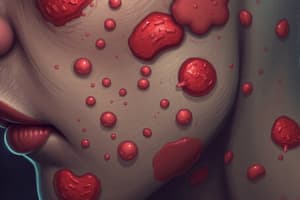
Toxic Epidermal Necrolysis (TEN)
- TEN is a delayed-type hypersensitivity reaction to drugs
- Symptoms include:
- More than 30% of the body surface area is affected, similar to an extensive burn
- A painful, red to purple area that spreads quickly
- The skin may peel without blistering, resulting in raw areas
- Discomfort and fever
- Condition spreads to eyes, mouth/throat, and genitals/urethra/anus
- Later stages can lead to toxic shock syndrome
Steven Johnson Syndrome (SJS)
- SJS is a delayed-type hypersensitivity reaction to drugs.
- Symptoms include:
- Less than 10% of the body surface area is affected
- Presents with nonspecific upper respiratory tract infection in a 1- to 14-day prodrome.
- Sore throat, chills, headache, fever, body aches (malaise), red rash, and cough
- Blisters and sores on the skin and mucous membranes of the mouth, throat, eyes, genitals, and anus
- Peeling skin; Drooling because closing the mouth is painful
- Eyes sealed shut due to blisters and swelling.
- Painful urination due to blistered mucous membranes
- SJS Prognosis: Factors associated with a poor prognosis
- old age (> 70 years)
- intestinal involvement
- pulmonary involvement
SJS/TEN Overlap
- SJS/TEN Overlap is a delayed-type hypersensitivity reaction to drugs
- Used to describe cases in which 10%-30% of the body surface area is detached.
- Long-term complications include ocular issues (including blindness), cutaneous changes (pigmentary changes and scarring), and renal involvement.
- Mucosal involvement with blisters and erosions can lead to strictures and scarring.
Overview of SJS
- SJS involves cell-mediated hypersensitivity and clinical presentation.
- Precipitating factors include drugs (antibiotics, anticonvulsants) and infective agents (Mycoplasma).
- Multiorgan/system involvement: eye, kidney, liver
Etiology
- Drugs precipitate over 50% of SJS cases and up to 95% of TEN cases.
- Common drug causes include:
- Sulfa drugs (e.g., cotrimoxazole, sulfasalazine)
- Other antibiotics (e.g., aminopenicillins [ampicillin, amoxicillin], fluoroquinolones, cephalosporins)
- Antiepileptics (e.g., phenytoin, carbamazepine, phenobarbital, valproate, lamotrigine)
- Miscellaneous individual drugs (less common causes)
- Analgesics (pain killers)
- Cough and cold medication
- NSAIDs
- Psycho-epileptics
- Antigout drugs
- Cases not caused by drugs can be attributed to:
- Infection (mostly with Mycoplasma pneumoniae, Cytomegalovirus)
- HIV/ Systemic Lupus Erythematosus
- Vaccination
- Graft-vs-host disease
- Rarely, a cause cannot be identified.
Symptoms
- SJS typically begins with a nonspecific upper respiratory tract infection.
- This is usually part of a 1- to 14-day prodrome, with fever, sore throat, chills, headache, and malaise.
- Vomiting and diarrhea may occur.
- The typical lesion has the appearance of a target and is considered pathognomonic.
- Typical prodromal symptoms include:
- Cough productive of a thick purulent sputum
- Headache; Malaise
- Arthralgia
Pathophysiology of SJS
- The exact mechanism is unknown
- Altered drug metabolism triggers a T-cell-mediated cytotoxic reaction to drug antigens in keratinocytes.
- CD8+ T cells are important mediators of blister formation.
- Recent findings suggest that granulysin released from cytotoxic T cells and natural killer cells may play a role in keratinocyte death.
- Granulysin concentration in blister fluid correlates with disease severity.
- Interactions between Fas and its ligand lead to cell death and blister formation; a genetic predisposition has been suggested.
Diagnosis
- Physical exam (clinical evaluation), doctors can often identify Stevens-Johnson syndrome, toxic epidermal necrolysis, and SJS/TEN overlap based on medical history and physical exam.
- Steps to conform the diagnosis include:
- Skin biopsy with showing necrotic epithelium
- Skin or oral culture may be taken to confirm/rule out infection; Imaging depending on symptoms to check for pneumonia
- Blood tests confirm other possible causes
Treatments
- Early recognition: treated in an inpatient dermatologic or ICU setting; treatment in a burn unit may be needed for severe disease.
- Ophthalmology consultation and specialized eye care are mandatory for patients with ocular involvement.
- Potentially causative drugs should be stopped immediately.
- Supportive care:
- Patients are isolated to minimize exposure to infection and are given fluids, electrolytes, blood products, and nutritional supplements as needed.
- Skin care includes prompt treatment of secondary bacterial infections and daily wound care as for severe burns.
- Prophylactic systemic antibiotics are controversial and often avoided.
- Controversial treatments:
- Cyclosporine inhibits CD8 cells and slows progression, possibly decreasing mortality.
- Plasma exchange or intravenous immunoglobulin treatment. -High-dose corticosteroid therapy may induce SJS/TEN in certain patients.
Studying That Suits You
Use AI to generate personalized quizzes and flashcards to suit your learning preferences.