Podcast
Questions and Answers
What is the approximate length of the spinal cord?
What is the approximate length of the spinal cord?
- 12 inches
- 36 inches
- 24 inches
- 18 inches (correct)
Where does the spinal cord end?
Where does the spinal cord end?
- Between vertebrae L2 and L3
- Between vertebrae L1 and L2 (correct)
- Between vertebrae L3 and L4
- Between vertebrae T12 and L1
Which groove separates the spinal cord into left and right sides?
Which groove separates the spinal cord into left and right sides?
- Lateral sulcus
- Posterior median sulcus (correct)
- Anterior median fissure
- Central sulcus
Which of the following is a characteristic of spinal reflexes?
Which of the following is a characteristic of spinal reflexes?
Which of the following is NOT true about the spinal cord?
Which of the following is NOT true about the spinal cord?
What is the deeper groove on the anterior side of the spinal cord called?
What is the deeper groove on the anterior side of the spinal cord called?
Which of the following is a characteristic of the spinal cord?
Which of the following is a characteristic of the spinal cord?
What is primarily responsible for the cervical enlargement of the spinal cord?
What is primarily responsible for the cervical enlargement of the spinal cord?
Which layer of the meninges is the outermost layer surrounding the spinal cord?
Which layer of the meninges is the outermost layer surrounding the spinal cord?
What is the primary function of the anterior gray horns in the spinal cord?
What is the primary function of the anterior gray horns in the spinal cord?
What is contained within the epidural space?
What is contained within the epidural space?
What does the subarachnoid space contain?
What does the subarachnoid space contain?
Where are the lateral gray horns found in the spinal cord?
Where are the lateral gray horns found in the spinal cord?
Which meningeal layer is described as 'simple squamous epithelia'?
Which meningeal layer is described as 'simple squamous epithelia'?
Which structure contains myelinated and unmyelinated axons?
Which structure contains myelinated and unmyelinated axons?
What type of fibers make up the pia mater?
What type of fibers make up the pia mater?
The lumbar enlargement of the spinal cord is associated with which part of the body?
The lumbar enlargement of the spinal cord is associated with which part of the body?
What is the function of cerebrospinal fluid (CSF)?
What is the function of cerebrospinal fluid (CSF)?
Which of the following correctly describes the role of gray commissures?
Which of the following correctly describes the role of gray commissures?
Which of the following is NOT a characteristic of the dura mater?
Which of the following is NOT a characteristic of the dura mater?
What is the consequence of peripheral neuropathy?
What is the consequence of peripheral neuropathy?
Which nuclei are responsible for connecting to peripheral receptors?
Which nuclei are responsible for connecting to peripheral receptors?
In which white matter region do axons cross the spinal cord?
In which white matter region do axons cross the spinal cord?
Flashcards
Cervical Enlargement
Cervical Enlargement
Part of the spinal cord with nerves for shoulders and upper limbs.
Lumbar Enlargement
Lumbar Enlargement
Part of the spinal cord with nerves for pelvis and lower limbs.
Dura Mater
Dura Mater
Outer protective layer of the spinal cord, tough and fibrous.
Arachnoid Mater
Arachnoid Mater
Signup and view all the flashcards
Pia Mater
Pia Mater
Signup and view all the flashcards
Epidural Space
Epidural Space
Signup and view all the flashcards
Cerebrospinal Fluid (CSF)
Cerebrospinal Fluid (CSF)
Signup and view all the flashcards
Subarachnoid Space
Subarachnoid Space
Signup and view all the flashcards
Spinal Reflexes
Spinal Reflexes
Signup and view all the flashcards
Function of Spinal Cord
Function of Spinal Cord
Signup and view all the flashcards
Location of Spinal Cord End
Location of Spinal Cord End
Signup and view all the flashcards
Dimensions of Spinal Cord
Dimensions of Spinal Cord
Signup and view all the flashcards
Bilateral Symmetry
Bilateral Symmetry
Signup and view all the flashcards
Posterior Median Sulcus
Posterior Median Sulcus
Signup and view all the flashcards
Anterior Median Fissure
Anterior Median Fissure
Signup and view all the flashcards
Motor Output via Spinal Nerves
Motor Output via Spinal Nerves
Signup and view all the flashcards
White Matter
White Matter
Signup and view all the flashcards
Gray Matter
Gray Matter
Signup and view all the flashcards
Gray Horns
Gray Horns
Signup and view all the flashcards
Sensory Nuclei
Sensory Nuclei
Signup and view all the flashcards
Motor Nuclei
Motor Nuclei
Signup and view all the flashcards
Peripheral Neuropathy
Peripheral Neuropathy
Signup and view all the flashcards
Gray Commissures
Gray Commissures
Signup and view all the flashcards
Study Notes
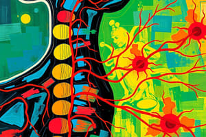
Spinal Cord, Spinal Nerves, and Spinal Reflexes
- Spinal reflexes are rapid, automatic nerve responses triggered by specific stimuli.
- They are controlled by the spinal cord, not the brain.
- The spinal cord is approximately 18 inches (45 cm) long and 1/2 inch (14 mm) wide.
- It extends between vertebrae L1 and L2.
- The spinal cord has bilateral symmetry.
- Grooves divide it into left and right sides.
- The posterior median sulcus is a groove on the posterior side.
- The anterior median fissure is a deeper groove on the anterior side.
Spinal Cord Enlargements
- Enlargements are caused by the amount of gray matter in a segment, and the involvement of the sensory and motor nerves of the limbs.
- The cervical enlargement contains nerves for the shoulders and upper limbs.
- The lumbar enlargement contains nerves for the pelvis and lower limbs.
Three Meningial Layers
- The dura mater is the outermost layer of the spinal cord.
- The arachnoid mater is the middle layer.
- The pia mater is the innermost layer.
Dura Mater
- The dura mater is tough and fibrous.
- Cranially, it fuses with the periosteum of the occipital bone.
- It's continuous with the cranial dura mater.
- Caudally, it tapers to a dense cord of collagen fibers that attach to the coccygeal ligament.
Epidural Space
- Located between the spinal dura mater and the walls of the vertebral canal.
- It contains loose connective tissue and adipose tissue.
- It's a common site for anesthetic injection.
Arachnoid Mater
- It's the middle meningeal layer.
- The arachnoid membrane is a simple squamous epithelium.
- It covers the arachnoid mater.
- The subdural space is between the arachnoid and dura mater.
- The subarachnoid space contains a network of collagen/elastin fibers (arachnoid trabeculae).
- The space is filled with cerebrospinal fluid (CSF).
Cerebrospinal Fluid (CSF)
- CSF carries dissolved gases, nutrients, and waste products.
- Lumbar puncture (spinal tap) withdraws CSF.
Pia Mater
- The pia mater is the innermost meningeal layer.
- It's a mesh of collagen and elastic fibers that adheres to the underlying neural tissue.
Sectional Anatomy of the Spinal Cord
- White matter is superficial and contains myelinated and unmyelinated axons.
- Gray matter surrounds the central canal of the spinal cord and contains neuron cell bodies, neuroglia, and unmyelinated axons.
- It has projections called gray horns.
Organization of Gray Matter
- Posterior gray horns contain somatic and visceral sensory nuclei.
- Anterior gray horns contain somatic motor nuclei.
- Lateral gray horns are located in the thoracic and lumbar segments and contain visceral motor nuclei.
- Gray commissures connect axons from one side of the spinal cord to the other side.
Organization of White Matter
- Posterior white columns are located between the posterior gray horns and the posterior median sulcus.
- Anterior white columns are between the anterior gray horns and the anterior median fissure.
- Anterior white commissure contains axons that cross to the other side of the spinal cord.
- Lateral white columns are on each side of the spinal cord.
Spinal Nerves and Plexuses
- Peripheral neuropathy is a regional loss of sensory or motor function, due to trauma or compression.
Paraplegia vs. Quadriplegia
- Paraplegia affects both legs and is caused by injury to the thoracic, lumbar, or sacral regions of the spinal cord.
- Quadriplegia affects both arms and legs, and is caused by injury to the cervical region of the spinal cord.
Neuronal Pools
- Neuronal pools are functional groups of interconnected neurons (interneurons).
- They have limited input sources and output destinations.
- They can stimulate or depress parts of the brain or spinal cord.
Five Patterns of Neural Circuits
- Divergence: Spreading stimulation to multiple neurons or pools.
- Convergence: Providing input to a single neuron from multiple sources.
- Serial processing: Neurons or pools working sequentially.
- Parallel processing: Neurons or pools processing the same information simultaneously.
Studying That Suits You
Use AI to generate personalized quizzes and flashcards to suit your learning preferences.