Podcast
Questions and Answers
Which spinal column region is characterized by having the highest degree of flexibility and is essential for head rotation?
Which spinal column region is characterized by having the highest degree of flexibility and is essential for head rotation?
- Thoracic
- Lumbar
- Sacral
- Cervical (correct)
What is the key functional significance of the transverse foramen found in cervical vertebrae?
What is the key functional significance of the transverse foramen found in cervical vertebrae?
- Allowing passage of the vertebral arteries (correct)
- Increasing flexibility of the spine
- Housing the spinal cord
- Providing attachment points for muscles
Which of the following is the correct number of vertebrae found in the thoracic region of the spine, and what is its primary functional characteristic?
Which of the following is the correct number of vertebrae found in the thoracic region of the spine, and what is its primary functional characteristic?
- 5; Supports flexibility and heavy loads
- 7; Allows for the greatest range of motion
- 5; Facilitates hip and pelvic attachments
- 12; Provides stability and rib articulation (correct)
What is the primary role of the nucleus pulposus within an intervertebral disc, and how does the annulus fibrosus contribute to this function?
What is the primary role of the nucleus pulposus within an intervertebral disc, and how does the annulus fibrosus contribute to this function?
Why is the lumbar region of the spine particularly susceptible to disc herniation, and which structural feature contributes most to this susceptibility?
Why is the lumbar region of the spine particularly susceptible to disc herniation, and which structural feature contributes most to this susceptibility?
Which of the following best describes the anatomical feature and functional significance of the atlas (C1) and axis (C2) vertebrae?
Which of the following best describes the anatomical feature and functional significance of the atlas (C1) and axis (C2) vertebrae?
In the context of spinal stability, how does the three-column model classify instability, and what is the significance of the middle column?
In the context of spinal stability, how does the three-column model classify instability, and what is the significance of the middle column?
A patient has a spinal injury that disrupts the posterior elements, leaving the anterior and middle columns intact. According to the three-column model, which type of instability is most likely present?
A patient has a spinal injury that disrupts the posterior elements, leaving the anterior and middle columns intact. According to the three-column model, which type of instability is most likely present?
What is the key difference between the two-column and three-column models for assessing spinal instability, and why did the three-column model emerge?
What is the key difference between the two-column and three-column models for assessing spinal instability, and why did the three-column model emerge?
Which ligament is responsible for connecting the cranium to the cervical spine and supporting the head and neck while also allowing for flexion, extension, and rotation?
Which ligament is responsible for connecting the cranium to the cervical spine and supporting the head and neck while also allowing for flexion, extension, and rotation?
After a neck trauma, why are the vertebral arteries, which run through the transverse foramina, particularly susceptible to injury, and what is a potential consequence of such an injury?
After a neck trauma, why are the vertebral arteries, which run through the transverse foramina, particularly susceptible to injury, and what is a potential consequence of such an injury?
What anatomical feature makes the thoracic spine less flexible compared to the cervical and lumbar regions, and how does this relate to its function?
What anatomical feature makes the thoracic spine less flexible compared to the cervical and lumbar regions, and how does this relate to its function?
If a surgeon performs a laminectomy, what specific anatomical structures are removed, and what is the primary goal of this procedure in the lumbar spine?
If a surgeon performs a laminectomy, what specific anatomical structures are removed, and what is the primary goal of this procedure in the lumbar spine?
What are the main characteristics of the sacral spinal column, and how does its structure contribute to its primary function?
What are the main characteristics of the sacral spinal column, and how does its structure contribute to its primary function?
Why is knowledge of the location of the conus medullaris important during spinal procedures, and at what vertebral level does it typically terminate?
Why is knowledge of the location of the conus medullaris important during spinal procedures, and at what vertebral level does it typically terminate?
Flashcards
Cervical Column
Cervical Column
Vertebral arteries, lordotic curve, and smallest vertebrae are found in this spinal column section.
Lumbar Column
Lumbar Column
Flexibility, heavy loads, and the largest vertebrae are characteristics of this spinal section.
Thoracic Column
Thoracic Column
Greater rigidity, less flexibility, and rib attachments characterize this spinal section.
Sacral Column
Sacral Column
Signup and view all the flashcards
External Craniocervical Ligaments
External Craniocervical Ligaments
Signup and view all the flashcards
Cervical Vertebrae Movement
Cervical Vertebrae Movement
Signup and view all the flashcards
Lumbar Vertebrae Features
Lumbar Vertebrae Features
Signup and view all the flashcards
Sacral Spinal Column
Sacral Spinal Column
Signup and view all the flashcards
Annulus Fibrosus
Annulus Fibrosus
Signup and view all the flashcards
Nucleus Pulposus
Nucleus Pulposus
Signup and view all the flashcards
Spinal Stability
Spinal Stability
Signup and view all the flashcards
2-Column Model
2-Column Model
Signup and view all the flashcards
3-Column Model
3-Column Model
Signup and view all the flashcards
Posterior Column
Posterior Column
Signup and view all the flashcards
1º Mechanical
1º Mechanical
Signup and view all the flashcards
Study Notes
- Spinal Column Anatomy
- Presented by Kevin McCarthy, CNIM, Director of Education and Training
- Presented by Kent Rice, REPT, CNIM, DABNM, Sr. Manager Education and Training
Learning Objectives
- List the major components of a vertebra
- List the unique features of the cervical vertebra and spine
- Describe the curvatures of the spine by spinal section
- Describe the number of vertebra by spinal section
- Describe the component of a vertebral disc
- Know the 3 column model of stability
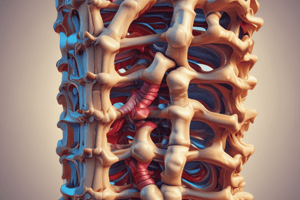
Spinal Column
-
Cervical Column: 7 vertebrae
-
Vertebral Arteries
-
Lordotic Curve
-
Smallest Vertebrae
-
Most Flexible for Rotation
-
Atlas (C1) and Axis (C2)
-
Thoracic Column: 12 vertebrae
-
Greater Rigidity and Less Flexible
-
Kyphotic Curve
-
Rib Attachments form a box
-
Lumbar Column: 5 vertebrae
-
Flexibility and Heavy Loads
-
Largest Vertebrae
-
Lordotic Curve
-
Sacral Column: 5 vertebrae
-
Hip and Pelvic Attachments
-
Fused Segments
Morphology and Section Size
- Sections vary by size to handle increasing loading forces
- Sections vary by shape specific to function (e.g., transverse foramen)
- Sections vary in rigidity and flexibility based on vertebral shape and function
Cervical Spinal Column
- 7 vertebra from C1 to C7
- Superiorly it joins the cranium, known as the occipital-cervical or atlanto-occipital junction
- Specialized components of the cervical spine include:
- Vertebral foramen of the transverse process
- Atlas (C1), axis (C2)
- The odontoid process of C2.
External Craniocervical Ligaments
- Connection between the cranium and the cervical spine
- Supports the head and neck; allows for flexion, extension, and rotation
- Vertebral arteries running through the transverse foramina are susceptible to injury from neck trauma, with potential for significant CNS ischemic event
Cervical Vertebrae – Atlas (C1) Axis (C2)
- The 2 most superior vertebrae are unique in their form and function
- Their form is derived from the necessary function of supporting and rotation of the skull
- Cervical vertebra 1 (C1) is the Atlas
- Cervical vertebra 2 (C2) is the Axis
Specific Features of the Atlas & Axis
- Superior and inferior articular facets form the rotating joint between C1 and C2
- Dens (odontoid process): the pivot center for rotation of the skull on the spinal column
Cervical Vertebrae
- The smallest bodies, lamina, and pedicles
- The transverse processes contain the transverse foramen (vertebral arteries)
- The most flexible region in anterior/posterior, lateral and rotational movement
Cervical Instrumentation
- Anterior fixation is achieved with a vertebral body screw and plate
- Posterior cervical screws are usually placed in the lateral mass
Thoracic Spinal Column
- 12 vertebrae from T1-T12
- A natural kyphosis of 20-40° to accommodate vital organs within the rib cage
- Rib attachments at each thoracic vertebra make the thoracic spine more rigid and less flexible than cervical or lumbar regions
Thoracic Vertebrae
- Facets are articulating surfaces for spinal joints (facet joints)
- Ribs attach at the body and transverse process
Lumbar Spinal Column
- 5 vertebra from L1 to L5
- The conus medullaris of the spinal cord ends at the level of L1
- The cauda equina, pelvic, and lower limb nerve roots, are present from L1 through the sacral regions
- Largest vertebra, separated by 5 lumbar discs
- Discs bear the most force and are at greatest risk for herniation
Lumbar Vertebrae
- Specific features of the lumbar vertebra
- The largest vertebral body and spinous process
- Largest pedicles of any vertebra
- Removal of the lamina and spinous process (called a laminectomy), is done to facilitate cauda equina decompression
Lumbar Pedicle Screw Instrumentation
- The pedicle is a secure location for fixation of the spine, especially in the lumbar and thoracic spine
- Medial malpositioning of the pedicle screw endangers nerve root or spinal cord
Important Ligaments of the Spinal Column
- The ligaments provide support and protection for the spine
- Anterior Longitudinal Ligament (ALL)
- Posterior Longitudinal Ligament (PLL)
- Supraspinous ligament
- Interspinous ligament
- Ligamentum Flavum
Sacral Spinal Column
- 5 fused vertebral levels S1 to S5
- No intervertebral disc
- Lumbosacral junction at L5-S1
- Coccyx bone at tip
Vertebral Discs
- Intervertebral discs are between each vertebra and cushion force load axially onto the spinal column
- The nucleus pulposus absorbs most force and is held in by the firm annulus fibrosus
- Thinning or tears in the annulus fibrosus can lead to bulging of the disc and compression of neurological structures
- The intervertebral disc is an avascular structure deriving its nutrients from the vertebral end plate
Nucleus and Annulus
- Nucleus Pulposus (NP) - granular, semi-gelatinous
- Annulus Fibrous (AF) - striated, multi-layered fibrous material
- Two cartilaginous vertebral end-plates (VEP)
Vertebral Discs
- The nucleus absorbs and distributes force evenly against the annulus.
- The annulus supports the loading force on the disc
- Abnormal forces overstress the annulus and can course herniation or rupture
- Common spinal surgery is for a HNP, herniated nucleus pulposus
Spinal Instability
- The loss of ability of the spine under physiological loads to maintain its pattern of displacement so that there is no initial or additional neurological deficit, no major deformity, and no incapacitating pain
- A stable spine should be one that can withstand axial forces anteriorly through the vertebral bodies, tension forces posteriorly, and rotational stresses
- Total of 5 or more = instability
2-Column Models
- Considers the anterior and posterior segments, grouping fractures as stable and unstable
- The posterior elements were considered the most important in spinal stability
- Failed to recognize "clinical instability" i.e., burst fractures, where the posterior elements (all structures posterior to PLL) are intact
- Ability to protect the neural elements is compromised
3-Column Models
- Classifying instability into three degrees, according to mechanical and/or neurological involvement
- First degree or Mechanical: disruption of posterior elements, neuro intact, potential for chronic instability "Spine can buckle" i.e. Seat-belt and distraction injuries
- Second degree or Neurological: still neurologically intact, but spinal canal has been violated, posing risk to its contents i.e. burst fractures
- Third degree or Mechanical and Neuro compromise: burst and fracture dislocations with neuro involvement
3-Column Model
- It is the current model used to determine spinal instability and spinal instrumentation construct The 3 columns are:
- Posterior: pedicles, facts, lamina, etc
- Middle: posterior half of vertebral body
- Anterior: anterior half of vertebral body
Linked to 3 degrees of instability
- 1º Mechanical: structural damage only to the posterior column
- 2° Neurological: spinal canal violated by posterior and/or middle column high risk of neurological injury
- 3° Mechanical & Neurological: ighly unstable involving anterior and middle column and possibly posterior column and neurological changes
Studying That Suits You
Use AI to generate personalized quizzes and flashcards to suit your learning preferences.